Matrix metalloproteinases (MMPs): Chemical–biological functions ...
Matrix metalloproteinases (MMPs): Chemical–biological functions ...
Matrix metalloproteinases (MMPs): Chemical–biological functions ...

Create successful ePaper yourself
Turn your PDF publications into a flip-book with our unique Google optimized e-Paper software.
2226 R. P. Verma, C. Hansch / Bioorg. Med. Chem. 15 (2007) 2223–2268<br />
86% between MMP-3 and MMP-10. 19 A comparison of<br />
such sequence similarity with the available 3D structure<br />
shows that the overall topology of the enzyme active-site<br />
is highly conserved between the different <strong>MMPs</strong>, with<br />
the two significant emerging features that are the depth<br />
of S1 0 pocket and the length as well as composition of<br />
the loop constituting the outside wall of the S1 0 pocket.<br />
20 A recent report describes the structure of MMP-2<br />
in which the catalytic domain possess a similar overall<br />
topology characterized by a twisted five-stranded<br />
b-sheet, containing four parallel strands and one antiparallel<br />
strand, and three long a-helices. 21<br />
It has also been observed that the conserved active-site<br />
sequence motif HEXXHXXGXXH coordinates the catalytic<br />
zinc(II) ion and contains the glutamic acid residue<br />
which facilitates catalysis. The substrate binding groove,<br />
which is relatively open at S3–S1 and S3 0 , narrow at S1 0<br />
and S2 0 with the S1 0 site being a well-defined pocket,<br />
penetrates the surface of the enzyme. The presence of<br />
second ‘structural’ zinc(II) ion and two or three calcium(II)<br />
ions has been confirmed. Thus, it has been difficult<br />
to obtain recombinant full-length enzymes suitable<br />
for structural determination. MMP-7, one of the smallest<br />
members of the MMP family, does not possess a<br />
C-terminal domain, whose inhibitor complexes possess<br />
broadly similar structures to that of the catalytic<br />
domains of MMP-1, MMP-3, and MMP-8. 7 This<br />
C-terminal domain is found to be present in almost all<br />
of the <strong>MMPs</strong> except MMP-7 and MMP-23, and seems<br />
to regulate the enzyme activity. 22<br />
A significant interaction between <strong>MMPs</strong> and their substrates/or<br />
inhibitors has been demonstrated between<br />
S1 0 subsite and the P1 0 residue. There is a variation between<br />
<strong>MMPs</strong> in the amino acid residues, which form the<br />
S1 0 pocket. 7 It has been shown that the modifications of<br />
the P1 0 portion of the molecule play a key role affecting<br />
both the potency and selectivity within the MMP family.<br />
Longer-chain aliphatic substituents in this region of the<br />
molecule tend to increase potency for MMP-3 and decrease<br />
potency for MMP-1, while aromatic substituents<br />
seem to generate broad-spectrum inhibition. 23<br />
From the X-ray crystallography and homology modeling<br />
<strong>MMPs</strong> may be classified into two broad structural<br />
classes depending on the depth of the S1 0 pocket. This<br />
selective pocket is relatively deep for most MMP<br />
His<br />
Glu<br />
O C O<br />
HOH<br />
H H<br />
C C N H C<br />
P '<br />
P1 O 1<br />
Zn 2+<br />
His His<br />
O<br />
Ala<br />
His<br />
Glu<br />
O C O<br />
HO H<br />
H<br />
C C N H H<br />
C<br />
'<br />
P1 P<br />
O 1<br />
Zn 2+<br />
His His<br />
O<br />
Ala<br />
His<br />
enzymes (e.g., MMP-2, MMP-3, MMP-8, MMP-9,<br />
MMP-13, etc.), but for certain MMP enzymes (e.g.,<br />
MMP-1, MMP-7, and MMP-11) it is partially or completely<br />
occluded due to an increase in the size of the side<br />
chain of the amino acid at position 193 (MMP-8 numbering)<br />
from leucine to arginine (MMP-1), tyrosine<br />
(MMP-7), glutamine (MMP-11), or one of the amino<br />
acid residues that form the pocket. It has also been<br />
shown that the mutation of S1 0 subunit tyrosine of<br />
MMP-7 to leucine changes the substrate specificity to<br />
be more like that of the deep pocket enzyme<br />
MMP-3. 24 Homology models for MMP-2 and MMP-9<br />
based on the structure of MMP-3 suggest that there<br />
may be differences in the shape of the bottom of the<br />
S1 0 subunit for the deep pocket enzymes. 25 In the case<br />
of MMP-2, the S1 0 pocket may be a channel with no<br />
bottom, whereas that for MMP-9 is said to be a pocket-like<br />
subsite. The subunit (S2 0 ) is a solvent-exposed<br />
cleft, with hydrophobic P2 0 residues in both substrates<br />
and inhibitors. The S3 0 subunit is a really ill-defined solvent-exposed<br />
region. While there are some variations in<br />
residues for this subsite for the various <strong>MMPs</strong>, the<br />
introduction of different P3 0 substituents in general<br />
tends to have only a modest effect on inhibitory<br />
selectivity. 7<br />
An NMR structural study of MMP-1 catalytic domain<br />
suggests that substantial structural changes occur in<br />
the active-site cleft on the binding of an inhibitor. 26<br />
Conformational changes have also been observed in<br />
the active site between X-ray structures of MMP-3 catalytic<br />
domain and different inhibitors bound. 27 Thus,<br />
structural information must be considered in the design<br />
of MMP inhibitors.<br />
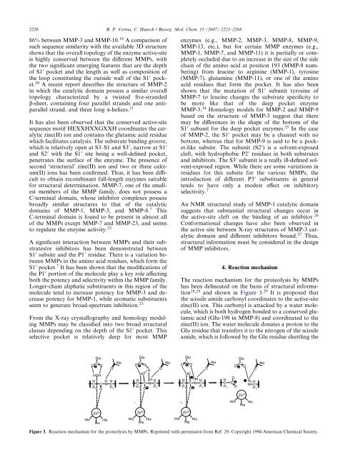
4. Reaction mechanism<br />
The reaction mechanism for the proteolysis by <strong>MMPs</strong><br />
has been delineated on the basis of structural information<br />
28,29 and shown in Figure 3. 29 It is proposed that<br />
the scissile amide carbonyl coordinates to the active-site<br />
zinc(II) ion. This carbonyl is attacked by a water molecule,<br />
which is both hydrogen bonded to a conserved glutamic<br />
acid (Glu-198 in MMP-8) and coordinated to the<br />
zinc(II) ion. The water molecule donates a proton to the<br />
Glu residue that transfers it to the nitrogen of the scissile<br />
amide, which is followed by the Glu residue shuttling the<br />
Glu<br />
O C O<br />
HO<br />
H<br />
C C N H HH<br />
C<br />
P '<br />
P1 O 1<br />
Figure 3. Reaction mechanism for the proteolysis by <strong>MMPs</strong>. Reprinted with permission from Ref. 29. Copyright 1994 American Chemical Society.<br />
Zn 2+<br />
His His<br />
O<br />
Ala<br />
H<br />
C<br />
P 1<br />
O<br />
C<br />
O<br />
His<br />
Glu<br />
O C O<br />
Zn 2+<br />
N H H3 C<br />
'<br />
P1<br />
His His<br />
O<br />
Ala