Matrix metalloproteinases (MMPs): Chemical–biological functions ...
Matrix metalloproteinases (MMPs): Chemical–biological functions ...
Matrix metalloproteinases (MMPs): Chemical–biological functions ...
Create successful ePaper yourself
Turn your PDF publications into a flip-book with our unique Google optimized e-Paper software.
2228 R. P. Verma, C. Hansch / Bioorg. Med. Chem. 15 (2007) 2223–2268<br />
substitutions at P2 0 position and that generally Trp is<br />
preferred for efficient cleavage. The same is true for amino<br />
acid changes at P3 0 . However, there is a contradiction<br />
between substrate and phage studies; the former<br />
suggests a strong preference of Met at P3 0 for selective<br />
cleavage by MMP-7, whereas the latter indicates that<br />
Met at P3 0 results in poor cleavage by MMP-7. 7,35,37<br />
The S3–S1 subsites from a shallow region bordered on<br />
one side by the b-strand IV that features a hydrophobic<br />
proline binding cleft at S3. Proline is a preferred P3<br />
group in MMP substrates and the structure of the lefthand<br />
side inhibitor Pro-Leu-Gly-NHOH ligated to the<br />
catalytic domain of MMP-8 illustrates the tight complementarity<br />
in proline binding at S3. This inhibitor binds<br />
anti-parallel to the b-strand IV by two backbone hydrogen<br />
bond interactions (via NH of P3–P2 amide and carbonyl<br />
of P2–P1 amide) with the P2 leucine residing in a<br />
shallow cleft and the carbonyl of the P3–P2 amide and<br />
NH of the P2–P1 amide exposed to solvent. Differences<br />
between the various <strong>MMPs</strong> in the S3–S1 region are relatively<br />
subtle. 7,31,38 It is interesting to note that the selective<br />
replacement of catalytic zinc of MMP-3 catalytic<br />
domain with other transition metals that are Co 2+ ,<br />
Mn 2+ ,Cd 2+ , and Ni 2+ results in the retention of protease<br />
activity. However, substitution of the catalytic metal<br />
influences the substrate specificity of enzyme, since the<br />
active-site geometry is altered and hence affects substrate<br />
binding. 39<br />
At present, the selectivity of the most known MMP<br />
inhibitors relies on the two dominant molecular features:<br />
(a) chelating moiety that interacts with the catalytic<br />
zinc ion and (b) hydrophobic extensions protruding<br />
from the catalytic site into the large and hydrophobic<br />
S1 0 pocket (P1 0 group). Since the structural differences<br />
between MMP families occur mainly in the S1 0 subsite,<br />
modifications of the P1 0 group have been utilized to<br />
introduce inhibitor selectivity. 2<br />
7. <strong>MMPs</strong> and apoptosis<br />
Apoptosis, also known as programmed cell death<br />
(PCD), is an extremely well-ordered process by which<br />
unwanted, defective, or damaged cells are rapidly and<br />
selectively eliminated from the body. <strong>MMPs</strong> play an<br />
intriguing role in PCD, showing both apoptotic and<br />
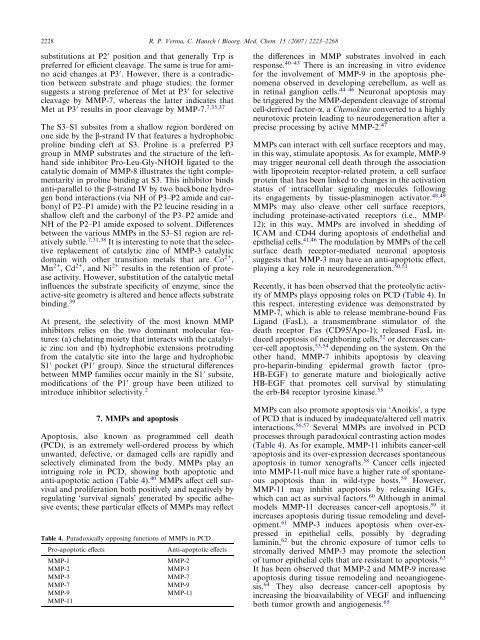
anti-apoptotic action (Table 4). 40 <strong>MMPs</strong> affect cell survival<br />
and proliferation both positively and negatively by<br />
regulating ‘survival signals’ generated by specific adhesive<br />
events; these particular effects of <strong>MMPs</strong> may reflect<br />
Table 4. Paradoxically opposing <strong>functions</strong> of <strong>MMPs</strong> in PCD<br />
Pro-apoptotic effects Anti-apoptotic effects<br />
MMP-1 MMP-2<br />
MMP-2 MMP-3<br />
MMP-3 MMP-7<br />
MMP-7 MMP-9<br />
MMP-9<br />
MMP-11<br />
MMP-11<br />
the differences in MMP substrates involved in each<br />
response. 40–43 There is an increasing in vitro evidence<br />
for the involvement of MMP-9 in the apoptosis phenomena<br />
observed in developing cerebellum, as well as<br />
in retinal ganglion cells. 44–46 Neuronal apoptosis may<br />
be triggered by the MMP-dependent cleavage of stromal<br />
cell-derived factor-a, aChemokine converted to a highly<br />
neurotoxic protein leading to neurodegeneration after a<br />
precise processing by active MMP-2. 47<br />
<strong>MMPs</strong> can interact with cell surface receptors and may,<br />
in this way, stimulate apoptosis. As for example, MMP-9<br />
may trigger neuronal cell death through the association<br />
with lipoprotein receptor-related protein, a cell surface<br />
protein that has been linked to changes in the activation<br />
status of intracellular signaling molecules following<br />
its engagements by tissue-plasminogen activator. 48,49<br />
<strong>MMPs</strong> may also cleave other cell surface receptors,<br />
including proteinase-activated receptors (i.e., MMP-<br />
12); in this way, <strong>MMPs</strong> are involved in shedding of<br />
ICAM and CD44 during apoptosis of endothelial and<br />
epithelial cells. 41,46 The modulation by <strong>MMPs</strong> of the cell<br />
surface death receptor-mediated neuronal apoptosis<br />
suggests that MMP-3 may have an anti-apoptotic effect,<br />
playing a key role in neurodegeneration. 50,51<br />
Recently, it has been observed that the proteolytic activity<br />
of <strong>MMPs</strong> plays opposing roles on PCD (Table 4). In<br />
this respect, interesting evidence was demonstrated by<br />
MMP-7, which is able to release membrane-bound Fas<br />
Ligand (FasL), a transmembrane stimulator of the<br />
death receptor Fas (CD95/Apo-1); released FasL induced<br />
apoptosis of neighboring cells, 52 or decreases cancer-cell<br />
apoptosis, 53,54 depending on the system. On the<br />
other hand, MMP-7 inhibits apoptosis by cleaving<br />
pro-heparin-binding epidermal growth factor (pro-<br />
HB-EGF) to generate mature and biologically active<br />
HB-EGF that promotes cell survival by stimulating<br />
the erb-B4 receptor tyrosine kinase. 55<br />
<strong>MMPs</strong> can also promote apoptosis via ‘Anoikis’, a type<br />
of PCD that is induced by inadequate/altered cell matrix<br />
interactions. 56,57 Several <strong>MMPs</strong> are involved in PCD<br />
processes through paradoxical contrasting action modes<br />
(Table 4). As for example, MMP-11 inhibits cancer-cell<br />
apoptosis and its over-expression decreases spontaneous<br />
apoptosis in tumor xenografts. 58 Cancer cells injected<br />
into MMP-11-null mice have a higher rate of spontaneous<br />
apoptosis than in wild-type hosts. 59 However,<br />
MMP-11 may inhibit apoptosis by releasing IGFs,<br />
which can act as survival factors. 60 Although in animal<br />
models MMP-11 decreases cancer-cell apoptosis, 59 it<br />
increases apoptosis during tissue remodeling and development.<br />
61 MMP-3 induces apoptosis when over-expressed<br />
in epithelial cells, possibly by degrading<br />
laminin, 62 but the chronic exposure of tumor cells to<br />
stromally derived MMP-3 may promote the selection<br />
of tumor epithelial cells that are resistant to apoptosis. 63<br />
It has been observed that MMP-2 and MMP-9 increase<br />
apoptosis during tissue remodeling and neoangiogenesis.<br />
64 They also decrease cancer-cell apoptosis by<br />
increasing the bioavailability of VEGF and influencing<br />
both tumor growth and angiogenesis. 65


