reSolution_LNT_No1_en - Leica Microsystems
reSolution_LNT_No1_en - Leica Microsystems
reSolution_LNT_No1_en - Leica Microsystems
You also want an ePaper? Increase the reach of your titles
YUMPU automatically turns print PDFs into web optimized ePapers that Google loves.
BIOLOGY<br />
Dry Ultrathin Sectioning Combined With High Pressure<br />
Freezing/Freeze-substitution Improves Ret<strong>en</strong>tion and<br />
Visualization of Calcium and Phosphorus Ions Prior to<br />
Nucleation of Mineral Crystals Within Osteoblastic Cultures<br />
Jeff P. Gorski1 , N.T. Huffman1 , T. Hillman-Marti2 , and Daniel Studer2 1Departm<strong>en</strong>t of Oral Biology and the UMKC C<strong>en</strong>ter of Excell<strong>en</strong>ce<br />
in Mineralized Tissue Research, School of D<strong>en</strong>tistry,<br />
Univ. Missouri-KC, Kansas City, Kansas City, MO and<br />
2Institute of Anatomy, University of Bern, Bern, Switzerland<br />
12 reSOLUTION<br />
We have used cultured UMR106-01 osteoblastic cells to<br />
investigate the process of bone mineralization. UMR106-<br />
01 cells as well as primary calvarial bone cells assemble<br />
spherical extracellular supramolecular protein-lipid complexes,<br />
termed biomineralization foci (BMF), in which the<br />
fi rst crystals of hydroxyapatite mineral are deposited (Midura<br />
et al., 2004; Wang et al., 2004). A major differ<strong>en</strong>ce<br />
betwe<strong>en</strong> these culture models is the speed with which<br />
mineralization occurs, ranging from 12-16 days after plating<br />
for primary osteoblastic cells to 88h for UMR106-01<br />
cells.<br />
If mineralization is blocked by omission of phosphate<br />
source or by addition of serine protease inhibitor AEBSF,<br />
BMF complexes are formed but no mineralization occurs.<br />
Interestingly, ultra structural studies have shown that prior<br />
to mineralization BMF contain numerous membrane limited<br />
vesicles ranging in size from 50 nm to 2 microns in diameter.<br />
However, the fi rst mineral crystals are not detected<br />
until 78 h after plating of UMR106-01 cells and are localized<br />
within spherical sites presumed to be vesicles.<br />
Specifi cally, confocal Raman spectral analyses have shown<br />
that mineralization within BMF is a progressive, multi-step<br />
process occurring simultaneously in all BMF within a culture<br />
fl ask (Wang et al., 2009). Importantly, several protein<br />
spectral changes are detectable within each BMF prior<br />
to the deposition of poorly crystalline hydroxyapatite and<br />
wh<strong>en</strong> mineralization was blocked, these changes did not<br />
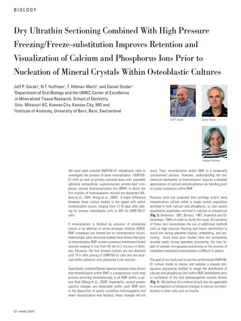
Jeff P. Gorski Daniel Studer<br />
Thérèse Hillmann<br />
occur. Thus, mineralization within BMF is a temporally<br />
synchronized process. However, understanding the biochemical<br />
mechanism of mineralization requires a detailed<br />
appreciation of calcium and phosphorus ion handling prior<br />
to crystal nucleation within BMF.<br />
Previous work has proposed that cartilage and/or bone<br />
mineralization utilizes either a single vesicle population<br />
<strong>en</strong>riched in both calcium and phosphorus, or, two vesicle<br />
populations separately <strong>en</strong>riched in calcium or phosphorus<br />
(Fig. 1) (Anderson, 1967; Bonucci, 1967; Ars<strong>en</strong>ault and Ott<strong>en</strong>smeyer,<br />
1984). In order to clarify this issue, the solubility<br />
of these ions necessitates the use of additional methods<br />
such as high pressure freezing and freeze substitution to<br />
avoid loss during specim<strong>en</strong> fi xation, embedding, and sectioning.<br />
Since most prior studies have not consist<strong>en</strong>tly<br />
avoided water during specim<strong>en</strong> processing, the true impact<br />
of pseudo non-aqueous processing on the process of<br />
osteoblast-mediated mineralization is diffi cult to assess.<br />
The goal of our study was to use the synchronized UMR106-<br />
01 culture model to devise and validate a pseudo nonaqueous<br />
processing method to image the distribution of<br />
calcium and phosphorus ions within BMF immediately prior<br />
to nucleation of the fi rst hydroxyapatite crystals therein<br />
(Fig. 1). We believe this method should also be applicable<br />
to investigations of temporal changes in calcium ion distributions<br />
in other cells such as muscle.


