reSolution_LNT_No1_en - Leica Microsystems
reSolution_LNT_No1_en - Leica Microsystems
reSolution_LNT_No1_en - Leica Microsystems

Create successful ePaper yourself
Turn your PDF publications into a flip-book with our unique Google optimized e-Paper software.
BIOLOGY<br />
Perusing alternatives for automated staining of TEM thin sections<br />
Substitutes for Uranyl Acetate in<br />
TEM Thin Section Post-Staining<br />
Nicole Fellner1,3 , Marl<strong>en</strong>e Brandstetter1,3 , Karin Trimmel1,2 , and Dr. Gu<strong>en</strong>ter P. Resch1,3 1IMP-IMBA-GMI Electron Microscopy Facility, Institute of Molecular Biotechnology, Vi<strong>en</strong>na, Austria<br />
2University of Applied Sci<strong>en</strong>ces, Wi<strong>en</strong>er Neustadt, Austria<br />
3Campus Sci<strong>en</strong>ce Support Facilities GmbH, Vi<strong>en</strong>na, Austria<br />
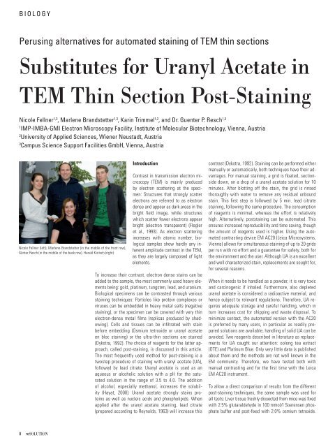
Nicole Fellner (left), Marl<strong>en</strong>e Brandstetter (in the middle of the front row),<br />
Günter Resch (in the middle of the back row), Harald Kotisch (right)<br />
8 reSOLUTION<br />
Introduction<br />
Contrast in transmission electron microscopy<br />
(TEM) is mainly produced<br />
by electron scattering at the specim<strong>en</strong>:<br />
Structures that strongly scatter<br />
electrons are referred to as electron<br />
d<strong>en</strong>se and appear as dark areas in the<br />
bright fi eld image, while structures<br />
which scatter fewer electrons appear<br />
bright (electron transpar<strong>en</strong>t) (Flegler<br />
et al., 1993). As electron scattering<br />
increases with atomic number, biological<br />
samples show hardly any inher<strong>en</strong>t<br />
amplitude contrast in the TEM,<br />
as they are largely composed of light<br />
elem<strong>en</strong>ts.<br />
To increase their contrast, electron d<strong>en</strong>se stains can be<br />
added to the sample, the most commonly used heavy elem<strong>en</strong>ts<br />
being: gold, platinum, tungst<strong>en</strong>, lead, and uranium.<br />
Biological specim<strong>en</strong>s can be contrasted through various<br />
staining techniques: Particles like protein complexes or<br />
viruses can be embedded in heavy metal salts (negative<br />
staining), or the specim<strong>en</strong> can be covered with very thin<br />
electron-d<strong>en</strong>se metal fi lms (replicas produced by shadowing).<br />
Cells and tissues can be infi ltrated with stain<br />
before embedding (Osmium tetroxide or uranyl acetate<br />
<strong>en</strong> bloc staining) or the ultra-thin sections are stained<br />
(Dykstra, 1992). The choice of reag<strong>en</strong>ts for the latter approach,<br />
called post-staining, is discussed in this article.<br />
The most frequ<strong>en</strong>tly used method for post-staining is a<br />
twostep procedure of staining with uranyl acetate (UA),<br />
followed by lead citrate. Uranyl acetate is used as an<br />
aqueous or alcoholic solution with a pH for the saturated<br />
solution in the range of 3.5 to 4.0. The addition<br />
of alcohol, especially methanol, increases the solubility<br />
(Hayat, 2000). Uranyl acetate strongly stains proteins<br />
as well as nucleic acids and phospholipids. Wh<strong>en</strong><br />
applied after the uranyl acetate staining, lead citrate<br />
(prepared according to Reynolds, 1963) will increase this<br />
contrast (Dykstra, 1992). Staining can be performed either<br />
manually or automatically, both techniques have their advantages.<br />
For manual staining, a grid is fl oated, sectionside<br />
down, on a drop of a uranyl acetate solution for 10<br />
minutes. After blotting off the stain, the grid is rinsed<br />
thoroughly with water to remove any residual unbound<br />
stain. This fi rst step is followed by 5 min. lead citrate<br />
staining, following the same procedure. The consumption<br />
of reag<strong>en</strong>ts is minimal, whereas the effort is relatively<br />
high. Alternatively, poststaining can be automated. This<br />
<strong>en</strong>sures increased reproducibility and time saving, though<br />
the amount of reag<strong>en</strong>ts used is higher. Using the automated<br />
contrasting device EM AC20 (<strong>Leica</strong> <strong>Microsystems</strong>,<br />
Vi<strong>en</strong>na) allows for simultaneous staining of up to 20 grids<br />
per run with no effort and a guarantee for safety, both for<br />
the <strong>en</strong>vironm<strong>en</strong>t and the user. Although UA is an excell<strong>en</strong>t<br />
and well characterized stain, replacem<strong>en</strong>ts are sought for,<br />
for several reasons.<br />
Wh<strong>en</strong> it needs to be handled as a powder, it is very toxic<br />
and carcinog<strong>en</strong>ic if inhaled. Furthermore, also depleted<br />
uranyl acetate is considered a radioactive material, and<br />
h<strong>en</strong>ce subject to relevant regulations. Therefore, UA requires<br />
adequate storage and careful handling, which in<br />
turn increases cost for shipping and waste disposal. To<br />
minimize contact, the automated version with the AC20<br />
is preferred by many users, in particular as readily prepared<br />
solutions are available, handling of solid UA can be<br />
avoided. Two reag<strong>en</strong>ts described in literature as replacem<strong>en</strong>ts<br />
for UA caught our att<strong>en</strong>tion: oolong tea extract<br />
(OTE) and Platinum Blue. Only very little data is published<br />
about them and the methods are not well known in the<br />
EM community. Therefore, we have tested both with<br />
manual contrasting and for the fi rst time with the <strong>Leica</strong><br />
EM AC20 instrum<strong>en</strong>t.<br />
To allow a direct comparison of results from the differ<strong>en</strong>t<br />
post-staining techniques, the same sample was used for<br />
all tests: Liver tissue freshly dissected from mice was fi xed<br />
with 2.5% glutaraldehyde in 100 mmol/l Soer<strong>en</strong>s<strong>en</strong> phosphate<br />
buffer and post-fi xed with 2.0% osmium tetroxide.