reSolution_LNT_No1_en - Leica Microsystems
reSolution_LNT_No1_en - Leica Microsystems
reSolution_LNT_No1_en - Leica Microsystems

You also want an ePaper? Increase the reach of your titles
YUMPU automatically turns print PDFs into web optimized ePapers that Google loves.
INDUSTRY<br />
A Word on Cathodoluminesc<strong>en</strong>ce<br />
Cathy Johnson, Nanotechnology Division, Leider Lane, Buffalo Grove, IL<br />
Cathodoluminesc<strong>en</strong>ce microanalysis is an emerging technique that is fast gaining popularity in the world of materials<br />
sci<strong>en</strong>ce. CL is a light emission ph<strong>en</strong>om<strong>en</strong>a resulting from the electron beam excitation of a luminesc<strong>en</strong>t material. As electronic<br />
transitions occur betwe<strong>en</strong> the conduction and val<strong>en</strong>ce bands, CL photons are g<strong>en</strong>erated and detected. Electronic<br />
transitions due to defect levels within the band gap, particularly in the case of semiconductors and devices, can also<br />
infl u<strong>en</strong>ce CL data. Data acquisition results in a mapping of the optical activity for a specim<strong>en</strong>.<br />
Cathy Johnson, <strong>Leica</strong> <strong>Microsystems</strong><br />
CL data can indicate defects such as imperfections or impurities<br />
within the microstructure of a material phase. These<br />
defects can have an effect on the material’s optical, electrical<br />
and mechanical properties. Utilizing the high resolution<br />
capability of a SEM or STEM, a spectrum can be acquired<br />
at each point location (i.e. hyperspectral imaging). As such,<br />
it serves as an important spectroscopy and imaging technique<br />
in the characterization of materials. Image resolution<br />
is dep<strong>en</strong>d<strong>en</strong>t on instrum<strong>en</strong>t confi guration, experim<strong>en</strong>tal parameters<br />
and specim<strong>en</strong> interaction, but can range from < 10<br />
nanometers to the micron level.<br />
The fi rst MAS Cathodoluminesc<strong>en</strong>ce Topical Confer<strong>en</strong>ce<br />
was hosted October 24-28, 2011 by the National Institute<br />
of Standards and Technology (NIST) in Gaithersburg, MD.<br />
This confer<strong>en</strong>ce was sponsored by the Microbeam Analysis<br />
Society (MAS), and was co-sponsored by the Australian<br />
Microbeam Analysis Society (AMAS). The four day program<br />
included a pre-confer<strong>en</strong>ce tutorial targeted for the CL<br />
novice on October 24 th . The remaining three days included<br />
a combination of technical pres<strong>en</strong>tations, hands-on laboratory<br />
demonstrations and a contributed poster session. Pres<strong>en</strong>tation<br />
topics included: CL theory, data quantifi cation,<br />
advances in instrum<strong>en</strong>tation, analysis and databases. Applications<br />
in geological, semiconductor and nanomaterial<br />
disciplines including sample preparation and Correlative<br />
CL in conjunction with complem<strong>en</strong>tary techniques such as<br />
EBIC and EBSD were also addressed.<br />
Instrum<strong>en</strong>ts rel related to this sample preparation:<br />
<strong>Leica</strong> EM TXP<br />
Target Surfacing System<br />
<strong>Leica</strong> EM TIC 3X<br />
Ion Beam Slope Cutter<br />
<strong>Leica</strong> EM RES101<br />
Ion Milling System<br />
Contact<br />
Cathy Johnson<br />
<strong>Leica</strong> <strong>Microsystems</strong><br />
Nanotechnology Division<br />
1700 Leider Lane<br />
Buffalo Grove, IL 60089<br />
Cathy.Johnson@leica-microsystems.com<br />
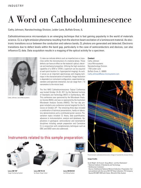
Panchromatic cathodoluminesc<strong>en</strong>ce image of cassiterite mineral (SnO 2 )<br />
Image Credits<br />
Scott Wight 1 , Ed Vic<strong>en</strong>zi 2 , Doug Meier 1 , and Kurt B<strong>en</strong>kstein 1<br />
1 National Institute of Standards and Technology<br />
2 Smithsonian Institution<br />
Source: Smithsonial National Mineral Collection, Preparation: Cut<br />
and polished with the <strong>Leica</strong> EM TXP Data Collection: FEI Company<br />
QuantaTM 200F SEM with Gatan, MonoCL4 Elite System