AVOIDING FURNACE SLIP IN THE ERA OF SHALLOW ... - MEMC
AVOIDING FURNACE SLIP IN THE ERA OF SHALLOW ... - MEMC
AVOIDING FURNACE SLIP IN THE ERA OF SHALLOW ... - MEMC

Create successful ePaper yourself
Turn your PDF publications into a flip-book with our unique Google optimized e-Paper software.
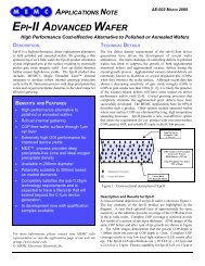
Fig. 3: The LEFT photo shows slip steps at the edge of a P{100} wafer as revealed by an<br />
interference contrast microscope. The RIGHT photo shows dislocation etch pits at a P{100}<br />
wafer surface after HF strip-back and 3 min Schimmel etch (6).<br />
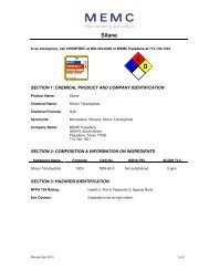
Fig. 4: The photo shows rows of diamond-shaped dislocation etch pits at the cleaved {110}<br />
surface in the center of a P/P+{100} epi wafer. The cleaved surface was etched for 3 min in<br />
Leo’s etch (7). Many large oxygen precipitation defects are also observed in the P+.<br />
Other slip characterization methods include Makyoh (magic mirror) topography (8),<br />
minority carrier recombination lifetime mapping (9), and scanning Lang transmission xray<br />
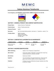
topography (10-12). Figure 5 shows an x-ray topography image.<br />
Fig. 5: X-ray topography image of one edge of an in-process P{100} wafer after HF stripback<br />
and 3 min Schimmel etch. The black spots are damage sites on the back side of the<br />
wafer and the many dark lines are dislocations in the wafer bulk.<br />
Page 777