Il trattamento delle fratture laterali del collo femorale con il ... - Giot.it
Il trattamento delle fratture laterali del collo femorale con il ... - Giot.it
Il trattamento delle fratture laterali del collo femorale con il ... - Giot.it

You also want an ePaper? Increase the reach of your titles
YUMPU automatically turns print PDFs into web optimized ePapers that Google loves.
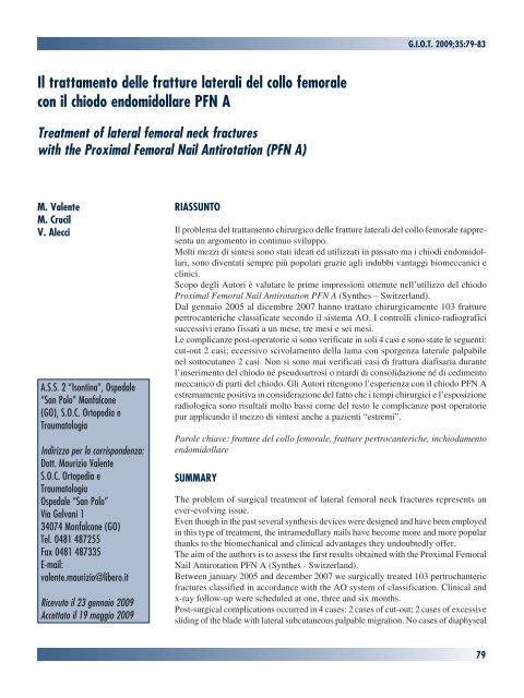
<strong>Il</strong> <strong>trattamento</strong> <strong><strong>del</strong>le</strong> <strong>fratture</strong> <strong>laterali</strong> <strong>del</strong> <strong>collo</strong> <strong>femorale</strong><br />
<strong>con</strong> <strong>il</strong> chiodo endomidollare PFN A<br />
Treatment of lateral femoral neck fractures<br />
w<strong>it</strong>h the Proximal Femoral Na<strong>il</strong> Antirotation (PFN A)<br />
M. Valente<br />
M. Cruc<strong>il</strong><br />
V. Alecci<br />
A.S.S. 2 “Isontina”, Ospedale<br />
“San Polo” Monfal<strong>con</strong>e<br />
(GO), S.O.C. Ortopedia e<br />
Traumatologia<br />
Indirizzo per la corrispondenza:<br />
Dott. Maurizio Valente<br />
S.O.C. Ortopedia e<br />
Traumatologia<br />
Ospedale “San Polo”<br />
Via Galvani 1<br />
34074 Monfal<strong>con</strong>e (GO)<br />
Tel. 0481 487255<br />
Fax 0481 487335<br />
E-ma<strong>il</strong>:<br />
valente.maurizio@libero.<strong>it</strong><br />
Ricevuto <strong>il</strong> 23 gennaio 2009<br />
Accettato <strong>il</strong> 19 maggio 2009<br />
RIASSUNTO<br />
<strong>Il</strong> problema <strong>del</strong> <strong>trattamento</strong> chirurgico <strong><strong>del</strong>le</strong> <strong>fratture</strong> <strong>laterali</strong> <strong>del</strong> <strong>collo</strong> <strong>femorale</strong> rappresenta<br />
un argomento in <strong>con</strong>tinuo sv<strong>il</strong>uppo.<br />
Molti mezzi di sintesi sono stati ideati ed ut<strong>il</strong>izzati in passato ma i chiodi endomidollari,<br />
sono diventati sempre più popolari grazie agli indubbi vantaggi biomeccanici e<br />
clinici.<br />
Scopo degli Autori è valutare le prime impressioni ottenute nell’ut<strong>il</strong>izzo <strong>del</strong> chiodo<br />
Proximal Femoral Na<strong>il</strong> Antirotation PFN A (Synthes – Sw<strong>it</strong>zerland).<br />
Dal gennaio 2005 al dicembre 2007 hanno trattato chirurgicamente 103 <strong>fratture</strong><br />
pertrocanteriche classificate se<strong>con</strong>do <strong>il</strong> sistema AO. I <strong>con</strong>trolli clinico-radiografici<br />
successivi erano fissati a un mese, tre mesi e sei mesi.<br />
Le complicanze post-operatorie si sono verificate in soli 4 casi e sono state le seguenti:<br />
cut-out 2 casi; eccessivo scivolamento <strong>del</strong>la lama <strong>con</strong> sporgenza laterale palpab<strong>il</strong>e<br />
nel sottocutaneo 2 casi. Non si sono mai verificati casi di frattura diafisaria durante<br />
l’inserimento <strong>del</strong> chiodo né pseudoartrosi o r<strong>it</strong>ardi di <strong>con</strong>solidazione né di cedimento<br />
meccanico di parti <strong>del</strong> chiodo. Gli Autori r<strong>it</strong>engono l’esperienza <strong>con</strong> <strong>il</strong> chiodo PFN A<br />
estremamente pos<strong>it</strong>iva in <strong>con</strong>siderazione <strong>del</strong> fatto che i tempi chirurgici e l’esposizione<br />
radiologica sono risultati molto bassi come <strong>del</strong> resto le complicanze post operatorie<br />
pur applicando <strong>il</strong> mezzo di sintesi anche a pazienti “estremi”.<br />
Parole chiave: <strong>fratture</strong> <strong>del</strong> <strong>collo</strong> <strong>femorale</strong>, <strong>fratture</strong> pertrocanteriche, inchiodamento<br />
endomidollare<br />
SUMMARY<br />
G.I.O.T. 2009;35:79-83<br />
The problem of surgical treatment of lateral femoral neck fractures represents an<br />
ever-evolving issue.<br />
Even though in the past several synthesis devices were designed and have been employed<br />
in this type of treatment, the intramedullary na<strong>il</strong>s have become more and more popular<br />
thanks to the biomechanical and clinical advantages they undoubtedly offer.<br />
The aim of the authors is to assess the first results obtained w<strong>it</strong>h the Proximal Femoral<br />
Na<strong>il</strong> Antirotation PFN A (Synthes - Sw<strong>it</strong>zerland).<br />
Between january 2005 and december 2007 we surgically treated 103 pertrochanteric<br />
fractures classified in accordance w<strong>it</strong>h the AO system of classification. Clinical and<br />
x-ray follow-up were scheduled at one, three and six months.<br />
Post-surgical complications occurred in 4 cases: 2 cases of cut-out; 2 cases of excessive<br />
sliding of the blade w<strong>it</strong>h lateral subcutaneous palpable migration. No cases of diaphyseal<br />
79
<strong>Il</strong> <strong>trattamento</strong> <strong><strong>del</strong>le</strong> <strong>fratture</strong> <strong>laterali</strong> <strong>del</strong> <strong>collo</strong> <strong>femorale</strong> <strong>con</strong> <strong>il</strong> chiodo endomidollare PFN A<br />
fractures during insertion of the na<strong>il</strong> have occurred, as well<br />
as no pseudoarthrosis or <strong>del</strong>ays in bone <strong>con</strong>solidation, or<br />
even mechanical loosening of the na<strong>il</strong> have been noticed.<br />
In <strong>con</strong>clusion, the authors <strong>con</strong>sider the employment of<br />
the PFN A na<strong>il</strong> as extremely pos<strong>it</strong>ive in <strong>con</strong>sideration of<br />
the fact that surgical time and radiological exposure have<br />
decreased significantly as intra- and post-surgery complications<br />
though having employed this synthesis device even<br />
on very unstable fractures.<br />
Key words: femoral neck fractures, pertrochanteric<br />
fractures, intramedullary na<strong>il</strong>ing<br />
INTRODUZIONE<br />
<strong>Il</strong> problema <strong>del</strong> <strong>trattamento</strong> chirurgico <strong><strong>del</strong>le</strong> <strong>fratture</strong> <strong>laterali</strong><br />
<strong>del</strong> <strong>collo</strong> <strong>femorale</strong> da sempre rappresenta una sfida giornaliera<br />
per <strong>il</strong> traumatologo.<br />
Molti mezzi di sintesi sono stati ideati ed ut<strong>il</strong>izzati in passato.<br />
Fino ad alcuni anni fa, <strong>il</strong> golden standard era rappresentato<br />
dalla v<strong>it</strong>e-placca a scivolamento (DHS) ma, fin dalla loro<br />
introduzione, i chiodi endomidollari, sono diventati sempre<br />
più popolari grazie agli indubbi vantaggi biomeccanici e<br />
clinici che offrono 1-4 .<br />
Peraltro, in letteratura esistono numerosi lavori che documentano<br />
importanti complicanze correlate a questi mezzi<br />
di sintesi: <strong>fratture</strong> diafisarie intraoperatorie fino al 17%<br />
8 , cedimento <strong>del</strong>la sintesi in oltre <strong>il</strong> 7% 8-10 , problemi nel<br />
bloccaggio distale in oltre <strong>il</strong> 10% 6 7 11 12 .<br />
Da pochi anni è stato immesso sul mercato <strong>il</strong> Proximal<br />
Femoral Na<strong>il</strong> Antirotation PFN A (Synthes - Sw<strong>it</strong>zerland)<br />
che in linea teorica dovrebbe rappresentare una rivoluzione<br />
nell’inchiodamento endomidollare <strong>del</strong> femore prossimale.<br />
Scopo <strong>del</strong> nostro lavoro è valutare le prime impressioni<br />
ottenute nell’ut<strong>il</strong>izzo di questo mezzo di sintesi.<br />
MATERIALI E METODO<br />
Dal gennaio 2005 al dicembre 2007, presso la nostra U.O.<br />
abbiamo trattato chirurgicamente 103 <strong>fratture</strong> pertrocanteriche.<br />
In 87 casi si trattava di pazienti di sesso femmin<strong>il</strong>e<br />
mentre in 16 casi si trattava di pazienti di sesso masch<strong>il</strong>e.<br />
L’età andava da un max di 96 anni ad un min di 57 anni <strong>con</strong><br />
una età media di 87,7 anni. <strong>Il</strong> lato coinvolto era <strong>il</strong> destro in<br />
47 casi mentre <strong>il</strong> sinistro era coinvolto in 56 casi.<br />
Lo studio pre-operatorio prevedeva l’anamnesi, la valutazione<br />
generale, la valutazione anestesiologica (ASA),<br />
le rxgrafie nelle proiezioni <strong>del</strong> bacino, antero-posteriore<br />
ed assiale <strong>del</strong>l’anca interessata. Per quanto riguarda lo<br />
studio <strong><strong>del</strong>le</strong> <strong>fratture</strong> abbiamo ut<strong>il</strong>izzato <strong>il</strong> sistema di clas-<br />
80<br />
1 5-<br />
sificazione AO 13 ed abbiamo quindi suddiviso i pazienti<br />
in tre gruppi:<br />
• 31-A1: <strong>fratture</strong> pertrocanteriche semplici 24 pazienti;<br />
• 31-A2: <strong>fratture</strong> pertrocanteriche pluriframmentarie 52<br />
pazienti;<br />
• 31-A3: <strong>fratture</strong> pertrocanteriche complesse altamente<br />
instab<strong>il</strong>i 27 pazienti.<br />
TECNICA CHIRURGICA<br />
Tutti i pazienti sono stati sottoposti ad intervento chirurgico<br />
entro le 24 ore previa somministrazione perioperatoria di<br />
2 gr. di Cefazolina. Su letto traumatologico nella classica<br />
posizione supina, si è provveduto alla riduzione <strong>del</strong>la<br />
frattura per manovre esterne e quindi ad inchiodamento<br />
endomidollare ut<strong>il</strong>izzando <strong>il</strong> chiodo PFN A.<br />
Attraverso una piccola incisione per<strong>it</strong>rocanterica si è giunti<br />
sull’apice <strong>del</strong> gran trocantere e, <strong>con</strong> appos<strong>it</strong>o preparatore si<br />
è praticato un opercolo sufficiente ad inserire manualmente<br />
<strong>il</strong> chiodo. In nessun caso è stato necessario l’alesaggio<br />
<strong>del</strong> canale midollare. Una volta in sede, grazie alla guida<br />
esterna in materiale radiotrasparente, si è quindi proceduto<br />
<strong>con</strong> la preparazione <strong>del</strong>la sede per la lama cefalica <strong>con</strong> la<br />
tecnica <strong>con</strong>solidata <strong>del</strong> f<strong>il</strong>o guida. L’ut<strong>il</strong>izzo di frese dedicate<br />
m<strong>il</strong>limetrate <strong>con</strong>sentiva di calcolare <strong>con</strong> precisione la<br />
lunghezza di tale lama. Indi l’intervento si chiudeva <strong>con</strong> <strong>il</strong><br />
bloccaggio distale mediante una v<strong>it</strong>e, la sutura e le rxgrafie<br />
di <strong>con</strong>trollo.<br />
<strong>Il</strong> <strong>trattamento</strong> post operatorio, compatib<strong>il</strong>mente <strong>con</strong> le <strong>con</strong>dizioni<br />
psico-fisiche precedenti all’intervento, prevedeva<br />
l’assunzione <strong>del</strong>la posizione seduta in prima giornata e<br />
l’inizio <strong>del</strong> training deambulatorio dalla se<strong>con</strong>da giornata<br />
<strong>con</strong> carico totale o parziale per i casi più complessi.<br />
I <strong>con</strong>trolli clinico-radiografici successivi erano fissati a un<br />
mese, tre mesi e sei mesi (Fig. 1A, B, C).<br />
CARATTERISTICHE DEL MEZZO DI SINTESI<br />
<strong>Il</strong> chiodo endomidollare PFN A è l’evoluzione <strong>del</strong> <strong>con</strong>solidato<br />
PFN dal quale si differenzia essenzialmente per <strong>il</strong><br />
bloccaggio prossimale.<br />
Come è noto nel PFN, alla v<strong>it</strong>e cefalica principale era associata<br />
una se<strong>con</strong>da v<strong>it</strong>e più sott<strong>il</strong>e a scopo antirotatorio. <strong>Il</strong><br />
PFN A, diversamente, ut<strong>il</strong>izza una lama elicoidale che,<br />
durante l’inserimento, grazie al suo movimento avv<strong>it</strong>ante,<br />
risulta <strong>con</strong>temporaneamente stab<strong>il</strong>izzante e antirotatoria<br />
(Figg. 2-3). Inoltre, si risparmia una notevole quant<strong>it</strong>à d’osso<br />
in quanto, l’inserimento ne determinana una compattazione<br />
anziché una asportazione, come avviene fresando per<br />
preparare l’inserimento di una v<strong>it</strong>e (Figg. 4A, B).
A<br />
B<br />
C<br />
Fig. 1. A: Frattura pertrocanterica destra tipo 31-A1. B: Controllo radiografico postoperatorio.<br />
C: Controllo radiografico a tre mesi.<br />
Fig. 2. <strong>Il</strong> chiodo PFNA completo di lama elicoidale e v<strong>it</strong>e distale.<br />
A tali vantaggi innovativi<br />
rispetto a tutti i chiodi<br />
endomidollari presenti<br />
sul mercato si associano<br />
quelli già standardizzati<br />
<strong>con</strong> <strong>il</strong> PFN come l’uso<br />
<strong>del</strong> T<strong>it</strong>anio, la possib<strong>il</strong><strong>it</strong>à<br />
di scelta tra vari diametri<br />
(9, 10, 11, 12 mm), la<br />
lunghezza dei chiodi (170<br />
mm, 200 mm, 240 mm), <strong>il</strong><br />
M. Valente, et al.<br />
Fig. 3. Particolare <strong>del</strong>la lama elicoidale.<br />
81
<strong>Il</strong> <strong>trattamento</strong> <strong><strong>del</strong>le</strong> <strong>fratture</strong> <strong>laterali</strong> <strong>del</strong> <strong>collo</strong> <strong>femorale</strong> <strong>con</strong> <strong>il</strong> chiodo endomidollare PFN A<br />
Fig. 4. Sezioni di testa <strong>femorale</strong> che mostrano <strong>il</strong> passaggio creato da un v<strong>it</strong>one cefalico<br />
(A) e dalla lama elicoidale <strong>del</strong> PFN A (B).<br />
bloccaggio distale statico o dinamico <strong>con</strong> una singola v<strong>it</strong>e<br />
e <strong>il</strong> tutto ut<strong>il</strong>izzando lo stesso strumentario guidato.<br />
RISULTATI<br />
<strong>Il</strong> tempo operatorio medio “skin to skin” è stato di 30 minuti<br />
<strong>con</strong> un minimo di 20 e un massimo di 45 minuti <strong>con</strong> un<br />
ut<strong>il</strong>izzo medio <strong>del</strong> fluoroscopio di 25 se<strong>con</strong>di.<br />
<strong>Il</strong> bloccaggio distale è sempre stato esegu<strong>it</strong>o in modal<strong>it</strong>à<br />
statica anche nei casi di <strong>fratture</strong> <strong>del</strong> gruppo 31-A1 e non si<br />
82<br />
A<br />
B<br />
è mai ricorsi all’alesaggio intramidollare e l’intervento è<br />
sempre stato esegu<strong>it</strong>o rigorosamente a “cielo chiuso”.<br />
Grazie ad uno strumentario molto preciso, non si sono<br />
verificate complicanze intraoperatorie di r<strong>il</strong>ievo. Le uniche<br />
difficoltà le abbiamo talvolta in<strong>con</strong>trate nello stab<strong>il</strong>ire la<br />
lunghezza <strong>del</strong>la lama elicoidale, problema a dire <strong>il</strong> vero<br />
legato più ad un errore di ut<strong>il</strong>izzo <strong>del</strong>lo strumentario in fase<br />
iniziale <strong>del</strong>la curva di apprendimento che ad un difetto <strong>del</strong><br />
chiodo stesso.<br />
Tutti i pazienti sono stati in grado di seguire <strong>il</strong> proto<strong>collo</strong> di<br />
ripresa funzionale post operatoria <strong>con</strong> inizio <strong>del</strong>la deambulazione<br />
assist<strong>it</strong>a da personale specializzato fornendo all’arto<br />
operato, nelle <strong>fratture</strong> stab<strong>il</strong>i (31-A1) un carico totale e<br />
progressivo nei casi più complessi (31-A2;3).<br />
La dimissione è avvenuta in media alla 10° giornata sempre<br />
in un ambiente protetto, dove i pazienti hanno prosegu<strong>it</strong>o<br />
la riab<strong>il</strong><strong>it</strong>azione.<br />
Nei sei mesi di osservazione post operatoria 8 pazienti sono<br />
deceduti per cause non legate direttamente all’intervento<br />
chirurgico. Del totale dei pazienti valutati a sei mesi di<br />
distanza dall’intervento, 53 erano in grado di deambulare<br />
autonomamente, 34 erano in grado di deambulare <strong>con</strong><br />
aus<strong>il</strong>i,16 non erano in grado di deambulare.<br />
Una guarigione in accorciamento sensib<strong>il</strong>e (circa 2 cm) si<br />
è verificata in 5 casi.<br />
Le complicanze post operatorie si sono verificate in soli<br />
4 casi e sono state le seguenti: cut-out 2 casi; eccessivo<br />
scivolamento <strong>del</strong>la lama <strong>con</strong> sporgenza laterale palpab<strong>il</strong>e<br />
nel sottocutaneo 2 casi. Non si sono mai verificati casi<br />
di frattura diafisaria durante l’inserimento <strong>del</strong> chiodo né<br />
pseudoartrosi o r<strong>it</strong>ardi di <strong>con</strong>solidazione, né di cedimento<br />
meccanico di parti <strong>del</strong> chiodo.<br />
DISCUSSIONE<br />
<strong>Il</strong> <strong>con</strong>tinuo sv<strong>il</strong>uppo di materiali e tecniche chirurgiche da<br />
un lato e l’età sempre più avanzata dei pazienti dall’altro,<br />
fanno sì che, nella pratica quotidiana <strong>del</strong>l’ortopedico, la<br />
frattura laterale <strong>del</strong> <strong>collo</strong> <strong>femorale</strong> rappresenta una sfida<br />
<strong>con</strong>tinua.<br />
L’avvento dei chiodi endomidollari per <strong>il</strong> <strong>trattamento</strong> di<br />
tali <strong>fratture</strong> ha rappresentato un vero salto di qual<strong>it</strong>à e da<br />
allora le prospettive di guarigione per questi pazienti sono<br />
indubbiamente aumentate.<br />
Permangono, purtroppo, vari problemi che appaiono ancor<br />
oggi di diffic<strong>il</strong>e soluzione. Cut-out, <strong>fratture</strong> intraoperatorie,<br />
cedimenti <strong><strong>del</strong>le</strong> componenti <strong>del</strong> chiodo, sporgenza laterale<br />
<strong><strong>del</strong>le</strong> v<strong>it</strong>i, pseudoartrosi, infezioni sono le complicanze più<br />
frequenti riportate in letteratura da cui nessun mo<strong>del</strong>lo di<br />
chiodo sembra essere esente 1 5-12 .
Nella nostra modesta esperienza si sono verificati 2 cut-out<br />
nei primi 30 impianti. Si trattava di pazienti <strong>con</strong> qual<strong>it</strong>à<br />
ossea molto scadente e <strong>fratture</strong> molto instab<strong>il</strong>i. Abbiamo<br />
<strong>con</strong>siderato questi problemi essere derivati da errori di tecnica<br />
chirurgica legati alla curva di apprendimento piuttosto<br />
che ad un problema correlato al mezzo di sintesi.<br />
È nostra opinione che <strong>il</strong> chiodo endomidollare prossimale<br />
PFN A sia un ulteriore passo avanti nel <strong>trattamento</strong> <strong><strong>del</strong>le</strong><br />
<strong>fratture</strong> <strong>laterali</strong> <strong>del</strong> <strong>collo</strong> <strong>femorale</strong>.<br />
Infatti presenta dei vantaggi rispetto ai principali chiodi<br />
presenti sul mercato.<br />
Esperienza più lunga, risalente ai tempi <strong>del</strong> PFN, nell’uso<br />
<strong>del</strong> T<strong>it</strong>anio, che come è noto, presenta un modulo di elastic<strong>it</strong>à<br />
circa la metà <strong>del</strong>l’acciaio garantendo una maggiore<br />
flessib<strong>il</strong><strong>it</strong>à.<br />
Ampia scelta nei diametri (9, 10, 11, 12 mm) e di lunghezza<br />
dei chiodi (170 mm, 200 mm, 240 mm). <strong>Il</strong> diametro più<br />
sott<strong>il</strong>e diminuisce <strong>il</strong> rischio di <strong>fratture</strong> diafisarie durante<br />
l’inserimento <strong>del</strong> chiodo e di <strong>fratture</strong> all’apice <strong>del</strong> chiodo in<br />
segu<strong>it</strong>o al fenomeno di stress shielding 8 14-16 . La lunghezza<br />
diversa, ut<strong>il</strong>izzando lo stesso strumentario guidato, permette<br />
di allargare notevolmente le possib<strong>il</strong><strong>it</strong>à di <strong>trattamento</strong> <strong><strong>del</strong>le</strong><br />
<strong>fratture</strong> <strong>del</strong> distretto prossimale <strong>del</strong> femore, <strong>con</strong> notevole<br />
risparmio e<strong>con</strong>omico, di magazzino e di apprendimento.<br />
Ma la vera prerogativa <strong>del</strong> PFN A è senza dubbio rappresentata<br />
dalla lama elicoidale cefalica. Questa soluzione supera<br />
i v<strong>it</strong>oni cefalici che nell’osso porotico non riuscivano ad<br />
espletare grossa presa e comunque creavano problemi nel<br />
<strong>con</strong>trollo <strong>del</strong>la rotazione <strong>del</strong>la testa <strong>femorale</strong>.<br />
Supera anche <strong>il</strong> doppio avv<strong>it</strong>amento cefalico, introdotto<br />
per <strong>con</strong>trollare la rotazione <strong>del</strong>la testa, <strong>con</strong>sentendo<br />
un netto risparmio di tempo chirurgico e di esposizione<br />
radiologica.<br />
La lama elicoidale garantisce risparmio di osso poiché la sua<br />
sezione è inferiore ai v<strong>it</strong>oni tradizionali e non asporta osso<br />
durante la preparazione <strong>con</strong> frese ma, essendo introdotta<br />
<strong>con</strong> un martello, compatta l’osso trabecolare <strong>con</strong> un netto<br />
vantaggio nei <strong>con</strong>fronti <strong>del</strong> problema cut-out 17-19 .<br />
In <strong>con</strong>clusione, r<strong>it</strong>eniamo la nostra esperienza <strong>con</strong> <strong>il</strong> chiodo<br />
PFN A estremamente pos<strong>it</strong>iva in <strong>con</strong>siderazione <strong>del</strong> fatto<br />
che i tempi chirurgici e l’esposizione radiologica sono<br />
diminu<strong>it</strong>i come <strong>del</strong> resto le complicanze post operatorie pur<br />
applicando <strong>il</strong> mezzo di sintesi anche a pazienti “estremi”.<br />
BIBLIOGRAFIA<br />
1 Bridle SH, Patel AD, Bircher M, Calvert PT. Fixation of intertrochanteric<br />
fractures of the femur: a randomised prospective<br />
comparison of the gamma na<strong>il</strong> and the dynamic hip screw. J<br />
Bone Joint Surg Br 1991;73:330-4.<br />
M. Valente, et al.<br />
2 Davis TR, Sher JL, Horsman A, Simpson M, Porter BB, Checketts<br />
RG. Intertrochanteric femoral fractures: mechanical fa<strong>il</strong>ure after<br />
internal fixation. J Bone Joint Surg Br1990;72:26-31.<br />
3 David A, von der Heyde D, Pommer A. Therapeutic possib<strong>il</strong><strong>it</strong>ies<br />
in trochanteric fractures: safe-fast-stable. Orthopade<br />
2000;29:294-301.<br />
4 Schwab E, Hontzsch K, Weise K. Versorgung instab<strong>il</strong>er per<br />
und subtrochantarer Femurfrakturen m<strong>it</strong> dem Proximalen<br />
Femurnagel (PFN). Akt Traumatol 1998;28:56-60.<br />
5 Halder SC. The Gamma na<strong>il</strong> for per<strong>it</strong>rochanteric fractures. J<br />
Bone Joint Surg Br 1992;74:340-4.<br />
6 Leung KS, So WS, Shen WY, Hui PW. Gamma na<strong>il</strong>s and<br />
dynamic hip screws for per<strong>it</strong>rochanteric fractures: a randomised<br />
prospective study in elderly patients. J Bone Joint Surg Br<br />
1992;74:345-51.<br />
7 Albareda J, Laderiga A, Palanca D, Paniagua L, Seral F.<br />
Complications and technical problems w<strong>it</strong>h gamma na<strong>il</strong>. Int<br />
Orthop 1996;20:47-50.<br />
8 Boriani S, De Iure F, Bettelli G, Specchia L, Bungaro P,<br />
Montanari G, et al. The results of a multicenter <strong>it</strong>alian study on<br />
the use of the Gamma na<strong>il</strong> for the treatment of pertrochanteric<br />
and subtrochanteric fractures: a review of 1181 cases. Chir<br />
Organi Mov 1994;79:193-203.<br />
9 Heinz T, Vecsei V. Complications and errors in use of gamma<br />
na<strong>il</strong>: causes and prevention. Chirurg 1994;65:943-52.<br />
10 Valverde JA, Alonso MG, Porro JG, Rueda D, Larrauri PM,<br />
Soler JJ. Use of the Gamma na<strong>il</strong> in the treatment of fractures<br />
of the proximal femur. Clin Orthop 1998;350:56-61.<br />
11 Friedl W, Colombo M, Dockter S, Machens HG, Mieck U.<br />
Gamma na<strong>il</strong> osteosynthesis of per and subtrochanteric femoral<br />
fractures: 4 years’ experience and their <strong>con</strong>sequences for<br />
further implant development. Chirurg 1994;65:953-63.<br />
12 Sturmer KM, Dresing K. Pertrochanteric fractures. Zentralb<br />
Chir 1995;120:862-72.<br />
13 Muller ME, Nazarian S, Koch P, Schatzker J, Heim U. The<br />
comprehensive classification of fractures of the long bones.<br />
Berlin: Springer-Verlag 1990.<br />
14 Ingman AM. Percutaneous intramedullary fixation of trochanteric<br />
fractures of the femur. Clinical trial of a new hip<br />
na<strong>il</strong>. Injury 2000;31:483-7.<br />
15 Rebuzzi E, Pannone A, Schiavetti S, Santoriello P, de Nicola U,<br />
Fancellu G, et al. IMHS clinical experience in the treatment of<br />
per<strong>it</strong>rochanteric fractures. The results of a multicentric <strong>it</strong>alian<br />
study of 981 cases. Injury 2002;33:407-12.<br />
16 Alvarez DB, Aparicio JP, Fernandez EL, Mugica IG, Batalla DN,<br />
Jimenez JP. Implant breakage, a rare complication w<strong>it</strong>h the Gamma<br />
na<strong>il</strong>. A review of 843 fractures of the proximal femur treated w<strong>it</strong>h<br />
a Gamma na<strong>il</strong>. Acta Orthop Belg 2004;70:435-43.<br />
17 Gardner MJ, Bhandari M, Lawrence BD, Helfet DL, Lorich<br />
DG. Treatment of intertrochanteric hip fractures w<strong>it</strong>h the AO<br />
trochanteric fixation na<strong>il</strong>. Orthopaedics 2005;28:117-22.<br />
18 Strauss E, Frank J, Lee J, Kummer F, Tejwani N. Helical blade vs.<br />
sliding hip screw for treatment of unstable intertrochanteric hip<br />
fractures: a biomechanical evaluation. Injury 2006;37:984-988.<br />
19 Sommers MB, Roth C, Hall H, Kam BC, Ehmke LW, Krieg<br />
JC, et al. A laboratory mo<strong>del</strong> to evaluate cutout resistence of<br />
implants for pertrochanteric fracture fixation. J Orthop Trauma<br />
2004;18:361-8.<br />
83