Riparazione di lesioni inveterate del tendine d'Achille con innesto ...
Riparazione di lesioni inveterate del tendine d'Achille con innesto ...
Riparazione di lesioni inveterate del tendine d'Achille con innesto ...

You also want an ePaper? Increase the reach of your titles
YUMPU automatically turns print PDFs into web optimized ePapers that Google loves.
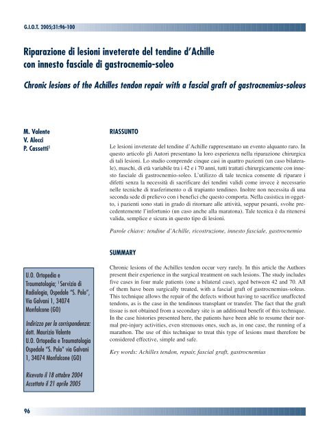
G.I.O.T. 2005;31:96-100<br />
<strong>Riparazione</strong> <strong>di</strong> <strong>lesioni</strong> <strong>inveterate</strong> <strong>del</strong> ten<strong>di</strong>ne d’Achille<br />
<strong>con</strong> <strong>innesto</strong> fasciale <strong>di</strong> gastrocnemio-soleo<br />
Chronic lesions of the Achilles tendon repair with a fascial graft of gastrocnemius-soleus<br />
M. Valente<br />
V. Alecci<br />
P. Cassetti 1<br />
U.O. Ortope<strong>di</strong>a e<br />
Traumatologia; 1 Servizio <strong>di</strong><br />
Ra<strong>di</strong>ologia, Ospedale “S. Polo”,<br />
Via Galvani 1, 34074<br />
Monfal<strong>con</strong>e (GO)<br />
In<strong>di</strong>rizzo per la corrispondenza:<br />
dott. Maurizio Valente<br />
U.O. Ortope<strong>di</strong>a e Traumatologia<br />
Ospedale “S. Polo” via Galvani<br />
1, 34074 Monfal<strong>con</strong>e (GO)<br />
Ricevuto il 18 ottobre 2004<br />
Accettato il 21 aprile 2005<br />
96<br />
RIASSUNTO<br />
Le <strong>lesioni</strong> <strong>inveterate</strong> <strong>del</strong> ten<strong>di</strong>ne d’Achille rappresentano un evento alquanto raro. In<br />
questo articolo gli Autori presentano la loro esperienza nella riparazione chirurgica<br />
<strong>di</strong> tali <strong>lesioni</strong>. Lo stu<strong>di</strong>o comprende cinque casi in quattro pazienti (un caso bilaterale),<br />
maschi, <strong>di</strong> età variabile tra i 42 e i 70 anni, tutti trattati chirurgicamente <strong>con</strong> <strong>innesto</strong><br />
fasciale <strong>di</strong> gastrocnemio-soleo. L’utilizzo <strong>di</strong> tale tecnica <strong>con</strong>sente <strong>di</strong> riparare i<br />
<strong>di</strong>fetti senza la necessità <strong>di</strong> sacrificare dei ten<strong>di</strong>ni vali<strong>di</strong> come invece è necessario<br />
nelle tecniche <strong>di</strong> trasferimento o <strong>di</strong> trapianto ten<strong>di</strong>neo. Inoltre non necessita <strong>di</strong> una<br />
se<strong>con</strong>da sede <strong>di</strong> prelievo <strong>con</strong> i benefici che questo comporta. Nella casistica in oggetto,<br />
i pazienti sono stati in grado <strong>di</strong> ritornare alle attività, seppur pesanti, svolte precedentemente<br />
l’infortunio (un caso anche alla maratona). Tale tecnica è da ritenersi<br />
valida, semplice e sicura in questo tipo <strong>di</strong> <strong>lesioni</strong>.<br />
Parole chiave: ten<strong>di</strong>ne d’Achille, ricostruzione, <strong>innesto</strong> fasciale, gastrocnemio<br />
SUMMARY<br />
Chronic lesions of the Achilles tendon occur very rarely. In this article the Authors<br />
present their experience in the surgical treatment on such lesions. The study includes<br />
five cases in four male patients (one a bilateral case), aged between 42 and 70. All<br />
of them have been surgically treated, with a fascial graft of gastrocnemius-soleus.<br />
This technique allows the repair of the defects without having to sacrifice unaffected<br />
tendons, as is the case in the ten<strong>di</strong>nous transplant or transfer. The fact that the graft<br />
tissue is not obtained from a se<strong>con</strong>dary site is an ad<strong>di</strong>tional benefit of this technique.<br />
In the case histories presented here, the patients have been able to resume their normal<br />
pre-injury activities, even strenuous ones, such as, in one case, the running of a<br />
marathon. The use of this technique to treat this type of lesions must therefore be<br />
<strong>con</strong>sidered effective, simple and safe.<br />
Key words: Achilles tendon, repair, fascial graft, gastrocnemius
INTRODUZIONE<br />
Le rotture <strong>inveterate</strong> <strong>del</strong> ten<strong>di</strong>ne d’Achille rappresentano<br />
un evento alquanto raro ma molto <strong>di</strong>fficile da trattare. Di<br />
norma, sono il risultato <strong>di</strong> una <strong>di</strong>agnosi errata, mis<strong>con</strong>osciuta<br />
o più spesso, sono semplicemente legate al mancato<br />
<strong>con</strong>sulto me<strong>di</strong>co dopo il trauma da parte <strong>del</strong> paziente<br />
stesso.<br />
Diversamente dalle rotture acute, possono mancare segni<br />
come dolore, soluzione <strong>di</strong> <strong>con</strong>tinuo palpabile nella sede <strong>di</strong><br />
rottura, instabilità. Generalmente è l’impotenza funzionale<br />
perdurante accompagnata dall’atrofia muscolare <strong>del</strong><br />
tricipite surale la causa che <strong>con</strong>duce il paziente al <strong>con</strong>sulto<br />
ortope<strong>di</strong>co.<br />
La RM, o più semplicemente l’ecografia, metteranno in<br />
evidenza un <strong>di</strong>fetto <strong>con</strong> alterazione <strong>del</strong> normale segnale<br />
ten<strong>di</strong>neo.<br />
Mentre è ancora in corso il <strong>di</strong>battito tra trattamento <strong>con</strong>servativo,<br />
trattamento chirurgico mini-invasivo o trattamento<br />
chirurgico a cielo aperto per le <strong>lesioni</strong> acute, è unanimamente<br />
ri<strong>con</strong>osciuto che le <strong>lesioni</strong> <strong>inveterate</strong> necessitano<br />
<strong>di</strong> trattamento chirurgico.<br />
In seguito alla retrazione ten<strong>di</strong>nea ed alla <strong>con</strong>seguente<br />
sostituzione <strong>del</strong> <strong>di</strong>fetto da parte <strong>di</strong> materiale fibrotico, il<br />
debridment chirurgico necessario lascia un <strong>di</strong>fetto molto<br />
ampio che nella gran parte dei casi non può essere colmato<br />
da una sutura termino-terminale.<br />
A tal proposito sono state proposte molte varianti chirurgiche<br />
<strong>del</strong>le seguenti tecniche principali: a) <strong>innesto</strong> libero<br />
<strong>di</strong> ten<strong>di</strong>ne (fascia lata; ten<strong>di</strong>ni donatori come semiten<strong>di</strong>noso,<br />
gracile, rotuleo, alloinnesti) 1-3 ; b) avanzamento<br />
fasciale (plastica a V-Y; <strong>innesto</strong> fasciale <strong>di</strong> gastrocnemiosoleo<br />
ruotato in basso) 4-14 ; c) trasferimento <strong>di</strong> ten<strong>di</strong>ne<br />
locale 15-19 ; d) materiali sintetici 20-22 .<br />
MATERIALI E METODO<br />
Gabel e Manoli 23 hanno definito rotture <strong>inveterate</strong> <strong>del</strong><br />
ten<strong>di</strong>ne d’Achille quelle <strong>lesioni</strong> che datavano almeno<br />
quattro settimane. Nel biennio 2000-2001 sono giunti alla<br />
nostra osservazione quattro pazienti che presentavano<br />
tale lesione, in un caso bilateralmente. Tutti erano maschi,<br />
età compresa tra 42 e 70 anni. Il lato interessato dalla<br />
lesione era il destro in 3 casi; in 2 casi era interessato il<br />
sinistro. Un caso era bilaterale. Il tempo intercorso tra la<br />
lesione e il trattamento chirurgico è variato da un minimo<br />
<strong>di</strong> 4 settimane ad un massimo <strong>di</strong> 30 settimane.<br />
M. Valente et al.<br />
In tutti i casi abbiamo adottato la medesima tecnica<br />
operatoria. Somministrazione <strong>di</strong> due grammi <strong>di</strong><br />
Cefazolina nell’imme<strong>di</strong>ato pre-operatorio, anestesia<br />
spinale, decubito prono <strong>con</strong> laccio pneumoischemico<br />
alla ra<strong>di</strong>ce <strong>del</strong>la coscia, incisione longitu<strong>di</strong>nale paraten<strong>di</strong>nea<br />
me<strong>di</strong>ale, <strong>di</strong>eresi per strati <strong>con</strong> particolare attenzione<br />
al paratenonio, sbrigliamento generoso <strong>del</strong> tessuto<br />
fibroso interposto tra i capi ten<strong>di</strong>nei retratti, avvicinamento<br />
degli stessi <strong>con</strong> filo riassorbibile <strong>di</strong> grosse<br />
<strong>di</strong>mensioni e preparazione <strong>di</strong> lembo fasciale centrale<br />
dal gastrocnemio <strong>di</strong> circa due centimetri <strong>di</strong> larghezza<br />
che viene in seguito ribaltato <strong>di</strong> 180° a colmare la soluzione<br />
<strong>di</strong> <strong>con</strong>tinuo e suturato sul mon<strong>con</strong>e <strong>di</strong>stale <strong>con</strong><br />
piede in flessione plantare <strong>di</strong> circa 20°. Quin<strong>di</strong> sutura<br />
per strati, me<strong>di</strong>cazione e apparecchio gessato femoropodalico<br />
in equinismo per 30 giorni sostituito da gambaletto<br />
per ulteriori 30 giorni. Il carico è stato <strong>con</strong>cesso alla<br />
rimozione <strong>del</strong>l’apparecchio gessato e <strong>con</strong>temporaneamente<br />
è iniziata la FKT.<br />
Il follow-up è variato da un minimo <strong>di</strong> 33 mesi ad un massimo<br />
<strong>di</strong> 54 mesi (me<strong>di</strong>a 43,2). Tutti i pazienti sono stati<br />
sottoposti ad esame anamnestico e clinico.<br />
In due casi abbiamo sottoposto i pazienti a risonanza<br />
magnetica. Tali esami sono stati eseguiti me<strong>di</strong>ante apparecchio<br />
GE Ovation da 0.35 T <strong>con</strong> bobina de<strong>di</strong>cata per<br />
tibio-tarsica ottenendo sequenze pesate in T1, sequenze<br />
STIR e sequenze ad eco <strong>di</strong> gra<strong>di</strong>ente <strong>con</strong>dotte se<strong>con</strong>do<br />
piani assiali e sagittali per una durata complessiva <strong>del</strong>l’esame<br />
<strong>di</strong> circa 35 minuti.<br />
RISULTATI<br />
In due casi, dopo circa 2 settimane dall’intervento, si è<br />
verificata una necrosi cutanea in corrispondenza <strong>del</strong> sito<br />
<strong>di</strong> rottura ten<strong>di</strong>nea. Questa zona cutanea, al momento <strong>del</strong>l’intervento<br />
appariva già sofferente e, nonostante l’accortezza<br />
<strong>del</strong>l’atto chirurgico e <strong>del</strong>le successive me<strong>di</strong>cazioni,<br />
è esitata in una per<strong>di</strong>ta <strong>di</strong> sostanza profonda che ci ha visti<br />
costretti ad eseguire una copertura <strong>con</strong> un lembo fascioa<strong>di</strong>poso<br />
peduncolato ottenendo il risultato sperato.<br />
Al momento <strong>del</strong>la valutazione nessun paziente presenta<br />
dolore importante in sede <strong>di</strong> lesione. Tutti i pazienti sono<br />
stati in grado <strong>di</strong> ritornare alle attività svolte prima <strong>del</strong>la<br />
lesione anche se in tutti permane il timore <strong>di</strong> eseguire<br />
quei gesti che hanno determinato la rottura ten<strong>di</strong>nea.<br />
Tutti presentano un passo simmetrico e <strong>con</strong> buona propulsione.<br />
Inoltre, ciascun paziente è in grado <strong>di</strong> sollevar-<br />
97
<strong>Riparazione</strong> <strong>di</strong> <strong>lesioni</strong> <strong>inveterate</strong> <strong>del</strong> ten<strong>di</strong>ne d’Achille <strong>con</strong> <strong>innesto</strong> fasciale <strong>di</strong> gastrocnemio-soleo<br />
si sulla punta dei pie<strong>di</strong> e camminare in tale modo; <strong>di</strong> saltare<br />
<strong>con</strong> un solo piede (anche se <strong>con</strong> un certo timore).<br />
L’arco <strong>di</strong> movimento, <strong>con</strong>frontato <strong>con</strong> il <strong>con</strong>trolaterale,<br />
non ha mai mostrato significative variazioni.<br />
Due pazienti hanno accettato <strong>di</strong> sottoporsi a RM.<br />
Nel primo caso, R.A. maschio 53 anni, si apprezzano gli<br />
esiti <strong>del</strong>l’intervento, soprattutto nella sequenza assiale<br />
(Foto 1a), a livello <strong>del</strong> cellulare a<strong>di</strong>poso sottocutaneo in<br />
prossimità <strong>del</strong> ten<strong>di</strong>ne, ma non si rilevano alterazioni <strong>del</strong>l’intensità<br />
<strong>del</strong> segnale, soprattutto nella sequenza STIR<br />
(Foto 1b).<br />
Nel se<strong>con</strong>do caso, P.F. maschio 43 anni, non si rilevano<br />
significative alterazioni <strong>del</strong>l’intensità <strong>del</strong> segnale in nessuna<br />
<strong>del</strong>le sequenze, in particolare nella sequenza T1<br />
Foto 1a e 1b. Nella sequenza assiale (a), a livello <strong>del</strong> cellulare a<strong>di</strong>poso sottocutaneo<br />
in prossimità <strong>del</strong> ten<strong>di</strong>ne, si apprezzano gli esiti <strong>del</strong>l’intervento <strong>di</strong> plastica cutanea ma<br />
non si rilevano alterazioni <strong>del</strong>l’intensità <strong>del</strong> segnale, in particolare nella sequenza<br />
STIR(b).<br />
98<br />
Foto 2a e 2b. Non si rilevano significative alterazioni <strong>del</strong>l’intensità <strong>del</strong> segnale ten<strong>di</strong>neo<br />
in nessuna <strong>del</strong>le sequenze, in particolare nella sequenza T1 pesata sagittale (a) ed<br />
in quella sagittale ad eco <strong>di</strong> gra<strong>di</strong>ente (b).<br />
pesata sagittale (Foto 2a) ed in quella sagittale ad eco <strong>di</strong><br />
gra<strong>di</strong>ente (Foto 2b). Come reperto accessorio, nelle<br />
sequenze sagittali, si rileva una alterazione <strong>del</strong>l’intensità<br />
<strong>del</strong> segnale <strong>del</strong>l’osso sub-<strong>con</strong>drale a livello tibio-astragalico<br />
riferibile ad iniziale patologia osteo-<strong>con</strong>drale.<br />
DISCUSSIONE<br />
In caso <strong>di</strong> rotture sottocutanee <strong>del</strong> ten<strong>di</strong>ne d’Achille, la<br />
scelta <strong>del</strong> trattamento è stata a lungo <strong>di</strong>battuta ed attualmente<br />
il favore va al trattamento chirurgico 23 .<br />
Naturalmente, l’intervento andrebbe eseguito quanto
prima per evitare la retrazione dei capi ten<strong>di</strong>nei e quin<strong>di</strong><br />
<strong>con</strong>sentire una sutura non in tensione. Se la lesione passa<br />
mis<strong>con</strong>osciuta per <strong>di</strong>versi giorni o, peggio, per settimane,<br />
ripristinare l’integrità ten<strong>di</strong>nea <strong>di</strong>venta impossibile.<br />
Molte tecniche sono state proposte per la riparazione<br />
<strong>del</strong>le rotture <strong>inveterate</strong> <strong>del</strong> ten<strong>di</strong>ne d’Achille.<br />
L’uso <strong>del</strong>l’<strong>innesto</strong> fasciale <strong>di</strong> gastrocnemio-soleo nel caso<br />
<strong>di</strong> impossibilità a ripristinare una <strong>con</strong>tinuità tra i capi ten<strong>di</strong>nei<br />
presenta degli specifici vantaggi.<br />
Innanzitutto non richiede il prelievo da un’altra sede <strong>di</strong><br />
materiale ten<strong>di</strong>neo autologo evitando così l’indebolimento<br />
<strong>di</strong> altri muscoli <strong>del</strong>l’arto inferiore e l’allestimento <strong>di</strong> un<br />
se<strong>con</strong>do campo operatorio.<br />
Un ulteriore vantaggio <strong>con</strong>siste nell’evitare l’utilizzo <strong>di</strong><br />
materiali omologhi <strong>con</strong> i relativi problemi <strong>di</strong> carattere<br />
immunologico.<br />
È indubbia una certa <strong>di</strong>minuzione <strong>del</strong>la forza in plantarflessione<br />
se<strong>con</strong>daria al prelievo <strong>del</strong>la parte centrale <strong>del</strong><br />
gastrocnemio. In ogni caso, eseguendo un accurato prelievo<br />
evitando <strong>di</strong> intaccare il ventre muscolare sottostante,<br />
la riparazione anatomica <strong>del</strong> ten<strong>di</strong>ne <strong>con</strong>sente un recupero<br />
<strong>del</strong>la funzione muscolare pressoché completa.<br />
Naturalmente, a completamento <strong>del</strong>l’<strong>innesto</strong>, è in<strong>di</strong>spensabile<br />
eseguire una sutura tra i capi ten<strong>di</strong>nei ed applicare<br />
un’immobilizzazione gessata <strong>con</strong> femoro-podalico per un<br />
tempo adeguato per proteggere la sede <strong>di</strong> <strong>innesto</strong>.<br />
L’immobilizzazione protratta anche per 60 giorni non ha<br />
mai provocato rigi<strong>di</strong>tà gravi né comparsa <strong>di</strong> sindrome<br />
algo-<strong>di</strong>strofica.<br />
In due casi, dopo circa 2 settimane dall’intervento, si è<br />
verificata una necrosi cutanea in corrispondenza <strong>del</strong> sito<br />
<strong>di</strong> rottura ten<strong>di</strong>nea che ci ha visti costretti ad eseguire una<br />
copertura <strong>con</strong> un lembo fascioa<strong>di</strong>poso peduncolato. Al<br />
momento <strong>del</strong> ri<strong>con</strong>oscimento <strong>del</strong>la lesione ten<strong>di</strong>nea, tale<br />
zona appariva molto sofferente. È nostra opinione che<br />
questa complicanza sia legata alla ritardata <strong>di</strong>agnosi <strong>con</strong><br />
<strong>con</strong>seguente utilizzo <strong>del</strong>l’arto (entrambi i pazienti erano<br />
<strong>con</strong>vinti <strong>di</strong> essere vittime <strong>di</strong> <strong>di</strong>storsioni <strong>di</strong> caviglia in<br />
corso <strong>di</strong> guarigione!) e quin<strong>di</strong> al coinvolgimento <strong>del</strong>la<br />
cute nel mancato processo riparativo piuttosto che all’insulto<br />
chirurgico stesso.<br />
I risultati ottenuti sono stati incoraggianti. Tutti i pazienti<br />
si sono <strong>di</strong>chiarati sod<strong>di</strong>sfatti dei risultati ottenuti e sono<br />
stati in grado <strong>di</strong> ritornare alle attività, seppur pesanti,<br />
svolte precedentemente l’infortunio (in un caso anche alla<br />
maratona). Tale tecnica è da ritenersi valida, semplice e<br />
sicura nelle <strong>lesioni</strong> <strong>inveterate</strong> dove, dopo sbrigliamento<br />
dei tessuti cicatriziali ed avvicinamento dei capi ten<strong>di</strong>nei,<br />
M. Valente et al.<br />
permane un <strong>di</strong>fetto al massimo <strong>di</strong> circa quattro/cinque<br />
centimetri. In quei casi in cui il <strong>di</strong>fetto risulti maggiore è<br />
<strong>con</strong>sigliabile utilizzare <strong>del</strong>le tecniche alternative come<br />
una plastica tipo V-Y oppure l’utilizzo <strong>di</strong> ten<strong>di</strong>ni autologhi<br />
od omologhi <strong>di</strong> banca.<br />
BIBLIOGRAFIA<br />
1 Zadek I. Repair of old rupture of the teno-achilles by means<br />
of fascia lata: report of a case. J Bone Joint Surg<br />
1941;22A:1070-1.<br />
2 Besse JL, Lerat JL, Moyen B, Brunet-Guedj E. Achilles tendon<br />
repair using a bone tendon graft harvested from the knee<br />
extensor system: three cases. J Foot Ankle Surg 1999;38:70-4.<br />
3 Nellas ZJ, Loder BG, Weirtheimer SJ. Re<strong>con</strong>struction of an<br />
Achilles tendon defect utilizing an achilles tendon allograft. J<br />
Foot Surg 1996;35:144-8.<br />
4 Christensen I. Rupture of the Achilles tendon: analysis of 57<br />
cases. Acta Chir Scand 1953;106:50-60.<br />
5 Bosworth DM. Repair of defects in the tenoachilles. J Bone<br />
Joint Surg 1956;38A:111-4.<br />
6 Ralston E, Schmidt E. Repair of ruptured Achilles tendon. J<br />
Trauma 1971;11:15-21.<br />
7 Lynn TA. Repair of the torn achilles tendon, using the plantaris<br />
tendon as a reinforcing membrane. J Bone Joint Surg<br />
1966;48A:268-72.<br />
8 Arner O, Lindholm A. Subcutaneous rupture of the Achilles<br />
tendon. Acta Chir Scand 1959;239(Suppl):1-47.<br />
9 Gerdes MH, Brown TD, Bell AL, Baker JA, Levson M, Layer<br />
S. A flap augmentation tecnique for Achilles tendon repair.<br />
Clin Orthop 1992;280:241-6.<br />
10 Abraham E, Pankovich A. Neglected rupture of the Achilles<br />
tendon. J Bone Joint Surg 1975;57A:253-5.<br />
11 Leitner A, Voigt C, Rahamanzadeh R. Treatment of extensive<br />
aseptic defects in old Achilles tendon ruptures. Foot Ankle<br />
1992;13:176-80.<br />
12 Bugg EI, Boyd BM. Repair of neglected rupture or laceration<br />
of the Achilles tendon. Clin Orthop 1968;56:73-5.<br />
13 Men<strong>di</strong>cino SS. Repair of neglected Achilles tendon ruptures<br />
with a triceps surae muscle tendon advancement. J Foot<br />
Ankle Surg 1996;35:13-8.<br />
14 Lindholm A. A new method of operation in subcutaneous rupture<br />
of the Achilles tendon. Acta Chir Scand 1959;117:261-3.<br />
15 White RK, Kraynick BM. Surgical uses of peroneus brevis<br />
tendon. Surg Gynecol Obstet 1959;108:117-21.<br />
16 Perez Teuffer A. Traumatic rupture of the Achilles tendon.<br />
Re<strong>con</strong>struction by Transplant and graft of the lateral peroneus<br />
brevis. Orthop Clin North Am 1974;5:89-93.<br />
17 Mann RA, Holmes GB Jr, Seale KS, Collins DN. Chronic<br />
rupture of the Achilles tendon: a new tecnique of repair. J<br />
Bone Joint Surg 1991;73A:214-9.<br />
99
<strong>Riparazione</strong> <strong>di</strong> <strong>lesioni</strong> <strong>inveterate</strong> <strong>del</strong> ten<strong>di</strong>ne d’Achille <strong>con</strong> <strong>innesto</strong> fasciale <strong>di</strong> gastrocnemio-soleo<br />
18 Wapner KL, Pavlock GS, Hecht PJ, Naselli F, Walther R.<br />
Repair of chronic Achilles tendon rupture with flexor hallucis<br />
longus tendon transfer. Foot Ankle 1993;14:443-9.<br />
19 Pintore E, Barra V, Pintore R, Maffulli N. Peroneus brevis<br />
tendon transfer in neglected tears of the Achilles tendon. J<br />
Trauma 2001;50:71-8.<br />
20 Howard CB, Winston I, Bell W, Mackie I, Jenkins DH. Late<br />
repair of the calcaneal tendon with carbon fibre. J Bone Joint<br />
Surg 1984;66B:206-8.<br />
100<br />
21 Ozaki J, Fujiki J, Sugimoto K, Tamai S, Masuhara K.<br />
Re<strong>con</strong>struction of neglected Achilles tendon rupture with<br />
Marlex mesh. Clin Orthop 1989;238:204-8.<br />
22 Choksey A, Soonawalla D, Murray J. Repair of neglected<br />
Achilles tendon ruptures with Marlex mesh. Injury<br />
1996;27:215-7.<br />
23 Gabel S, Manoli A. Neglected rupture of the Achilles tendon.<br />
Foot Ankle Int 1994;15:512-7.