iv. mezioborové setkênè mladðch biologů ... - Chemické listy
iv. mezioborové setkênè mladðch biologů ... - Chemické listy
iv. mezioborové setkênè mladðch biologů ... - Chemické listy

You also want an ePaper? Increase the reach of your titles
YUMPU automatically turns print PDFs into web optimized ePapers that Google loves.
Chem. Listy 98, 271 – 314 (2004) IV. Amerika 2004.<br />
ASSOCIATION OF EF-Tu WITH BIOLOGICAL<br />
MEMBRANES AND ITS AGGREGATION INCREASES<br />
FACTOR’S NANO-SWITCH POTENTIAL<br />
MARTIN HOLUB, LADISLAVA KALACHOVÁ, SILVIE<br />
BEZOUŠKOVÁ, and JAROSLAV WEISER<br />
Mikrobiologický ústav AV ČR, Vídeňská 1083, 142 20 Praha 4<br />
holub@biomed.cas.cz<br />
Protein synthesis elongation factor Tu (EF-Tu) represents<br />
one of the major components of translation system in<br />
prokaryotes. It participates on correct positioning of the<br />
incoming aminoacyl-tRNA on the ribosome where polypeptide<br />
chain is synthesised. Besides this, EF-Tu is proposed to<br />
function in other parts of the cell metabolism as well. The<br />
protein is represented by three-domain structure and behaves<br />
like a typical G (guanine nucleotide binding) protein.<br />
Interaction of flexible domain 1 containing GDP/GTP binding<br />
pocket with more rigid domains 2 and 3 allows it to work as a<br />
molecular switch changing between “on” and “off”<br />
conformation upon binding of GDP or GTP. There are<br />
available specific inhibitors of EF-Tu, which are able to<br />
“freeze “the protein in either “on”, or “off” conformation as an<br />
example can be mentioned kirromycin or pulvomycin.<br />
There have been described several post-translation<br />
modifications of the protein some of them playing the role in<br />
translation, others important for its potential functions outside<br />
of the elongation cycle. In E.coli, Bacillus subtilis and Bacillus<br />
licheniformis a part of EF-Tu population, which is located on<br />
the membrane, can be methylated in response to starvation for<br />
an essential nutrient.<br />
E.coli and T. thermophilus EF-Tu were found to be<br />
phosphorylated in v<strong>iv</strong>o, and the phosphorylated fraction<br />
remained stable under different conditions. Since the<br />
phosphorylated residue (Thr-382) is conserved in all known<br />
EF-Tu corresponding sequences from other species, the<br />
phosphorylation might be a common phenomenon and the<br />
phosphorylated form of EF-Tu might play a fundamental role<br />
in the physiology of all organisms. EF-Tu was also described<br />
recently as a major component of cytoskeleton like structures<br />
in Mycoplasma pneumoniae cells and most importantly it was<br />
identified there as a protein binding fibronectin which is a<br />
multifunctional protein interacting with molecular motor like<br />
structures in eukaryotes.<br />
In order to study potential role(s) of EF-Tu posttranslation<br />
modifications in Streptomyces we studied its<br />
heterogeneity, content and distribution in several Streptomyces<br />
strains and compared it with that in Mycobacterium<br />
smegmatis, Escherichia coli and Bacillus subtilis. For that<br />
purpose we have prepared membrane, ribosomal and soluble<br />
cell fractions by differential centrifugation and EF-Tu was<br />
detected in the fractions by western blot technique. Using 2Delectrophoresis<br />
of proteins we analysed cell membrane<br />
proteomes in several Streptomyces strains during development<br />
and compared it with that of non-differentiating<br />
Mycobacterium.<br />
We described previously a spontaneous polymerisation<br />
of EF-Tu from Streptomyces aureofaciens, which might serve<br />
as a protect<strong>iv</strong>e mechanism for EF-Tu present in spores or<br />
enables the protein to play a structural role. Aggregates are<br />
formed under physiological conditions and g<strong>iv</strong>e raise to<br />
filamentous structures large enough to be visible in the light<br />
microscope. We have developed simple and effect<strong>iv</strong>e method<br />
for purification of large amounts of the aggregated protein,<br />
which retains its nucleotide binding act<strong>iv</strong>ity. We found that<br />
two closely related strains of Streptomyces aureofaciens<br />
contain EF-Tu capable of spontaneous aggregation. We<br />
purified EF-Tu from both strains using above mentioned<br />
method and use them in comparat<strong>iv</strong>e studies in order to<br />
understand better the structural and functional basis of this<br />
phenomenon. Using 2D electrophoresis of purified proteins<br />
and their hydrolysis products we analysed their structural<br />
differences and heterogeneity resulting from their posttranslation<br />
modifications.<br />
NOVÉ MOŽNOSTI ON-LINE PREKONCENTRACE<br />
ANALYTŮ V KAPILÁRNÍ ELEKTROFORÉZE<br />
JANA HORÁKOVÁ, VÍTĚZSLAV MAIER a JURAJ<br />
ŠEVČÍK<br />
Katedra analytické chemie Přírodovědecká Fakulta Un<strong>iv</strong>estita<br />
Palackého, Třída Svobody 8, 771 46 Olomouc<br />
Metody kapilární elektroforézy (CE) se dnes stávají čím<br />
dále častěji využívanějšími separačními technikami. Jejími<br />
hlavními výhodami jsou nenáročná instrumentace, nízká<br />
spotřeba vzorku a zejména vysoká separační účinnost.<br />
Slabinou CE je nedostatečná koncentrační citl<strong>iv</strong>ost, zvláště při<br />
použití UV-VIS detekce. K částečné eliminaci tohoto<br />
problému slouží on-line prekoncentrační techniky založené na<br />
využití principů přechodné izotachoforézy, stacking nebo<br />
sweeping efektu.<br />
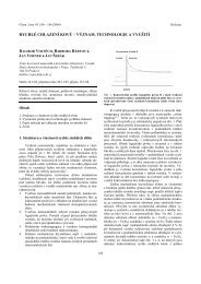
Prezentovaná nová kapilárně elektroforetická on-line<br />
prekoncentrační technika umožňuje stanovovat nanomolární<br />
koncentrace slabých elektrolytů pomocí klasických UV-VIS<br />
detektorů. Vhodnost navrženého postupu je demonstrovaná na<br />
stanovení benzoové a sorbové kyseliny ve vzorcích stolního<br />
oleje a ovocné šťávy. Filozofie on-line prekoncentrace je<br />
založená na vhodném experimentálním uspořádání dvou<br />
pracovních elektrolytů, které se liší svým složením a použitým<br />
pH. Na elektrolytickém rozhraní těchto dvou pracovních elektrolytů<br />
dochází k elektroforetické imobilizaci stanovovaných<br />
analytů. Po dostatečném „nakoncentrování“ analytů je jejich<br />
následná mobilizace a separace umožněna namigrováním<br />
micelotvorních látek do separační kapiláry.<br />
Vlastní metoda se může z metodického hlediska rozdělit<br />
na dvě fáze: akumulační a mobilizační. V námi demonstrovaném<br />
případě jsou ve fázi akumulační použité modelové<br />
analyty (benzoová a sorbová kyselina) rozpuštěny v borátovém<br />
pufru o pH 9,5. Při tomto pH dochází k jejich ionizaci. Protože<br />
obě kyseliny jsou slabými elektrolyty, jsou záporně nábité a<br />
migrují ke kladně nabité elektrodě. Při této migraci „narazí“<br />
molekuly kyselin (disociované v borátovém elektrolytu) na<br />
rozhraní fosfátového pufru o pH 2,5. Nízká hodnota pH tohoto<br />
elektrolytu způsobí jejich deionizaci (ztratí náboj), a tudíž<br />
přestanou v kapiláře v tomto místě migrovat. Po dostatečně<br />
dlouhé době elektrokinetického dávkování vzorku dochází k<br />
281