- Page 2 and 3:
10 ib
- Page 7:
THE ZYGNEMATACEAE
- Page 10 and 11:
GRADUATE SCHOOL MONOGRAPHS Contribu
- Page 13 and 14:
INTRODUCTION The absence of a moder
- Page 15:
INTRODUCTION ix Illinois State Teac
- Page 19 and 20:
Plat€ ILLUSTRATIONS Plates Page I
- Page 21:
THE ZYGNEMATACEAE
- Page 24 and 25:
4 ZYGNEMATACEAE gametes is by means
- Page 26 and 27:
6 ZYGNEMATACEAE are common but are
- Page 28 and 29:
8 ZYGNEMATACEAE between cells, the
- Page 30 and 31:
10 ZYGNEMATACEAE sporiferous cells
- Page 32 and 33:
12 ZYGNEMATACEAE TABLE I Summary of
- Page 34 and 35:
14 ZYGNEMATACEAE In higher latitude
- Page 36 and 37:
i6 ZYGNEMATACEAE specimens with mat
- Page 38 and 39:
1 ZYGNEMATACEAE II. Spores usually
- Page 40 and 41:
20 ZYGNEMATACEAE 32. Diameter veget
- Page 42 and 43:
22 ZYGNEMATACEAE REPRODUCTION BY AP
- Page 44 and 45:
24 ZYGNEMATACEAE spores formed in t
- Page 46 and 47:
26 ZYGNEMATACEAE pitted; pits 5-6 /
- Page 48 and 49:
28 ZYGNEMATACEAE spores are formed
- Page 50 and 51:
30 ZYGNEMATACEAE spores formed in t
- Page 52 and 53:
32 ZYGNEMATACEAE biculate; pits 4.5
- Page 54 and 55:
34 ZYGNEMATACEAE United States: Ill
- Page 56 and 57:
36 ZYGNEMATACEAE zygospores globose
- Page 58 and 59:
38 ZYGNEMATACEAE spores formed in o
- Page 60 and 61:
40 ZYGNEMATACEAE spores in one of t
- Page 62 and 63:
42 ZYGNEMATACEAE an outer pectic la
- Page 64 and 65:
44 ZYGNEMATACEAE irregular in form
- Page 66 and 67:
46 ZYGNEMATACEAE cylindncum Transea
- Page 69 and 70:
CHAPTER THREE THE GENUS ZYGNEMOPSIS
- Page 71 and 72:
ZYGNEMOPSIS 51 6. Median spore wall
- Page 73 and 74:
ZYGNEMOPSIS 53 22-25 jLi X 25-30 /"
- Page 75 and 76:
ZYGNEMOPSIS 55 12. Zygnemopsis sple
- Page 77 and 78:
ZYGNEMOPSIS 57 23-50 Ai, with a mor
- Page 79:
quadrata Jao 1935 ZYGNEMOPSIS 59 si
- Page 83 and 84:
CHAPTER FIVE THE GENUS ZYGOGONIUM K
- Page 85 and 86:
ZYGOGONIUM 65 3. Zygospores 15-26/U
- Page 87 and 88:
ZYGOGONIUM 67 ospores ellipsoid to
- Page 89:
ZYGOGONIUM 69 14. Zygogonium stephe
- Page 93 and 94:
CHAPTER SEVEN THE GENUS MOUGEOTIOPS
- Page 95 and 96:
CHAPTER RIGHT THE GENUS DEBARYA WIT
- Page 97 and 98:
DEBARYA 77 3. Debarya ackleyana Tra
- Page 99 and 100:
CHAPTER NINE THE GENUS MOUGEOTIA C.
- Page 101 and 102:
MOUGEOTIA 8i not be cited as proof
- Page 103 and 104:
7. Spores globose to ovoid, variabl
- Page 105 and 106:
MOUGEOTIA 85 30. Spores ovoid 40-54
- Page 107 and 108:
MOUGEOTIA 87 52. Zygosporangia regu
- Page 109 and 110:
MOUGEOTIA 89 74. Spore wall smooth
- Page 111 and 112:
MOUGEOTIA 91 6. MouGEOTiA ELLiPsoiD
- Page 113 and 114:
MOUGEOTIA 93 tending somewhat into
- Page 115 and 116:
MOUGEOTIA 95 conjugation scalarifor
- Page 117 and 118:
MOUGEOTIA 97 pyrenoids; gametangia
- Page 119 and 120:
MOUGEOTIA 99 ous scattered pyrenoid
- Page 121 and 122:
MOUGEOTIA loi cylindric, with conca
- Page 123 and 124:
MOUGEOTIA 103 56. MouGEOTiA ATUBULO
- Page 125 and 126:
MOUGEOTIA 105 aplanospores occupyin
- Page 127 and 128:
MOUGEOTIA 107 with hornlike process
- Page 129 and 130:
MOUGEOTIA 109 77. MouGEOTiA viRiDis
- Page 131 and 132:
MOUGEOTIA III 85. MouGEOTiA QUADRAN
- Page 133 and 134:
MOUGEOTIA 113 94. MouGEOTiA MAYORi
- Page 135 and 136:
MOUGEOTIA 115 craterophora Bohlin 1
- Page 137 and 138:
CHAPTER TEN THE GENUS TEMNOGAMETUM
- Page 139:
CHAPTER ELEVEN THE GENUS SIROCLADIU
- Page 143 and 144:
CHAPTER THIRTEEN THE GENUS SPIROGYR
- Page 145 and 146:
SPIROGYRA 125 in thickness, and eac
- Page 147 and 148:
SPIROGYRA 127 may be widened and ne
- Page 149 and 150:
SPIROGYRA 129 Of the 2 species with
- Page 151 and 152:
SPIROGYRA 131 II. Most of the spore
- Page 153 and 154:
31. Diameter vegetative cells more
- Page 155 and 156:
SPIROGYRA 135 END WALLS PLANE (one-
- Page 157 and 158:
66. Spores 34-48 i". x 48-54^1 93-
- Page 159 and 160:
82. Median spore wall double, outer
- Page 161 and 162:
loi. Fertile cells cylindric, chrom
- Page 163 and 164:
115. Diameter vegetative cells 130-
- Page 165 and 166:
SPIROGYRA 145 133. Reproducing by a
- Page 167 and 168:
SPIROGYRA 147 157. Median spore wal
- Page 169 and 170:
SPIROGYRA 149 178. Chromatophores i
- Page 171 and 172:
SPIROGYRA 151 3. Spirogyra juercens
- Page 173 and 174:
SPIROGYRA 153 China, Szechwan. The
- Page 175 and 176:
SPIROGYRA 155 matophore; conjugatio
- Page 177 and 178:
SPIROGYRA 157 23. Spirogyra affinis
- Page 179 and 180:
SPIROGYRA 159 cells to globose, 28-
- Page 181 and 182:
SPIROGYRA i6i qucntly misapplied th
- Page 183 and 184:
SPIROGYRA 163 scalariform; tubes fo
- Page 185 and 186:
SPIROGYRA 165 ellipsoid, ^P-^^i^ x
- Page 187 and 188:
Germany; Latvia; Yugoslavia; Finlan
- Page 189 and 190:
Coll.). SPIROGYRA 169 Dutch New Gui
- Page 191 and 192:
SPIROGYRA 171 formed by both gameta
- Page 193 and 194:
SPIROGYRA 173 This imperfectly desc
- Page 195 and 196:
SPIROGYRA 175 Vegetative cells 35-4
- Page 197 and 198:
SPIROGYRA 177 loi. Spirogyra polyta
- Page 199 and 200:
SPIROGYRA 179 formed by both gameta
- Page 201 and 202:
SPIROGYRA i8i is thin, brownish and
- Page 203 and 204:
SPIROGYRA 183 PI. 8, Figs. 87-88. A
- Page 205 and 206:
SPIROGYRA 185 United States: Widely
- Page 207 and 208:
SPIROGYRA 187 65-83 /x X 75-1 00 /x
- Page 209 and 210:
SPIROGYRA 189 150. Spirogyra reinha
- Page 211 and 212:
SPIROGYRA 191 cylindric or slightly
- Page 213 and 214:
SPIROGYRA 193 by both gametangia; f
- Page 215 and 216:
SPIROGYRA 195 although the tubes be
- Page 217 and 218:
wall. United States: Texas, Karnac,
- Page 219 and 220:
SPIROGYRA 199 found the earlier sta
- Page 221 and 222:
SPIROGYRA 201 matophore, making 3 t
- Page 223 and 224:
SPIROGYRA 203 199. Spirogyra unduli
- Page 225 and 226:
SPIROGYRA 205 United States: Iowa;
- Page 227 and 228:
SPIROGYRA 207 spores ellipsoid, 20-
- Page 229 and 230:
SPIROGYRA 209 iform; tubes formed b
- Page 231 and 232:
SPIROGYRA 211 tubes formed by both
- Page 233 and 234:
SPIROGYRA 213 forma gracilior). Czu
- Page 235 and 236:
SPIROGYRA 215 244. Spirogyra gratia
- Page 237 and 238:
SPIROGYRA 217 252. Spirogyra incons
- Page 239 and 240:
SPIROGYRA 219 iform; tubes formed b
- Page 241 and 242:
SPIROGYRA 221 zygospores ellipsoid
- Page 243 and 244:
SPIROGYRA 223 274. Spirogyra verruc
- Page 245 and 246: SPIROGYRA 225 elliptica Jao 1935 83
- Page 247 and 248: SPIROGYRA 227 mirabilis (Hassall) K
- Page 249: SPIROGYRA 229 siibsalina Cedercreut
- Page 252 and 253: 232 ZYGNEMATACEAE Usually the matur
- Page 254 and 255: 234 ZYGNEMATACEAE cncd and rcilexcd
- Page 256 and 257: 236 ZYGNEMATACEAE Figs. H to K. Sir
- Page 259 and 260: INDEX Roman numerals refer to plate
- Page 261 and 262: grossii (Spirogyra), XXX, 6; 185 Ha
- Page 263 and 264: punctiformis (Spirogyra), XXXIV, 5-
- Page 265 and 266: PLATES
- Page 267 and 268: PLATE 1 247.
- Page 269 and 270: PLATE ir 249
- Page 271 and 272: PLATE III 251
- Page 273 and 274: PLATE IV 253
- Page 275 and 276: PLATE V 255
- Page 277 and 278: PLATE VI 257
- Page 279 and 280: PLATE VII 259
- Page 281 and 282: PLATE VIII 261
- Page 283 and 284: PLATE IX 263
- Page 285 and 286: PLATE X 265 '^=S
- Page 287 and 288: PLATE XI 267
- Page 289 and 290: PLATE XII 269
- Page 291 and 292: PLATE XIII 271
- Page 293 and 294: PLATE XIV 273
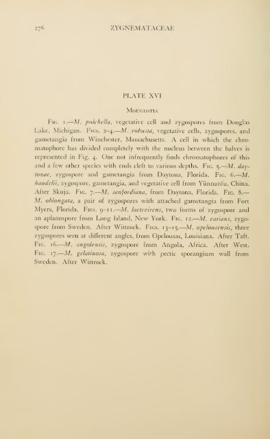
- Page 295: PLATE XV 275
- Page 299 and 300: PLATE XVII 279
- Page 301 and 302: PLATE XVIII 281
- Page 303 and 304: PLATE XIX 283
- Page 305 and 306: PLATE XX 285
- Page 307 and 308: PLATE XXI 287
- Page 309 and 310: PLATE XXII 289
- Page 311 and 312: PLATE XXIII 291
- Page 313 and 314: PLATE XXIV 293
- Page 315 and 316: PLATE XXV 295
- Page 317 and 318: PLATE XXVI 297
- Page 319 and 320: PLATE XXVII 299
- Page 321 and 322: PLATE XXVIII 301
- Page 323 and 324: PLATE XXIX 303
- Page 325 and 326: PLATE XXX 305
- Page 327 and 328: PLATE XXXI 307
- Page 329 and 330: PLATE XXXII 309
- Page 331 and 332: PLATE XXXIII 311
- Page 333 and 334: PLATE XXXIV 313
- Page 335 and 336: PLATE XXXV 315
- Page 337 and 338: PLATE XXXVI 317
- Page 339 and 340: PLATE XXXVII 319
- Page 341 and 342: PLATE XXXVIII 321
- Page 343 and 344: PLATE XXXIX 323
- Page 345 and 346: PLATE XL 325
- Page 347:
PLATE XLI 327


