- Page 2 and 3:
10 ib
- Page 7:
THE ZYGNEMATACEAE
- Page 10 and 11:
GRADUATE SCHOOL MONOGRAPHS Contribu
- Page 13 and 14:
INTRODUCTION The absence of a moder
- Page 15:
INTRODUCTION ix Illinois State Teac
- Page 19 and 20: Plat€ ILLUSTRATIONS Plates Page I
- Page 21: THE ZYGNEMATACEAE
- Page 24 and 25: 4 ZYGNEMATACEAE gametes is by means
- Page 26 and 27: 6 ZYGNEMATACEAE are common but are
- Page 28 and 29: 8 ZYGNEMATACEAE between cells, the
- Page 30 and 31: 10 ZYGNEMATACEAE sporiferous cells
- Page 32 and 33: 12 ZYGNEMATACEAE TABLE I Summary of
- Page 34 and 35: 14 ZYGNEMATACEAE In higher latitude
- Page 36 and 37: i6 ZYGNEMATACEAE specimens with mat
- Page 38 and 39: 1 ZYGNEMATACEAE II. Spores usually
- Page 40 and 41: 20 ZYGNEMATACEAE 32. Diameter veget
- Page 42 and 43: 22 ZYGNEMATACEAE REPRODUCTION BY AP
- Page 44 and 45: 24 ZYGNEMATACEAE spores formed in t
- Page 46 and 47: 26 ZYGNEMATACEAE pitted; pits 5-6 /
- Page 48 and 49: 28 ZYGNEMATACEAE spores are formed
- Page 50 and 51: 30 ZYGNEMATACEAE spores formed in t
- Page 52 and 53: 32 ZYGNEMATACEAE biculate; pits 4.5
- Page 54 and 55: 34 ZYGNEMATACEAE United States: Ill
- Page 56 and 57: 36 ZYGNEMATACEAE zygospores globose
- Page 58 and 59: 38 ZYGNEMATACEAE spores formed in o
- Page 60 and 61: 40 ZYGNEMATACEAE spores in one of t
- Page 62 and 63: 42 ZYGNEMATACEAE an outer pectic la
- Page 64 and 65: 44 ZYGNEMATACEAE irregular in form
- Page 66 and 67: 46 ZYGNEMATACEAE cylindncum Transea
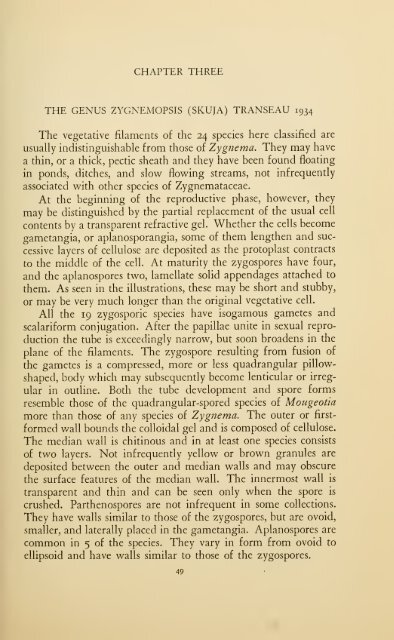
- Page 70 and 71: 50 ZYGNEMATACEAE When the zygospore
- Page 72 and 73: 52 ZYGNEMATACEAE Descriptions of Sp
- Page 74 and 75: 54 ZYGNEMATACEAE scrobiculate, usua
- Page 76 and 77: 56 ZYGNEMATACEAE i6. Zygnemopsis qu
- Page 78 and 79: 58 ZYGNEMATACEAE 23. Zygnemopsis pe
- Page 81: CHAPTER FOUR THE GENUS HALLASIA ROS
- Page 84 and 85: 64 ZYGNEMATACEAE soils, rocks, and
- Page 86 and 87: 66 ZYGNEMATACEAE igan, GDlorado, an
- Page 88 and 89: 68 ZYGNEMATACEAE globose bodies; re
- Page 91: CHAPTER SIX THE GENUS PLEURODISCUS
- Page 94 and 95: 74 ZYGNEMATACEAE among the Zygnemat
- Page 96 and 97: 76 ZYGNEMATACEAE Key to the Species
- Page 98 and 99: 78 ZYGNEMATACEAE Vegetative cells 8
- Page 100 and 101: 8o ZYGNEMATACEAE occupying only a p
- Page 102 and 103: 82 ZYGNEMATACEAE 2^^i in thickness.
- Page 104 and 105: 84 ZYGNEMATACEAE 17. Diameter veget
- Page 106 and 107: 86 ZYGNEMATACEAE 41. Spore wall scr
- Page 108 and 109: 88 ZYGNEMATACEAE 66. Zygospores col
- Page 110 and 111: 90 ZYGNEMATACEAE Vegetative cells 8
- Page 112 and 113: 92 ZYGNEMATACEAE 10. MouGEOTiA DROU
- Page 114 and 115: 94 Philippine Islands; Africa. ZYGN
- Page 116 and 117: 96 ZYGNEMATACEAE 27. MouGEOTiA MECA
- Page 118 and 119:
98 ZYGNEMATACEAE 36. MouGEOTiA GLOB
- Page 120 and 121:
100 ZYGNEMATACEAE United States: Wi
- Page 122 and 123:
102 ZYGNEMATACEAE outer pectic laye
- Page 124 and 125:
104 ZYGNEMATACEAE 60. MoucEOTiA TRA
- Page 126 and 127:
io6 ZYGNEMATACEAE 22-26 /i.; spore
- Page 128 and 129:
io8 ZYGNEMATACEAE with concave side
- Page 130 and 131:
no ZYGNEMATACEAE Differs from A/, p
- Page 132 and 133:
112 ZYGNEMATACEAE 90. MoucEOTiA TRO
- Page 134 and 135:
114 ZYGNEMATACEAE 98. MouGEOTiA coT
- Page 136 and 137:
ii6 ZYGNEMATACEAE pahidosa G. S. We
- Page 138 and 139:
ii8 ZYGNEMATACEAE Descriptions ok S
- Page 141:
CHAPTER TWELVE THE GENUS ENTRANSIA
- Page 144 and 145:
124 ZYGNEMATACEAE insists that cell
- Page 146 and 147:
126 ZYGNEMATACEAE cells, or the thi
- Page 148 and 149:
128 ZYGNEMATACEAE Spore form, spore
- Page 150 and 151:
130 ZYGNEMATACEAE END WALLS PLANE U
- Page 152 and 153:
132 ZYGNEMATACEAE 20. Chromatophore
- Page 154 and 155:
134 ZYGNEMATACEAE 39. Diameter vege
- Page 156 and 157:
136 ZYGNEMATACEAE 57. Spore diamete
- Page 158 and 159:
138 ZYGNEMATACEAE 8i 8i SPORES NOT
- Page 160 and 161:
140 ZYGNEMATACEAE 92. Fertile cells
- Page 162 and 163:
142 ZYGNEMATACEAE no. Vegetative ce
- Page 164 and 165:
144 ZYGNEMATACEAE 124. Median spore
- Page 166 and 167:
146 ZYGNEMATACEAE 144. Zygospores 2
- Page 168 and 169:
148 ZYGNEMATACEAE 169. Zygospores e
- Page 170 and 171:
150 ZYGNEMATACEAE 186. With 6 to 8
- Page 172 and 173:
152 ZYGNEMATACEAE 6. Spirogyra cond
- Page 174 and 175:
154 ZYGNEMATACEAE nation of dimensi
- Page 176 and 177:
156 Czechoslovakia; Tibet; China. Z
- Page 178 and 179:
158 ZYGNEMATACEAE United States: Mi
- Page 180 and 181:
i6o ZYGNEMATACEAE Vegetative cells
- Page 182 and 183:
1 62 ZYGNEMATACEAE 41. Spirogyra oc
- Page 184 and 185:
164 ZYGNEMATACEAE by both gametangi
- Page 186 and 187:
i66 ZYGNEMATACEAE 34-38^1 X 58-68 /
- Page 188 and 189:
1 68 ZYGNEMATACEAE aplanospores bro
- Page 190 and 191:
170 ZYGNEMATACEAE 73. Spirogyra hya
- Page 192 and 193:
172 ZYGNEMATACEAE Vegetative cells
- Page 194 and 195:
174 ZYGNEMATACEAE 89. Spirocyra spl
- Page 196 and 197:
176 ZYGNEMATACEAE spores and 1-2 ch
- Page 198 and 199:
178 ZYGNEMATACEAE Vegetative cells
- Page 200 and 201:
i8o ZYGNEMATACEAE 114. Spirogyra sc
- Page 202 and 203:
1 82 ZYGNEMATACEAE 122. Spirogyra s
- Page 204 and 205:
184 ZYGNEMATACEAE 130. Spirogyra mi
- Page 206 and 207:
1 86 ZYGNEMATACEAE 137. Spirocyra o
- Page 208 and 209:
i88 ZYGNEMATACEAE 146. Spirogyra cy
- Page 210 and 211:
190 ZYGNEMATACEAE 154. Spirogyra si
- Page 212 and 213:
192 ZYGNEMATACEAE chromatophores, m
- Page 214 and 215:
194 ZYGNEMATACEAE Most of the early
- Page 216 and 217:
196 ZYGNEMATACEAE iform; tubes form
- Page 218 and 219:
198 ZYGNEMATACEAE in which the tube
- Page 220 and 221:
20O ZYGNEMATACEAE 187. Spirckiyra e
- Page 222 and 223:
202 ZYGNEMATACEAE Vegetative cells
- Page 224 and 225:
204 ZYGNEMATACEAE and scalariform;
- Page 226 and 227:
2o6 ZYGNEMATACEAE United States: Io
- Page 228 and 229:
2o8 ZYGNEMATACEAE chromatophore, ma
- Page 230 and 231:
210 ZYGNEMATACEAE formed by the mal
- Page 232 and 233:
212 ZYGNEMATACEAE ctangia; fertile
- Page 234 and 235:
214 ZYGNEMATACEAE 2 chromatophores,
- Page 236 and 237:
2i6 ZYGNEMATACEAE United States: Il
- Page 238 and 239:
2i8 ZYGNEMATACEAE 254. Spirogyra na
- Page 240 and 241:
220 ZYGNEMATACEAE subglobose, 41-48
- Page 242 and 243:
222 ZYGNEMATACEAE 269. Spirogyra iN
- Page 244 and 245:
224 ZYGNEMATACEAE braziliensis (Nor
- Page 246 and 247:
226 ZYGNEMATACEAE inflata (Vaucher)
- Page 248 and 249:
228 ZYGNEMATACEAE punctijormis Tran
- Page 251 and 252:
CHAPTER FOURTEEN THE GENUS SIROGONI
- Page 253 and 254:
SIROGONIUM 233 Fig. G.—Conjugatio
- Page 255 and 256:
Coll.). SIROGONIUM 235 United State
- Page 257:
INDEX
- Page 260 and 261:
240 crassa (Mougcotia), 95 crassa (
- Page 262 and 263:
242 ZYGNEMATACEAE minor (Spirogyra)
- Page 264 and 265:
244 succica (Spirogyra), XXIII, 7;
- Page 266 and 267:
246 ZY(;\KMATACEAE PLATE I Ri PRoiu
- Page 268 and 269:
248 ZYGNEMATACEAE PLATE II Zygnema
- Page 270 and 271:
250 ZYGNEMATACEAE PLATE III Zygnema
- Page 272 and 273:
252 zygnemataceaf: PL ATI-. IV ZvGN
- Page 274 and 275:
ZYCiNEMATACHAE PLATE V Zygnema Fig.
- Page 276 and 277:
256 ZVGNEMATACEAE PLATE \'I ZvCNliN
- Page 278 and 279:
2
- Page 280 and 281:
26o ZYCJNEMATACEAE PLATE VIII Zygne
- Page 282 and 283:
262 ZYGNEMATACHAF. PLATH IX Zygnemo
- Page 284 and 285:
264 ZYGNEMATACEAE PLATE X Hallasia
- Page 286 and 287:
266 ZYGNEMATACEAE PLATE XI Zygogoni
- Page 288 and 289:
268 ZYC;.\HMATACEAE PLATE XII ZVGOG
- Page 290 and 291:
zy(;nemataceae PLATE XIII MoUGEOTIA
- Page 292 and 293:
272 ZYGNEMATACEAE PLATE XI\' MoUGEO
- Page 294 and 295:
274 ZYCINEMATACEAE PLATE X\' Mot'GE
- Page 296 and 297:
276 ZYCNFAfATACEAE PLATE XVI MOUGEO
- Page 298 and 299:
.78 ZYGNEMATACEAE PLATE XVII MOUGEO
- Page 300 and 301:
28o ZYGNEMATACEAE PLATK XVIII MOUGE
- Page 302 and 303:
282 ZYGNEMATACEAE PLATE XIX MOUGEOT
- Page 304 and 305:
284 ZYGNEMATACEAE PLATE XX Temnogam
- Page 306 and 307:
i86 ZYGNEMATACEAE PLATE XXI Spirogy
- Page 308 and 309:
288 ZYGNEMATACEAE PLATE XXII Spirog
- Page 310 and 311:
290 zv(;nemataceae PLATE XXIir Spir
- Page 312 and 313:
292 ZYCINEMATACEAE PLATE XXIV Spiro
- Page 314 and 315:
294 ZYGNEMATACEAE PLATE XXV Spirogv
- Page 316 and 317:
296 ZYGNEMATACEAE PLATE XX\'I Spiro
- Page 318 and 319:
298 ZYGNEMATACEAE PLATE XXVII Spiro
- Page 320 and 321:
300 Z^'(;NE\fATACF.AH PLATE XXVIII
- Page 322 and 323:
302 zv(;nfmataceae PLATE XXIX Spiko
- Page 324 and 325:
304 ZYGNEMATACEAE PLATE XXX Spikogy
- Page 326 and 327:
3o6 ZYGNEMATACEAE PLATE XXXI Spikog
- Page 328 and 329:
3o8 ZYGNEMATACEAE PLATE XXXII Spiro
- Page 330 and 331:
3IO ZYCJNHMA'rACEAH PLATl'. XXXIII
- Page 332 and 333:
312 ZYGNEMATACEAE PLATE XXXIV Spiro
- Page 334 and 335:
314 ZYCJNEMATACEAE PLATE XXXV Spiko
- Page 336 and 337:
3i6 ZYCINEMATACEAE PLATE XXXVI Spir
- Page 338 and 339:
3i8 zv(;\i;mata(:i-..\k PLATE XXX \
- Page 340 and 341:
320 ZYGNEMATACEAE PLATE XXXVIII Spi
- Page 342 and 343:
322 ZVCXKMATACEAE PLATE XXXIX Spiro
- Page 344 and 345:
324 Z^'CNFMATACHAH PLATE XL SlROGON
- Page 346 and 347:
326 ZVCiNHMATACEAE PLATE XLI /yg(k7


