FORENSIC TOXICOLOGY - Bio Medical Forensics
FORENSIC TOXICOLOGY - Bio Medical Forensics
FORENSIC TOXICOLOGY - Bio Medical Forensics

You also want an ePaper? Increase the reach of your titles
YUMPU automatically turns print PDFs into web optimized ePapers that Google loves.
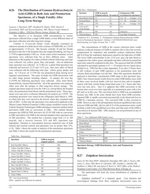
K26 The Distribution of Gamma-Hydroxybutyric<br />
Acid (GHB) in Both Ante and Postmortem<br />
Specimens, of a Single Fatality After<br />
Long-Term Storage<br />
Laureen J. Marinetti, MS*, Bradford R. Hepler, PhD, Daniel S.<br />
Isenschmid, PhD, and Sawait Kanluen, MD, Wayne County <strong>Medical</strong><br />
Examiner’s Office, 1300 East Warren Avenue, Detroit, MI<br />
The objective is to document GHB concentrations in various<br />
specimens collected from a single GHB fatality at four different times of<br />
specimen collection and after long-term storage.<br />
Content: A 15-year-old female victim allegedly consumed an<br />
unknown amount of a drink laced with a mixture of GHB/GBL on 1/16/99<br />
at approximately 11:30 p.m. She became violently ill and her friends<br />
decided to take her to the hospital when she stopped breathing, arriving on<br />
1/17/99 at approximately 5:00 a.m. In the course of her treatment, several<br />
specimens were collected from her at three different times. Upon<br />
admission to the hospital, five tubes of blood with the following color tops<br />
were collected: red, yellow, green, blue, and purple. Also an admission<br />
urine specimen was collected. At 11:00 a.m. a spinal fluid specimen was<br />
collected and between 12:35 and 12:55 p.m., four more tubes of blood<br />
were collected with the following color tops: yellow, green, purple, and<br />
gray. At 7:34 p.m. on 1/17/99 she was pronounced dead, having never<br />
regained consciousness. The cause of death was GHB intoxication with<br />
no other drugs present including ethanol. At autopsy the next day<br />
(1/18/99) the following specimens were collected: urine, heart blood,<br />
femoral blood, vitreous fluid and bile. All of the specimens were analyzed<br />
either soon after the autopsy or after long-term freezer storage. The<br />
original specimens analyzed were the 5:00 a.m. red top blood, the hospital<br />
urine, the postmortem heart blood, and the postmortem urine. These specimens<br />
were sent out to a reference laboratory for analysis on 1/19/99. The<br />
remaining specimens were stored in the refrigerator until March 6, 1999,<br />
at which time they were moved to the freezer where they remained until<br />
July of 2001. At this time the specimens were analyzed in duplicate at the<br />
Wayne County <strong>Medical</strong> Examiner’s Office using a modified version of the<br />
United Chemical Technologies GHB solid phase extraction method which<br />
derivatizes GHB with no conversion to GBL. This method directly<br />
measures the GHB di-TMS derivative by EI GC/MS with no conversion<br />
to GBL and utilizes d-6 GHB as the internal standard with a specimen size<br />
of 200 microliters. The method has a linearity range from 2.5 to 100<br />
mcg/mL, and a recovery between 35% and 60% (specimen type<br />
dependent). Variation in concentration between analytical runs of both<br />
internal and external controls and specimens was no greater than 15%,<br />
n=54. Table 1 shows the results of analysis of the antemortem specimens<br />
and Table 2 shows the results for the postmortem specimens.<br />
Table One - Antemortem<br />
Specimen GHB in mcg/ml<br />
original analysis re-analysis<br />
Red Top Bld 5:00a.m. 510 466<br />
Yellow Top Bld 5:00a.m. NP 566<br />
Yellow Top Bld 12:55p.m. NP 187<br />
Green Top Bld 5:00a.m. NP QNS<br />
Green Top Bld 12:35p.m. NP 232<br />
Purple Top Bld 5:00a.m. NP QNS<br />
Purple Top Bld 12:55p.m. NP 242<br />
Blue Top Bld 5:00a.m. NP 418<br />
Gray Top Bld 12:55p.m. NP 93<br />
Spinal Fluid 11:00a.m. NP 220<br />
Urine 5:00m<br />
NP = test not performed<br />
2300 QNS<br />
QNS = quantity not sufficient for analysis<br />
Table Two – Postmortem<br />
Specimen GHB in mcg/ml Endogenous GHB<br />
Original analysis Re-analysis in postmortem specimens*<br />
Heart Blood (NaFl) 15 12 1.6 – 36 Average = 12<br />
Femoral Blood NP 18 1.7 – 48 Average = 11<br />
Urine 150 102 0 – 14 Average = 4.6<br />
Urine at pH =<br />
14 for 30 min.<br />
NP 167 NA<br />
Bile NP 48 Unknown<br />
Vitreous Humor NP 127 Less than 10 mcg/ml<br />
*Anderson, D.T., Kuwahara, T., “Endogenous Gamma Hydroxybutyrate (GHB)<br />
levels in Postmortem Specimens”, Abstract CAT/NWAFS/SWAFS/SAT combined<br />
meeting, Las Vegas, Nevada, Nov. 7, 1997.<br />
The concentrations of GHB at the various collection times would<br />
indicate a reduced clearance of GHB in a patient who is alive but severely<br />
compromised by respiratory and metabolic acidosis (admission blood<br />
pH=6.8) from the respiratory depression caused by the high dose of GHB.<br />
The lower GHB concentration in the 12:55 p.m. gray top tube blood as<br />
compared to the yellow, green, and purple top tubes collected at around the<br />
same time cannot be explained at this time. The apparent half-life of GHB<br />
calculated in narcoleptic patients is 53 +/- 19 minutes for two 3gram doses<br />
(Scharf et al., 1998) but this is dose dependent. Even after 14 hours there<br />
is still evidence of past GHB ingestion in the postmortem urine and<br />
vitreous fluid and perhaps even the bile. More bile specimens should be<br />
analyzed to determine a postmortem GHB range in this specimen type.<br />
Table 3 has vitreous humor results from two other GHB fatality cases, both<br />
of these cases involved deaths with less survival time and no hospitalization<br />
with both victims being found dead some 5 to 7 hours after they<br />
were last seen alive. Also it is apparent that GHB conversion to the<br />
lactone does occur over time especially in a postmortem urine with a low<br />
pH such as in this case. The conversion back to GHB was accomplished<br />
because any GBL in the urine should have been from GHB excretion.<br />
Ingested GBL is rapidly converted enzymatically in the blood and liver to<br />
GHB by a lactonase enzyme. The converted GBL is then excreted as<br />
GHB. However, due to the pH dependent chemical equilibrium that exists<br />
between GHB and GBL, the low pH (4.2) of the postmortem urine would<br />
facilitate a shift in the equilibrium over time such that some of the GHB is<br />
converted to its lactone GBL. GBL analysis was not performed on any of<br />
these specimens.<br />
Table 3 – Two Additional GHB Fatality Cases<br />
Postmortem Specimen Case 1 GHB in mcg/mL Ethanol in g/dL<br />
Heart Blood 66 ND<br />
Femoral Blood 77 ND<br />
Vitreous Humor 85 ND<br />
Urine 1260 ND<br />
Postmortem Specimen Case 2<br />
Blood 400 .22<br />
Vitreous Humor 212 .12<br />
Specimen Storage, GHB Distribution, Ante and Postmortem<br />
Specimens<br />
K27 Zolpidem Intoxication<br />
Nancy B. Wu Chen PhD*, Edmund R. Donoghue MD, Aldo J. Fusaro, DO,<br />
Nancy L. Jones, MD, Barry D. Lifschultz, MD, Jennifer L. Jakalski, BS,<br />
Kathleen A. Mittel, BS, and James R. Reich Jr., MLT, Office of the <strong>Medical</strong><br />
Examiner, Cook County, 2121 West Harrison Street, Chicago, IL<br />
The participant will learn the tissue distribution of zolpidem in<br />
three cases.<br />
Zolpidem (Ambien® ) is a hypnotic agent. Case histories and<br />
toxicological findings from three zolpidem intoxication cases are<br />
271 * Presenting Author