Silica (crystalline, respirable) - OEHHA
Silica (crystalline, respirable) - OEHHA
Silica (crystalline, respirable) - OEHHA

You also want an ePaper? Increase the reach of your titles
YUMPU automatically turns print PDFs into web optimized ePapers that Google loves.
FINAL February 2005<br />
C. <strong>Silica</strong> exposure and lung cancer in workers<br />
In 1997, IARC classified <strong>respirable</strong> <strong>crystalline</strong> silica in Class 1, a Known Human Carcinogen,<br />
based on occupational epidemiologic studies. However, chronic RELs are not based on cancer<br />
endpoints. Further, there is no approved cancer potency factor for silica.<br />
V. Effects of Animal Exposures<br />
Several papers have reported that freshly fractured quartz, which has increased surface activity,<br />
causes greater inflammation than "aged" quartz. Vallyathan et al. (1991) reported that “fresh”<br />
silica was 4.2-fold more potent than silica aged for 1-2 days in decreasing the membrane<br />
integrity of male rat macrophages; 50% more potent in activating hydrogen peroxide secretion<br />
by macrophages; and 4.6-fold more potent in stimulating cellular chemiluminescence.<br />
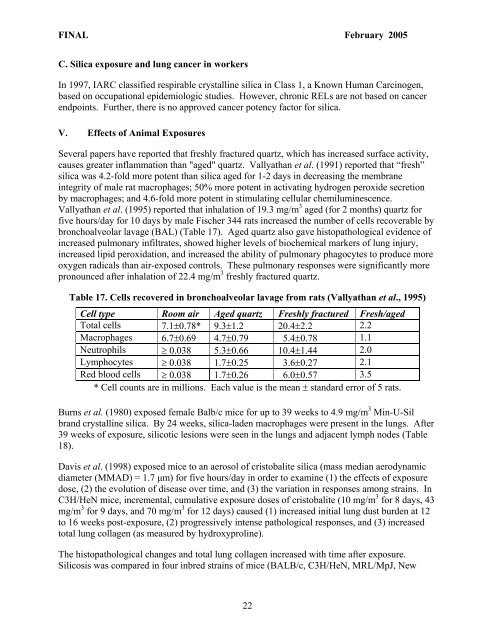
Vallyathan et al. (1995) reported that inhalation of 19.3 mg/m 3 aged (for 2 months) quartz for<br />
five hours/day for 10 days by male Fischer 344 rats increased the number of cells recoverable by<br />
bronchoalveolar lavage (BAL) (Table 17). Aged quartz also gave histopathological evidence of<br />
increased pulmonary infiltrates, showed higher levels of biochemical markers of lung injury,<br />
increased lipid peroxidation, and increased the ability of pulmonary phagocytes to produce more<br />
oxygen radicals than air-exposed controls. These pulmonary responses were significantly more<br />
pronounced after inhalation of 22.4 mg/m 3 freshly fractured quartz.<br />
Table 17. Cells recovered in bronchoalveolar lavage from rats (Vallyathan et al., 1995)<br />
Cell type Room air Aged quartz Freshly fractured Fresh/aged<br />
Total cells 7.1±0.78* 9.3±1.2 20.4±2.2 2.2<br />
Macrophages 6.7±0.69 4.7±0.79 5.4±0.78 1.1<br />
Neutrophils ≥ 0.038 5.3±0.66 10.4±1.44 2.0<br />
Lymphocytes ≥ 0.038 1.7±0.25 3.6±0.27 2.1<br />
Red blood cells ≥ 0.038 1.7±0.26 6.0±0.57 3.5<br />
* Cell counts are in millions. Each value is the mean ± standard error of 5 rats.<br />
Burns et al. (1980) exposed female Balb/c mice for up to 39 weeks to 4.9 mg/m 3 Min-U-Sil<br />
brand <strong>crystalline</strong> silica. By 24 weeks, silica-laden macrophages were present in the lungs. After<br />
39 weeks of exposure, silicotic lesions were seen in the lungs and adjacent lymph nodes (Table<br />
18).<br />
Davis et al. (1998) exposed mice to an aerosol of cristobalite silica (mass median aerodynamic<br />
diameter (MMAD) = 1.7 µm) for five hours/day in order to examine (1) the effects of exposure<br />
dose, (2) the evolution of disease over time, and (3) the variation in responses among strains. In<br />
C3H/HeN mice, incremental, cumulative exposure doses of cristobalite (10 mg/m 3 for 8 days, 43<br />
mg/m 3 for 9 days, and 70 mg/m 3 for 12 days) caused (1) increased initial lung dust burden at 12<br />
to 16 weeks post-exposure, (2) progressively intense pathological responses, and (3) increased<br />
total lung collagen (as measured by hydroxyproline).<br />
The histopathological changes and total lung collagen increased with time after exposure.<br />
Silicosis was compared in four inbred strains of mice (BALB/c, C3H/HeN, MRL/MpJ, New<br />
22