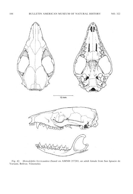
106 BULLETIN AMERICAN MUSEUM OF NATURAL HISTORY NO. 322 Fig. 42. Monodelphis brevicaudata (based on AMNH 257203, an adult female from San Ignacio de Yuruaní, Bolívar, Venezuela).
2009 VOSS AND JANSA: DIDELPHID MARSUPIALS 107 cealing nasal orifice from dorsal view), <strong>and</strong> conspicuously widened posteriorly near maxillary-frontal suture. Maxillary turbinals (viewed through the nasal orifice) simple or sparsely ornamented scrolls, not elaborately branched. Lacrimal foramina (usually two on each side) prominently exposed on orbital margin or on face anterior to orbit. Orbits small, interorbital region more or less parallel sided (usually without conspicuous constrictions); supraorbital margins smoothly rounded, without beads or distinct postorbital processes (but blunt, indistinct processes occasionally developed in large specimens <strong>of</strong> some species; e.g., M. emiliae). Parietal <strong>and</strong> alisphenoid in contact on lateral braincase (no frontal-squamosal contact). Sagittal crest absent (e.g., in M. theresa) or small (usually not extending to frontals; e.g., in M. brevicaudata). Petrosal not exposed laterally through fenestra in parietal-squamosal suture (fenestra absent). Parietal-mastoid contact usually present (interparietal seldom contacts squamosal). Maxillopalatine fenestrae present; palatine fenestrae usually absent; maxillary fenestrae absent; posterolateral palatal foramina small, not extending anteriorly between M4 protocones; posterior palatal morphology conforms to Didelphis morphotype (with moderately well-developed lateral corners, the choanae somewhat constricted behind). Maxillary <strong>and</strong> alisphenoid in contact on floor <strong>of</strong> orbit (not separated by palatine). Transverse canal foramen present. Alisphenoid tympanic process smoothly globular; posteromedial lamina forming secondary foramen ovale present in some species (e.g., M. theresa) or lamina <strong>and</strong> secondary foramen ovale absent (e.g., in M. brevicaudata). Anterior limb <strong>of</strong> ectotympanic suspended directly from basicranium. Stapes triangular with large obturator foramen (e.g., in M. brevicaudata), or columellar <strong>and</strong> microperforate or imperforate (e.g., in M. peruviana). Fenestra cochleae exposed in most species, but fenestra concealed in sinus formed by rostral <strong>and</strong> caudal tympanic processes <strong>of</strong> petrosal in M. emiliae. Paroccipital process small, rounded, <strong>and</strong> adnate to petrosal. Dorsal margin <strong>of</strong> foramen magnum bordered by supraoccipital <strong>and</strong> exoccipitals, incisura occipitalis present. Two mental foramina present on lateral surface <strong>of</strong> each hemim<strong>and</strong>ible; angular process acute <strong>and</strong> strongly inflected. Unworn crowns <strong>of</strong> I2–I5 symmetrically rhomboidal (‘‘premolariform’’), with subequal anterior <strong>and</strong> posterior cutting edges, <strong>and</strong> usually increasing in length (mesiodistal dimension) from I2 to I5. Upper canine (C1) alveolus in premaxillary-maxillary suture; C1 usually simple (without accessory cusps), but small posterior accessory cusp sometimes present (e.g., in M. peruviana). First upper premolar (P1) smaller than posterior premolars but well formed <strong>and</strong> not vestigial; third upper premolar (P3) taller than P2; P3 with posterior cutting edge only; upper milk premolar (dP3) large <strong>and</strong> molariform. Molars highly carnassialized (postmetacristae much longer than postprotocristae); relative widths consistently M1 , M2 , M3 , M4; centrocrista strongly inflected labially on M1– M3; ect<strong>of</strong>lexus shallow on M1, deeper on M2, <strong>and</strong> consistently deep on M3; anterolabial cingulum <strong>and</strong> preprotocrista discontinuous (anterior cingulum incomplete) on M3. Last upper tooth to erupt is P3 in some species (e.g., M. peruviana), or P3 <strong>and</strong> M4 erupt simultaneously (e.g., in M. brevicaudata). Lower incisors (i1–i4) with distinct lingual cusps. Second lower premolar (p2) subequal in height to p3 (e.g., in M. brevicaudata), or p3 taller than p2 (e.g., in M. emiliae); lower milk premolar (dp3) trigonid usually complete (tricuspid). Hypoconid lingual to protoconid (not labially salient) on m3; hypoconulid twinned with entoconid on m1–m3; entoconid subequal to or smaller than hypoconulid on m1–m3. DISTRIBUTION: Species <strong>of</strong> Monodelphis occur in lowl<strong>and</strong> <strong>and</strong> montane rain forests <strong>and</strong> dry forests from eastern Panama throughout most <strong>of</strong> tropical <strong>and</strong> subtropical South America to about 37uS in eastern Argentina (see range maps in Pine <strong>and</strong> H<strong>and</strong>ley, 2008); recorded elevations range from sea level to at least 2500 m (on the eastern slopes <strong>of</strong> the tropical Andes). The apparent absence <strong>of</strong> Monodelphis from the trans-Andean lowl<strong>and</strong>s <strong>of</strong> western South America is noteworthy, but this is possibly an artifact <strong>of</strong> inadequate collecting; a representative <strong>of</strong> the M. adusta complex is rumored to occur along the Pacific littoral