phylogenetic relationships and classification of didelphid marsupials ...
phylogenetic relationships and classification of didelphid marsupials ...
phylogenetic relationships and classification of didelphid marsupials ...
You also want an ePaper? Increase the reach of your titles
YUMPU automatically turns print PDFs into web optimized ePapers that Google loves.
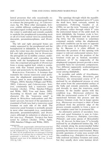
2009 VOSS AND JANSA: DIDELPHID MARSUPIALS 39<br />
lateral processes that only occasionally extend<br />
posteriorly into the mesopterygoid fossa<br />
to contact the pterygoids (e.g., in Caluromysiops;<br />
fig. 39). Most other <strong>marsupials</strong>, however,<br />
exhibit different configurations <strong>of</strong> the<br />
nasopharyngeal ro<strong>of</strong>ing bones. In particular,<br />
the vomer is undivided <strong>and</strong> extends caudally<br />
to underlie the presphenoid (concealing most<br />
or all <strong>of</strong> it from ventral view) in caenolestids,<br />
dasyurids, peramelemorphians, <strong>and</strong> Dromiciops.<br />
The left <strong>and</strong> right pterygoid bones are<br />
widely separated by the presphenoid <strong>and</strong> the<br />
basisphenoid in <strong>didelphid</strong>s. In other <strong>marsupials</strong>,<br />
the vomer may also extend between the<br />
left <strong>and</strong> right pterygoids, but in Dromiciops<br />
the pterygoids are in midline contact, underlying<br />
the presphenoid <strong>and</strong> concealing its<br />
suture with the basisphenoid from ventral<br />
view; the conjoined pterygoids <strong>of</strong> Dromiciops<br />
form a strong sagittal keel, which is continuous<br />
with that formed anteriorly by the<br />
vomer <strong>and</strong> posteriorly by the basisphenoid. 10<br />
BASICRANIAL FORAMINA: A foramen that<br />
transmits the venous transverse canal perforates<br />
the alisphenoid anterolateral to the<br />
carotid canal in most <strong>didelphid</strong>s (Sánchez-<br />
Villagra, 2001b; Sánchez-Villagra <strong>and</strong> Wible,<br />
2002). Caluromys <strong>and</strong> Caluromysiops, however,<br />
consistently lack a transverse canal<br />
foramen (Archer, 1976a; Sánchez-Villagra<br />
<strong>and</strong> Wible, 2002; Voss <strong>and</strong> Jansa, 2003).<br />
According to Kirsch <strong>and</strong> Archer (1982:<br />
character 24), the ‘‘transverse canal’’ is<br />
absent in Metachirus nudicaudatus <strong>and</strong> Phil<strong>and</strong>er<br />
opossum, but most <strong>of</strong> the specimens we<br />
examined <strong>of</strong> both taxa had a transverse canal<br />
foramen on each side <strong>of</strong> the skull. The<br />
transverse canal foramen is normally present<br />
in most other <strong>marsupials</strong> (e.g., caenolestids,<br />
dasyurids, peramelemorphians, <strong>and</strong> Dromiciops),<br />
although its reduction or absence in a<br />
few taxa (e.g., Planigale, Tarsipes) has been<br />
noted by authors (Archer, 1976a; Aplin,<br />
1990; Sánchez-Villagra <strong>and</strong> Wible, 2002).<br />
10 The obviously autapomorphic midventral basicranial keel<br />
<strong>of</strong> Dromiciops is usually described as formed by the presphenoid<br />
<strong>and</strong> the basisphenoid (e.g., by Hershkovitz, 1999; Giannini et<br />
al., 2004), but the presphenoid does not participate in forming<br />
this structure in any specimen that we examined. Note that the<br />
presphenoid is incorrectly labeled as the vomer in some<br />
illustrated ventral views <strong>of</strong> the <strong>didelphid</strong> skull (e.g., Novacek,<br />
1993: fig. 9.4B).<br />
The openings through which the m<strong>and</strong>ibular<br />
division <strong>of</strong> the trigeminal nerve (V 3 ) exits<br />
the skull have been variously named by<br />
systematists. Following Gaudin et al.<br />
(1996), we use the term ‘‘foramen ovale’’ for<br />
the primary orifice through which V 3 exits<br />
the endocranial lumen <strong>of</strong> the adult skull. In<br />
most <strong>didelphid</strong>s, the foramen ovale is bordered<br />
by the alisphenoid <strong>and</strong> the petrosal<br />
(fig. 16A), but the foramen is sometimes<br />
contained entirely within the alisphenoid,<br />
<strong>and</strong> both conditions can be seen on opposite<br />
sides <strong>of</strong> the same skull (Gaudin et al., 1996:<br />
fig. 6). Because it is <strong>of</strong>ten difficult to<br />
determine the position <strong>of</strong> this opening with<br />
respect to relevant endocranial sutures, however,<br />
we did not score the position <strong>of</strong> the<br />
foramen ovale per se. Instead, secondary<br />
enclosures <strong>of</strong> V 3 by outgrowths <strong>of</strong> the<br />
alisphenoid tympanic process provide a more<br />
accessible basis for taxonomic comparisons.<br />
Two different (apparently nonhomologous)<br />
conditions <strong>of</strong> secondary nerve enclosure<br />
occur among <strong>didelphid</strong>s.<br />
In juveniles <strong>and</strong> adults <strong>of</strong> Gracilinanus,<br />
Lestodelphys, Marmosops, Metachirus, <strong>and</strong><br />
Thylamys, the extracranial course <strong>of</strong> V 3 is<br />
enclosed by an alisphenoid process or strut<br />
that arises from the anteromedial surface <strong>of</strong><br />
the bulla <strong>and</strong> extends anteriorly, medially,<br />
<strong>and</strong> dorsally to span the transverse canal<br />
foramen. In the smaller species with this<br />
condition (e.g., Marmosops pinheiroi; fig.<br />
16B) the extracranial course <strong>of</strong> V 3 remains<br />
unenclosed between this process <strong>and</strong> the<br />
primary foramen ovale, but in larger species<br />
(e.g., Marmosops noctivagus) a sheet <strong>of</strong> bone<br />
produced from the posterior edge <strong>of</strong> the<br />
process extends caudally to form a more or<br />
less complete canal late in postnatal life.<br />
An alternative pattern <strong>of</strong> secondary foramen<br />
<strong>and</strong> canal formation is seen in Caluromysiops,<br />
Chironectes, Didelphis, Lutreolina,<br />
Phil<strong>and</strong>er, <strong>and</strong> some species <strong>of</strong> Monodelphis<br />
(e.g., M. theresa). In these taxa, V 3 is broadly<br />
enclosed by an alisphenoid lamina (fig. 16C)<br />
that extends along the posteromedial bullar<br />
surface, but there is no anteromedial process<br />
spanning the transverse canal foramen. Many<br />
juvenile specimens <strong>of</strong> some taxa scored with<br />
this condition (e.g., Didelphis) do not have a<br />
fully enclosed secondary foramen <strong>and</strong> canal,<br />
but they usually show some laminar devel-


