Final Risk Analysis - Biosecurity New Zealand
Final Risk Analysis - Biosecurity New Zealand
Final Risk Analysis - Biosecurity New Zealand

You also want an ePaper? Increase the reach of your titles
YUMPU automatically turns print PDFs into web optimized ePapers that Google loves.
Import <strong>Risk</strong> <strong>Analysis</strong>: Cattle<br />
from Australia, Canada, the<br />
European Union, and the<br />
United States of America<br />
FINAL<br />
ISBN 978-0-478-33814-0 (Print)<br />
ISBN 978-0-478-33815-7 (Online)<br />
13 February 2009
This page is intentionally blank
MAF <strong>Biosecurity</strong> <strong>New</strong> <strong>Zealand</strong><br />
Pastoral House<br />
25 The Terrace<br />
PO Box 2526<br />
Wellington 6011<br />
<strong>New</strong> <strong>Zealand</strong><br />
Tel: 64 4 894 0100<br />
Fax: 64 4 894 0731<br />
Policy and <strong>Risk</strong><br />
MAF <strong>Biosecurity</strong> <strong>New</strong> <strong>Zealand</strong><br />
Import <strong>Risk</strong> <strong>Analysis</strong>: Cattle from Australia, Canada, the European Union and the United<br />
States of America<br />
FINAL<br />
ISBN 978-0-478-33814-0 (Print)<br />
ISBN 978-0-478-33815-7 (Online)<br />
13 February 2009<br />
Approved for general release<br />
Christine Reed<br />
Manager, <strong>Risk</strong> <strong>Analysis</strong><br />
MAF <strong>Biosecurity</strong> <strong>New</strong> <strong>Zealand</strong>
This page is intentionally blank
Contents<br />
Executive Summary 1<br />
1. Introduction 2<br />
2. Scope 2<br />
3. Commodity Definition 2<br />
4. <strong>Risk</strong> <strong>Analysis</strong> Methodology 2<br />
5. Akabane and other Simbu Group Viruses 15<br />
6. Aujeszky’s Disease 18<br />
7. Bluetongue 20<br />
8. Borna Disease 23<br />
9. Bovine Calicivirus Infection 29<br />
10. Bovine Herpes Viruses 32<br />
11. Bovine Parvovirus Infection 37<br />
12. Bovine Rhinovirus Infection 39<br />
13. Bovine Viral Diarrhoea Virus 41<br />
14. Crimean Congo Haemorrhagic Fever 46<br />
15. Bovine Ephemeral Fever 50<br />
16. Foot and Mouth Disease 53<br />
17. Miscellaneous Arboviruses 57<br />
18. Palyam Group Viruses 61<br />
19. Rabies 64<br />
20. Ross River and Barmah Forest Viruses 68<br />
21. Tick Borne Encephalitis 70<br />
22. Vesicular Stomatitis 74<br />
23. West Nile Disease 79<br />
24. Bovine Spongifrom Encephalopathy 81<br />
25. Anthrax 86<br />
i
26. Brucellosis 89<br />
27. Bovine Tuberculosis 93<br />
28. Melioidosis 97<br />
29. Mollicutes Infections 99<br />
30. Haemorrhagic Septicaemia 107<br />
31. Salmonellosis 111<br />
32. Leptospirosis 116<br />
33. Lyme Disease 120<br />
34. Spirochaetosis (Borrelia theileri) 124<br />
35. Anaplasmosis (Anaplasma marginale, Anaplasma centrale and Anaplasma caudatum) 125<br />
36. Family Anaplasmataceae Infections (Ehrlichiosis/ Anaplasmosis ) 129<br />
37. Chlamydiosis (Chlamydophila abortus and Chlamydophila pecorum) 133<br />
38. Q Fever 137<br />
39. Haemobartonellosis 141<br />
40. Babesiosis 142<br />
41. Sarcocystosis 146<br />
42. Theileriosis (Theileria annulata) 148<br />
43. Lice 151<br />
44. Mange Mites 154<br />
45. Ticks 157<br />
46. Warble Fly 162<br />
47. Internal Parasites 165<br />
48. Weed Seeds 170<br />
ii
Contributors to this risk analysis<br />
1. Primary Authors<br />
Bob Worthington Contractor to <strong>Biosecurity</strong><br />
<strong>New</strong> <strong>Zealand</strong><br />
Stephen Cobb Senior Adviser, <strong>Risk</strong><br />
<strong>Analysis</strong> (Animal Kingdom)<br />
2. Internal Peer Review<br />
Stuart MacDiarmid Principal International<br />
Adviser, <strong>Risk</strong> <strong>Analysis</strong><br />
Howard Pharo Team Manager, <strong>Risk</strong><br />
<strong>Analysis</strong> (Animal Kingdom)<br />
Lincoln Broad Senior Adviser, <strong>Risk</strong><br />
<strong>Analysis</strong> (Animal Kingdom)<br />
José Derraik Senior Adviser, Pre-clearance<br />
risk analysis (Human Health)<br />
Sandy Toy Senior Adviser, <strong>Risk</strong><br />
<strong>Analysis</strong> (Animal Kingdom)<br />
Gillian Mylrea Team Manager, Import<br />
health standards (Animals)<br />
Leone Basher Senior Adviser, Import health<br />
standards (Animals)<br />
Joanne Thompson Contractor, Import health<br />
standards (Animals)<br />
3. External Scientific Review<br />
Geoff Ryan<br />
Tim Parkinson Professor, Institute of<br />
Veterinary, Animal and<br />
Biomedical Sciences<br />
iii<br />
<strong>Biosecurity</strong> <strong>New</strong> <strong>Zealand</strong>,<br />
Wellington<br />
<strong>Biosecurity</strong> <strong>New</strong> <strong>Zealand</strong>,<br />
Wellington<br />
<strong>Biosecurity</strong> <strong>New</strong> <strong>Zealand</strong>,<br />
Wellington<br />
<strong>Biosecurity</strong> <strong>New</strong> <strong>Zealand</strong>,<br />
Wellington<br />
<strong>Biosecurity</strong> <strong>New</strong> <strong>Zealand</strong>,<br />
Wellington<br />
<strong>Biosecurity</strong> <strong>New</strong> <strong>Zealand</strong>,<br />
Wellington<br />
<strong>Biosecurity</strong> <strong>New</strong> <strong>Zealand</strong>,<br />
Wellington<br />
<strong>Biosecurity</strong> <strong>New</strong> <strong>Zealand</strong>,<br />
Wellington<br />
<strong>Biosecurity</strong> <strong>New</strong> <strong>Zealand</strong>,<br />
Wellington<br />
<strong>Biosecurity</strong> <strong>New</strong> <strong>Zealand</strong>,<br />
Wellington<br />
Ruminant Section Manager Department of Agriculture,<br />
Fisheries and Forestry –<br />
Australia<br />
Massey University, <strong>New</strong><br />
<strong>Zealand</strong>
Executive Summary<br />
The risks associated with the importation of cattle from Australia, Canada, the European<br />
Union (27 countries), and the United States of America have been examined. Only risks<br />
associated with the importation of infectious organisms or parasites have been considered.<br />
Of an initial list of 93 micro organisms or groups of organisms, 43 disease agents or groups of<br />
disease agents/diseases that are exotic to <strong>New</strong> <strong>Zealand</strong> or are the subject of a national<br />
eradication campaign in <strong>New</strong> <strong>Zealand</strong>, were included in a preliminary hazard list. Thirty four<br />
of these were considered to be potential hazards and were subjected to a risk assessment.<br />
A non-negligible risk was identified with the following hazards:<br />
• Borna disease virus<br />
• Exotic bovine herpes viruses<br />
• Bovine viral diarrhoea virus type 2<br />
• Crimean Congo haemorrhagic fever virus<br />
• Bovine ephemeral fever virus<br />
• Foot and mouth disease virus<br />
• Rabies virus<br />
• Tick borne encephalitis viruses<br />
• Vesicular stomatitis virus<br />
• Bovine spongiform encephalopathy agent<br />
• Bacillus anthracis<br />
• Exotic Brucella spp.<br />
• Mycobacterium bovis<br />
• Exotic Mycoplasma spp.<br />
• Pasteurella multocida types B and E<br />
• Exotic Salmonella spp.<br />
• Exotic Leptospira spp.<br />
• Anaplasma spp.<br />
• Chlamydophila abortus<br />
• Coxiella burnetii<br />
• Babesia spp.<br />
• Theileria annulata<br />
• Exotic lice, mites, and ticks<br />
• Hypoderma spp.<br />
• Exotic internal parasites<br />
• Exotic weed seeds<br />
Options for risk management measures in order to effectively manage the risk associated with<br />
each of these hazards have been presented.<br />
MAF <strong>Biosecurity</strong> <strong>New</strong> <strong>Zealand</strong> Import <strong>Risk</strong> <strong>Analysis</strong>: Cattle from Australia, Canada, the EU & the USA ● 1
1. Introduction<br />
This risk analysis has been developed in response to a request from the Animals Import<br />
section of MAF <strong>Biosecurity</strong> <strong>New</strong> <strong>Zealand</strong>.<br />
2. Scope<br />
This risk analysis is limited to the description of the risks due to disease-causing organisms<br />
associated with the importation of cattle from the USA, Canada, Australia, and the European<br />
Union (27 countries). Other risk factors that may be of commercial importance to importers<br />
(e.g. genetic diseases) have not been considered in the analysis.<br />
The risk analysis does not consider speculative events that could occur in the future, such as<br />
the possible establishment of disease vectors such as Culicoides spp. due to climate change.<br />
MAF has the flexibility to modify any Import Health Standards based on this risk analysis<br />
when appropriate.<br />
The risk analysis is qualitative.<br />
3. Commodity Definition<br />
The commodity considered is cattle of the species Bos taurus and Bos indicus. This risk<br />
analysis does not apply to other bovids.<br />
4. <strong>Risk</strong> <strong>Analysis</strong> Methodology<br />
The methodology used in this risk analysis follows the guidelines as described in Import <strong>Risk</strong><br />
<strong>Analysis</strong>: Animals and Animal Products (Murray 2002) 1 and in section 1.3 of the Terrestrial<br />
Animal Health Code of the World Organisation for Animal Health (OIE 2006).<br />
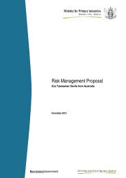
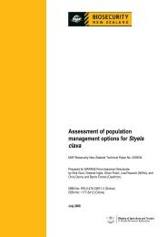
The risk analysis process used by the MAF is summarised in Figure 1.<br />
1 Import risk analysis projects which have commenced after 12 April 2006 follow guidelines described in<br />
<strong>Biosecurity</strong> <strong>New</strong> <strong>Zealand</strong> <strong>Risk</strong> <strong>Analysis</strong> Procedures – Verison 1. See www.biosecurity.govt.nz/files/pestsdiseases/surveillance-review/risk-analysis-procedures.pdf.<br />
2 ● Import <strong>Risk</strong> <strong>Analysis</strong>: Catlle from Australia, Canada, the EU & the USA MAF <strong>Biosecurity</strong> <strong>New</strong> <strong>Zealand</strong>
Figure 1. The risk analysis process.<br />
MAF <strong>Biosecurity</strong> <strong>New</strong> <strong>Zealand</strong> Import <strong>Risk</strong> <strong>Analysis</strong>: Cattle from Australia, Canada, the EU & the USA ● 3
4.1. PRELIMINARY HAZARD LIST<br />
The first step in the risk analysis is hazard identification. The process begins with the<br />
collation of a list of organisms potentially associated with cattle. The diseases of interest are<br />
those that could be transmitted by cattle and could infect domestic, feral or wild animals, or<br />
man in <strong>New</strong> <strong>Zealand</strong>. In this case an initial list was made of all the cattle diseases that are<br />
classified as listed diseases in the year 2005 edition of the OIE Terrestrial Animal Health<br />
Code and diseases mentioned in the following sources:<br />
• Veterinary Medicine. Blood DC and Radostits OM, 7th edition, 1989, published by<br />
Bailliere Tindall, ISBN 0-7020-1286-6.<br />
• Infectious Diseases of Livestock. Coetzer JAW and Tustin RC, Oxford University<br />
Press, Cape Town, Oxford, <strong>New</strong> York. 2004.<br />
• Foreign Animal Diseases “The Gray Book”<br />
http://www.vet.uga.edu/vpp/gray_book/FAD/SGP.htm<br />
• The MAF databases that contain complete listings of all diseases of cattle that appear<br />
in Import Health Standards (IHSs) and Overseas Market Access Requirements<br />
(OMARs) for all countries for which the information is available.<br />
As a result of internal review by representatives with a responsibility for diseases of interest to<br />
the Ministry of Health, Ross River and Barmah Forest viruses, miscellaneous arboviruses and<br />
Sarcosporidia spp. were included. A section on weed seeds has also been included.<br />
The diseases of cattle that were identified in these sources are listed in Table 1.<br />
Table 1. List of organisms and diseases of concern.<br />
ORGANISM<br />
VIRUSES<br />
OIE<br />
LIST<br />
ZOONOTIC NEW ZEALAND STATUS NOTES<br />
Akabane (Simbu group)<br />
viruses<br />
No No Exotic<br />
Aujeszky’s disease virus Yes No Exotic<br />
Adenovirus virus No No Endemic (Vermunt<br />
and Parkinson 2000b)<br />
Bluetongue virus Yes No Exotic 24 serotypes<br />
Borna disease virus No ? Exotic<br />
Bovine calicivirus No No Unknown<br />
Bovine corona virus No No Endemic (Durham et<br />
al 1979; Vermunt and<br />
Parkinson 2000a)<br />
Bovine herpes virus -1 Yes No BHV-1.2b endemic. Some 1.1 and<br />
(IBR/IPV)<br />
BHV-1.1 and 1.2a 1.2a strains are<br />
exotic<br />
abortifacient<br />
4 ● Import <strong>Risk</strong> <strong>Analysis</strong>: Catlle from Australia, Canada, the EU & the USA MAF <strong>Biosecurity</strong> <strong>New</strong> <strong>Zealand</strong>
ORGANISM<br />
OIE<br />
LIST<br />
ZOONOTIC NEW ZEALAND STATUS NOTES<br />
Bovine herpesvirus-2 No No Endemic (Vermunt<br />
and Parkinson 2000a;<br />
Vermunt and<br />
Parkinson 2000b)<br />
Bovine herpesvirus-5 No No Exotic<br />
Bovine parvovirus No No Unknown<br />
Bovine papular No No Endemic (Vermunt<br />
stomatitis virus<br />
and Parkinson 2000b)<br />
Bovine respiratory No No Endemic (Motha and<br />
syncitial disease virus<br />
Hansen 1997)<br />
Bovine rhinovirus No No Unknown<br />
Bovine virus diarrhoea No No BVDV1 endemic Two types<br />
virus<br />
BVDV 2 exotic<br />
(Horner 2000)<br />
Crimean Congo<br />
haemorrhagic fever virus<br />
No Yes Exotic<br />
Enzootic bovine leucosis Yes No Endemic Eradicated from<br />
virus<br />
the Dairy<br />
Industry<br />
Ephemeral fever virus No No Exotic<br />
Foot and mouth disease Yes No Exotic 7 serotypes<br />
virus<br />
multiple strains<br />
Ibaraki virus No No Exotic<br />
Jembrana disease virus No No Exotic<br />
Lumpy skin disease virus Yes No Exotic<br />
Malignant catarrhal fever<br />
virus (wildebeest<br />
associated)<br />
Yes No Exotic<br />
Malignant catarrhal fever<br />
virus (sheep associated)<br />
No No Endemic<br />
Palyam virus group No No Exotic Many strains<br />
Parainfluenza virus No No Endemic<br />
Pseudocowpox virus No No Endemic (Hill 1994)<br />
Rabies virus Yes Yes Exotic Related<br />
rhabdoviruses<br />
Rift Valley fever virus Yes Yes Exotic<br />
Rinderpest virus Yes No Exotic Strains vary in<br />
virulence<br />
Rotavirus No No Endemic (Durham et<br />
al 1979; Vermunt and<br />
Parkinson 2000a)<br />
Ross River virus No Yes Exotic<br />
Tick borne encephalitis No Yes Exotic Many related<br />
virus<br />
viruses in group<br />
Vesicular stomatitis virus Yes Yes Exotic 3 subtypes<br />
West Nile disease virus No Yes Exotic<br />
TRANSMISSIBLE SPONGIFORM ENCEPHALOPATHIES (TSEs)<br />
Bovine spongiform Yes Yes Exotic<br />
MAF <strong>Biosecurity</strong> <strong>New</strong> <strong>Zealand</strong> Import <strong>Risk</strong> <strong>Analysis</strong>: Cattle from Australia, Canada, the EU & the USA ● 5
ORGANISM<br />
encephalopathy (BSE)<br />
infective agent<br />
OIE<br />
LIST<br />
ZOONOTIC NEW ZEALAND STATUS NOTES<br />
BACTERIA INCLUDING MOLLICUTES<br />
Actinobacillus ligniersi No No Endemic<br />
Arcanobacter pyogenes No No Endemic<br />
Bacillus anthracis Yes Yes Exotic<br />
Brucella abortus Yes No Exotic<br />
Burkholderia<br />
pseudomallei<br />
No Yes Exotic<br />
Campylobacter fetus Yes No Endemic Subsp venerealis<br />
and fetus<br />
Campylobacter jejuni No Yes Endemic<br />
Clostridium spp. No No Endemic<br />
Corynebacterium renale No No Endemic<br />
Dermatophilus<br />
congolensis<br />
Yes Yes Endemic<br />
Escherichia coli No Yes Endemic Plasmid and<br />
virulence types<br />
Footrot associated<br />
organisms<br />
No No Endemic Various species<br />
Haemophilus<br />
somni(Haemophilus<br />
somnu, Histophilus<br />
somni)<br />
No No Endemic<br />
Klebsiella spp. No No Endemic<br />
Listeria monocytogenes No Yes Endemic<br />
Moraxella bovis No No Endemic<br />
Mycobacterium bovis Yes Yes Endemic/ eradication<br />
programme<br />
Mycobacterium avium<br />
subsp. avium<br />
Yes Yes Endemic<br />
Mycobacterium avium<br />
subsp. Paratuberculosis<br />
Yes No? Endemic<br />
Mycoplasma mycoides<br />
subsp. Mycoides SC<br />
Yes No Exotic<br />
Mollicutes various No No Some endemic<br />
species<br />
Nocardia spp. No No Endemic<br />
Pasteurella multocida B<br />
and E<br />
Yes No Exotic<br />
Pasteurella multocida<br />
other than B and E<br />
No No Endemic<br />
Pasteurella<br />
(Mannheimia)<br />
haemolytica<br />
No No Endemic<br />
Salmonella spp. No Yes Some serotypes<br />
exotic<br />
Staphylococcus spp. No Variable Endemic<br />
6 ● Import <strong>Risk</strong> <strong>Analysis</strong>: Catlle from Australia, Canada, the EU & the USA MAF <strong>Biosecurity</strong> <strong>New</strong> <strong>Zealand</strong>
ORGANISM<br />
OIE<br />
LIST<br />
ZOONOTIC NEW ZEALAND STATUS NOTES<br />
Streptococcus spp. No Variable Endemic<br />
Yersinia spp.<br />
No Yes Endemic<br />
SPIROCHAETES<br />
Leptospira spp. Yes Yes 6 serovars are<br />
endemic (Midwinter<br />
1999)<br />
Borrelia burgdorferi No Yes Exotic<br />
Borrelia theileri No No Exotic<br />
PROTOZOA<br />
Babesia spp. Yes No Exotic<br />
Besnoitia besnoiti No No Exotic<br />
Cryptosporidium parvum No Yes Endemic<br />
Eimeria spp. No No Endemic<br />
Neospora caninum No No Endemic<br />
Sarcocystis hirsuta. S. No S hominis S hominis exotic<br />
cruzi and S. hominis<br />
zoonotic<br />
Theileria parva Yes No Exotic.<br />
Theileria annulata Yes No Exotic<br />
Theileria spp. (nonpathogenic)<br />
No No Endemic<br />
Trichomonas foetus No No Endemic<br />
Trypanosoma evansi No No Exotic<br />
Trypanosoma spp. (tsetse<br />
fly-borne)<br />
Yes No Exotic<br />
RICKETTSIAS AND CHLAMYDIAS<br />
Anaplasma marginale,<br />
A. centrale, A. caudatum<br />
Yes No Exotic<br />
Anaplasma<br />
phagocytophilium<br />
No Yes Exotic<br />
Chlamydophila abortus Yes Yes Exotic<br />
Coxiella burnetii Yes Yes Exotic<br />
Ehrlichia ruminantium Yes No Exotic<br />
Eperythrozoon spp. No No Endemic<br />
Haemobartonella bovis<br />
PARASITES<br />
No No Unknown<br />
Ticks No Some spp. Mainly exotic<br />
Screwworm No Yes Exotic<br />
Lice (cattle species ) No No Some exotic<br />
Mites No Some spp. Some exotic<br />
Warbles No No Exotic<br />
Internal parasites No No Some exotic<br />
Over 200<br />
serovars<br />
Note: Organisms classified as endemic in <strong>New</strong> <strong>Zealand</strong> for which no reference is given are commonly identified<br />
and reported in the quarterly reports of diagnostic laboratories that are published in the MAF publication<br />
Surveillance. For less commonly diagnosed endemic organisms a reference is given to substantiate the<br />
classification.<br />
MAF <strong>Biosecurity</strong> <strong>New</strong> <strong>Zealand</strong> Import <strong>Risk</strong> <strong>Analysis</strong>: Cattle from Australia, Canada, the EU & the USA ● 7
Palyam viruses have been listed as exotic on the basis that they have not been recorded as occurring in <strong>New</strong><br />
<strong>Zealand</strong>. All other organisms listed as exotic have been classified by MAF as unwanted or notifiable organisms<br />
(Ministry of Agriculture and Forestry 2005).<br />
All organisms listed as exotic to, or of unknown status in, <strong>New</strong> <strong>Zealand</strong> in Table 1 were<br />
transferred to Table 2 (below) and classified as follows:<br />
• Those agents/diseases that are recorded in the OIE Handistatus II database were<br />
classified according their OIE status in the countries of concern. Where applicable the<br />
symbols used by OIE for the classification of organism/country status were used in<br />
Table 2. (see subscript to Table 2).<br />
• For those organism that do not occur in the Handistatus database, a search of the<br />
literature was made and disease agents that were found to occur in a country of<br />
concern were recorded as present in Table 2. Further information on the geographic<br />
distribution of the diseases/agents and references are given in the sections of the risk<br />
analysis pertaining to each disease. Organisms/diseases for which no information<br />
could be found to indicate that they occurred in a country of concern were classified as<br />
not present (NP) in Table 2.<br />
Table 2. Status of disease agents that are exotic to <strong>New</strong> <strong>Zealand</strong> in Australia, Canada, the<br />
European Union and the USA.<br />
Agent<br />
Australia<br />
Status<br />
Canada EU USA<br />
Akabane disease virus P NP NP P<br />
Aujeszky’s disease virus 0000 0000 +() +()P<br />
Bluetongue virus +?() (1988) +() +()<br />
Borna disease virus NP NP P NP<br />
Bovine calicivirus ?ww ?ww ?ww ?ww<br />
Bovine herpes virus 1.1 and 1.2 NP P P P<br />
Bovine herpes virus 5 P P P P<br />
Bovine parvo virus ?ww ?ww ?ww ?ww<br />
Bovine rhinovirus ?ww ?ww ?ww ?ww<br />
Bovine viral diarrhoea virus 2 NP P P P<br />
Crimean Congo haemorrhagic fever<br />
virus<br />
NP NP ? NP<br />
Ephemeral fever virus P NP NP NP<br />
Foot and mouth disease virus (1871) (1952) (2001) (1929)<br />
Ibaraki virus NP NP NP NP<br />
Jembrana virus NP NP NP NP<br />
Lumpy skin disease 0000 0000 0000 0000<br />
MCF (wildebeest associated) virus NP NP NP NP<br />
Miscellaneous arboviruses P(most) NP NP P(some)<br />
Palyam virus P NP NP P<br />
Rabies virus (1867) + +() +<br />
Rabies related rhabdovirus P NP P NP<br />
Rift Valley fever virus 0000 0000 0000 0000<br />
Rinderpest virus (1923) 0000 0000 0000<br />
Ross River and Barmah Forest viruses P(most) NP NP NP<br />
Tick borne encephalitis virus NP P P P<br />
Vesicular stomatitis virus 0000 (1949) 0000 +()<br />
West Nile disease virus NP P P P<br />
8 ● Import <strong>Risk</strong> <strong>Analysis</strong>: Catlle from Australia, Canada, the EU & the USA MAF <strong>Biosecurity</strong> <strong>New</strong> <strong>Zealand</strong>
Agent<br />
Australia<br />
Status<br />
Canada EU USA<br />
Bacillus anthracis +() + +() +()<br />
Brucella abortus (1989) (1989) +() +<br />
Burkholderia pseudomallei P NP NP NP<br />
Mycobacterium bovis 2002 + +() +<br />
Mycoplasma mycoides mycoides SC 1967 1897 (most by<br />
1900)<br />
1892<br />
Other Mycoplasma spp. P P P P<br />
Pasteurella multocida B and E 0000 0000 0000 +()<br />
Salmonella dublin & typhimurium<br />
DT104<br />
+ + + +<br />
Leptospira spp. + + + +<br />
Borrelia burgdorferi NP P P P<br />
Borrelia theileri P P P P<br />
Babesia spp. +() 0000 0000 to + +()<br />
Besnoitia besnoiti NP NP NP NP<br />
Theileria parva 0000 0000 0000 0000<br />
Theileria annulata NP NP P (Southern<br />
countries)<br />
NP<br />
Sarcocystis hominis ?ww ?ww ?ww ?ww<br />
Trypanosoma evansii NP NP NP NP<br />
Trypanosoma spp. (tsetse fly) 0000 0000 0000 0000<br />
Anaplasma marginale, A. centrale and<br />
A. caudatum<br />
+() +() +<br />
Anaplasma phagocytophilium NP P P P<br />
Chlamydophila abortus 0000 + + +<br />
Coxiella burnetii + + + +<br />
Ehrlichia ruminantium 0000 0000 0000 0000<br />
Ticks P P P P<br />
<strong>New</strong> World screwworm 0000 0000 0000 1982<br />
Old World screwworm 0000 0000 0000 0000<br />
Hypoderma spp.(warble flies) NP P P P<br />
Internal parasites P P P P<br />
0000 Never recorded (OIE)<br />
- not reported (date of last outbreak not known) (OIE)<br />
(date) Date of last occurrence (OIE)<br />
? Disease suspected but presence not confirmed (OIE)<br />
+ Reported present or know to be present (OIE)<br />
+? Serological evidence and/or isolation of organism but no clinical sign of disease (OIE)<br />
( ) Disease limited to specific zones (OIE). For the EU this may refer to zones or countries<br />
P No OIE records. Evidence in literature of presence (See relevant section of risk analysis for details)<br />
NP No OIE records. No evidence of presence found in literature (See relevant section of risk analysis for<br />
details)<br />
?ww Possible world-wide distribution<br />
NB. In the case of the European Union which includes 27 countries the information recorded in the table<br />
represents the predominant position in the EU but may vary in individual countries.<br />
Information attributed to OIE was obtained from Handistatus (OIE 2006)<br />
A preliminary hazard list constructed from the organisms in Table 2 was based on the<br />
following criteria:<br />
MAF <strong>Biosecurity</strong> <strong>New</strong> <strong>Zealand</strong> Import <strong>Risk</strong> <strong>Analysis</strong>: Cattle from Australia, Canada, the EU & the USA ● 9
Animal disease agents<br />
• All disease agents that are exotic to <strong>New</strong> <strong>Zealand</strong> and are present in any of the<br />
countries of concern (Australia, Canada, the 27 European Union countries and<br />
the USA) or about which there was some uncertainty.<br />
• In addition organisms that occur in <strong>New</strong> <strong>Zealand</strong> for which there are known<br />
sub-species or strains or host associations that do not occur in <strong>New</strong> <strong>Zealand</strong><br />
and are potentially harmful.<br />
• Organisms that occur in <strong>New</strong> <strong>Zealand</strong> but for which an eradication programme<br />
administered by a Pest Management Strategy under the <strong>Biosecurity</strong> Act is in<br />
place.<br />
Diseases that are of concern to human health<br />
• Disease agents that are already in <strong>New</strong> <strong>Zealand</strong> but because of the nature of the<br />
imports are likely to significantly increase existing hazards associated with<br />
them.<br />
• Disease agents that occur only in well defined geographically bounded areas of<br />
<strong>New</strong> <strong>Zealand</strong>.<br />
The preliminary list based on these criteria is shown below in table 3.<br />
Table 3. Preliminary hazard list.<br />
VIRUSES<br />
Akabane disease virus and other Simbu<br />
group viruses<br />
Ephemeral fever virus<br />
Aujeszky’s disease virus Foot and mouth disease virus<br />
Bluetongue virus Miscellaneous arboviruses<br />
Borna disease virus Palyam group viruses<br />
Bovine calicivirus Rabies virus<br />
Bovine herpes virus types 1.1 and 1.2a Rift Valley fever<br />
Bovine herpes virus 5 Rinderpest virus<br />
Bovine parvovirus Ross River and Barmah Forest viruses<br />
Bovine rhinovirus Tick-borne encephalitis virus group<br />
Bovine virus diarrhoea virus 2 Vesicular stomatitis virus<br />
Crimea Congo haemorrhagic disease virus West Nile disease virus<br />
TSE AGENTS<br />
Bovine spongiform encephalopathy (BSE)<br />
infective agent<br />
BACTERIA<br />
Bacillus anthracis Mollicutes of bovines<br />
Brucella abortus Pasteurella multocida B and E<br />
Burkholderia pseudomallei Salmonella dublin and typhimurium DT104<br />
Mycobacterium bovis<br />
10 ● Import <strong>Risk</strong> <strong>Analysis</strong>: Catlle from Australia, Canada, the EU & the USA MAF <strong>Biosecurity</strong> <strong>New</strong> <strong>Zealand</strong>
SPIROCHAETES<br />
Leptospira spp. Borrelia theileri<br />
Borrelia burgdorferi<br />
RICKETTSIAL AND CHLAMYDIAL ORGANISMS<br />
Anaplasma marginale, Anaplasma centrale<br />
and Anaplasma caudatum<br />
Coxiella burnetii<br />
Anaplasma phagocytophilum Haemobartonella<br />
Chlamydophila abortus<br />
PROTOZOAL ORGANISMS<br />
Babesia spp. Theileria annulata<br />
Sarcocystis hominis<br />
INTERNAL AND EXTERNAL PARASITES<br />
Lice Warble flies<br />
Mange mites Internal parasites<br />
Ticks<br />
4.2. HAZARD IDENTIFICATIION<br />
Organisms in the preliminary hazard list were subjected to further analysis to determine<br />
whether they were considered potential hazards in the commodity and organisms considered<br />
to be potential hazards were subjected to risk assessment.<br />
4.3. RISK ASSESSMENT<br />
Under the MAF <strong>Biosecurity</strong> <strong>New</strong> <strong>Zealand</strong> and OIE methodologies, risk assessment consists<br />
of:<br />
a) Entry assessment - the likelihood of the organism being imported in<br />
commodity.<br />
b) Exposure assessment - the likelihood of animals or humans in <strong>New</strong> <strong>Zealand</strong> being<br />
exposed to the potential hazard.<br />
c) Consequence assessment - the consequences of entry, exposure, establishment or spread<br />
of the organism.<br />
d) <strong>Risk</strong> estimation - a conclusion on the risk posed by the organism based on the<br />
release, exposure and consequence assessments.<br />
If the risk estimate is non-negligible, then the organism is<br />
classified as a hazard.<br />
It is important to understand that not all of the above steps may be necessary in all risk<br />
assessments. The MAF <strong>Biosecurity</strong> <strong>New</strong> <strong>Zealand</strong> and OIE methodologies makes it clear that<br />
if the likelihood of entry is negligible for a potential hazard, then the risk estimate is<br />
automatically negligible and the remaining steps of the risk assessment need not be carried<br />
out. The same situation arises where the likelihood of entry is non-negligible but the exposure<br />
MAF <strong>Biosecurity</strong> <strong>New</strong> <strong>Zealand</strong> Import <strong>Risk</strong> <strong>Analysis</strong>: Cattle from Australia, Canada, the EU & the USA ● 11
assessment concludes that the likelihood of exposure to susceptible species in the importing<br />
country is negligible, or where both entry and exposure are non-negligible but the<br />
consequences of introduction are concluded to be negligible.<br />
4.4. RISK MANAGEMENT<br />
For each organism classified as a hazard, a risk management step is carried out, which<br />
identifies the options available for managing the risk. Where the Code lists recommendations<br />
for the management of a hazard, these are described alongside options of similar, lesser, or<br />
greater stringency where available. In addition to the options presented, unrestricted entry or<br />
prohibition may also be considered for all hazards. Recommendations for the appropriate<br />
sanitary measures to achieve the effective management of risks are not made in this<br />
document. These will be determined when an import health standard (IHS) is drafted. As<br />
obliged under Article 3.1 of the WTO Agreement on Sanitary and Phytosanitary Measures<br />
(the SPS Agreement) the measures adopted in IHSs will be based on international standards,<br />
guidelines and recommendations where they exist, except as otherwise provided for under<br />
Article 3.3 (where measures providing a higher level of protection than international standards<br />
can be applied if there is scientific justification, or if there is a level of protection that the<br />
member country considers is more appropriate following a risk assessment).<br />
4.5. RISK COMMUNICATION<br />
MAF releases draft import risk analyses for a six-week period of public consultation to verify<br />
the scientific basis of the risk assessment and to seek stakeholder comment on the risk<br />
management options presented. Stakeholders are also invited to present alternative risk<br />
management options that they consider necessary or preferable.<br />
Following public consultation on the draft risk analysis, MAF produces a a review of<br />
submissions and determines whether any changes need to be made to the draft risk analysis as<br />
a result of public consultation, in order to make it a final risk analysis.<br />
Following this process of consultation and review, the Imports Standards team of MAF<br />
<strong>Biosecurity</strong> <strong>New</strong> <strong>Zealand</strong> decides on the appropriate combination of sanitary measures to<br />
ensure the effective management of identified risks. These are then presented in a draft IHS<br />
which is released for a six-week period of stakeholder consultation. Stakeholder submissions<br />
in relation to the draft IHS are reviewed before a final IHS is issued.<br />
4.6. SPECIAL CONSIDERATIONS<br />
The incubation period and the time for which an animal may remain infectious are critical<br />
parameters for determining quarantine periods. An animal could have been infected with a<br />
disease on the day it goes into quarantine. After the incubation period for the disease, it could<br />
then be infectious for a period that differs for each disease. In many acute diseases the<br />
infectious period may correspond with the period for which the animal remains viraemic or<br />
bacteraemic. However in cases of chronic diseases animals may be infectious for much longer<br />
periods. Animals could be kept in quarantine for a minimum of the incubation period and the<br />
time for which they remain infectious. Animals could be quarantined for the maximum known<br />
incubation period plus the maximum period for which they remain infectious. Ideally the<br />
maximum period would be the mean period plus three standard deviations. This would cover<br />
approximately 99% of cases. However, usually the true distribution of incubation period and<br />
12 ● Import <strong>Risk</strong> <strong>Analysis</strong>: Catlle from Australia, Canada, the EU & the USA MAF <strong>Biosecurity</strong> <strong>New</strong> <strong>Zealand</strong>
infectious period is not known because data are not available from a sufficiently large number<br />
of cases or because of technical difficulties in obtaining accurate data. Data quoted may be<br />
unreliable because of the small numbers of animals used in experiments or because analysis<br />
was done at discrete intervals and therefore exact end-points were not determined. The<br />
measurements are also dependent on the accuracy and sensitivity of the method used to detect<br />
the infectious agent. For these reasons a conservative margin of error may be added to the best<br />
available estimates when determining quarantine periods. The margin of error added cannot<br />
be scientifically determined but relies on judgement, taking into account such things as<br />
amount and perceived accuracy of the available data, type of disease and the analytical<br />
methods used. In some infectious diseases recovered animals remain carriers of the infectious<br />
agent for long periods or even for life, and in these cases quarantine is not useful. In this risk<br />
analysis quarantine periods are generally adjusted to whole weeks or months.<br />
Where animals for importation have been isolated as a group prior to export, the testing<br />
options within this risk analysis assume that any positive or inconclusive test results<br />
associated with any individual within that group will be notified to MAF <strong>Biosecurity</strong> <strong>New</strong><br />
<strong>Zealand</strong> for further consideration before any animal from that group is exported to <strong>New</strong><br />
<strong>Zealand</strong>.<br />
All risks associated with the importation of bull semen also apply to bulls. Therefore, when<br />
importing bulls, applicable risk management options presented in the risk analysis for the<br />
importation of semen and embryos from cattle could be considered.<br />
4.7. COUNTRY FREEDOM STATEMENTS<br />
Several important diseases have not been included in this risk analysis because they are not<br />
known to occur in any of the countries covered by this risk analysis. However, since the<br />
position could change, veterinary certificates provided by the exporting country should certify<br />
country freedom from the following disease agents/diseases:<br />
Besnoitia besnoiti (besnoitiosis)<br />
Ehrlichia ruminantium (heartwater)<br />
Ibaraki disease virus<br />
Jembrana disease virus<br />
Lumpy skin disease<br />
Acelaphine herpes virus-1(Malignant catarrhal fever virus, wildebeest type)<br />
Mycoplasma mycoides subsp. mycoides SC (Contagious bovine pleuropneumonia)<br />
Old and <strong>New</strong> World screwworm<br />
Rift Valley fever virus<br />
Rinderpest virus<br />
Theileria parva (East Coast fever and related theilerioses)<br />
Tsetse fly transmitted Trypanosoma spp.<br />
Trypanosoma evansi (Surra)<br />
For importation to be considered from further countries in the future, risk assessments for the<br />
relevant diseases from this list may need to be conducted.<br />
In addition, country freedom statements should be provided for any of the diseases in the risk<br />
analysis for which a country wishes to declare freedom and thereby obtain exemption from<br />
any relevant sanitary measures.<br />
MAF <strong>Biosecurity</strong> <strong>New</strong> <strong>Zealand</strong> Import <strong>Risk</strong> <strong>Analysis</strong>: Cattle from Australia, Canada, the EU & the USA ● 13
References<br />
Durham PJK, Stevenson BJ, Farquharson BC (1979). Rotavirus and coronavirus associated with diarrhoea in<br />
domestic animals. <strong>New</strong> <strong>Zealand</strong> Veterinary Journal, 27(2), 30-2.<br />
Hill F (1994). Zoonotic diseases of ruminants in <strong>New</strong> <strong>Zealand</strong>. Surveillance, 21(4), 25-7.<br />
Horner GW (2000). Typing of <strong>New</strong> <strong>Zealand</strong> strains of Pestivirus. Surveillance, 27(3), 16.<br />
Midwinter A (1999). Spirochaetes in <strong>New</strong> <strong>Zealand</strong>. Surveillance, 26(3), 10-2.<br />
Ministry of Agriculture and Forestry (2005). The Unwanted Organisms Register.<br />
http://mafuwsp6.maf.govt.nz/uor/searchframe.htm.<br />
Motha J, Hansen M (1997). A serological survey for bovine respiratory syncytial virus in <strong>New</strong> <strong>Zealand</strong>.<br />
Surveillance, 24(4), 28.<br />
Murray N (2002). Import <strong>Risk</strong> <strong>Analysis</strong>; Animals and Animal Products. <strong>New</strong> <strong>Zealand</strong> Ministry of Agriculture<br />
and Forestry, Wellington, ISBN 040-478-07660-6,<br />
OIE (2006). <strong>Risk</strong> analysis. In: OIE (ed). Terrestrial Animal Health Code.Chapter<br />
2.3.8http://www.oie.int/eng/normes/MCode/en_chapitre_2.3.8.htm. OIE, Paris.<br />
OIE (2006). Handistatus II. http://www.oie.int/hs2/report.asp.<br />
Vermunt JJ, Parkinson TJ (2000a). Infectious diseases of cattle in <strong>New</strong> <strong>Zealand</strong>. Part 1 - Calves and growing<br />
stock. Surveillance, 27(2), 3-8.<br />
Vermunt JJ, Parkinson TJ (2000b). Infectious diseases of cattle in <strong>New</strong> <strong>Zealand</strong>. Part 2 - adult animals.<br />
Surveillance, 27(3), 3-9.<br />
14 ● Import <strong>Risk</strong> <strong>Analysis</strong>: Catlle from Australia, Canada, the EU & the USA MAF <strong>Biosecurity</strong> <strong>New</strong> <strong>Zealand</strong>
5. Akabane and other Simbu Group Viruses<br />
5.1. HAZARD IDENTIFICATION<br />
5.1.1. Aetiological agent<br />
Family: Bunyaviridae; Genus: Bunyavirus, Serogroup Simbu. Akabane disease virus and<br />
related viruses belong to a group known collectively as Simbu viruses (St George and<br />
Kirkland 2004). The group includes viruses such as Aino, Tinaroo, Peaton and Cache Valley<br />
viruses that cause similar syndromes.<br />
5.1.2. OIE list<br />
Not listed.<br />
5.1.3. <strong>New</strong> <strong>Zealand</strong> status<br />
Listed on the unwanted organisms register as an exotic unwanted organism.<br />
5.1.4. Epidemiology<br />
Akabane and related viruses have been isolated from Culicoides spp. (midges) and<br />
mosquitoes. Culicoides spp. are assumed to be the vectors of these viruses (St George and<br />
Kirkland 2004). Cattle and other ruminants including sheep (Charles 1994; Haughey et al<br />
1988; St George and Kirkland 2004) and goats (Han and Du 2003) are susceptible.<br />
Viruses in the Simbu-group occur endemically in large areas of Africa, Asia, the Middle East<br />
and Australia (Charles 1994; Haughey et al 1988; St George and Kirkland 2004) and the<br />
related Cache Valley virus occurs in Texas (Edwards 1994; Edwards et al 1989). No reference<br />
was found to the occurrence of the virus group in Canada or the European Union.<br />
The incubation period (infection to start of viraemia) for Akabane virus is from 1-6 days (St<br />
George 1998) and the viraemic period lasts for 3-4 days (St George and Kirkland 2004). In<br />
non-pregnant animals infection does not lead to the development of any signs (Gard et al<br />
1989). Akabane virus crosses from maternal to foetal circulation in infected pregnant females<br />
and causes the development of malformed calves, particularly cases of arthrogryposis and<br />
hydroencephaly (Charles 1994; Parsonson et al 1977; Parsonson et al 1988; St George and<br />
Kirkland 2004). In cattle maximal damage occurs when infection takes place at about the 12th<br />
to 16th week of gestation (St George and Kirkland 2004). Once a foetus has become immunocompetent<br />
it can mount an immune reaction and damage is less apparent or does not occur.<br />
Infected calves are usually non viable (Charles 1994). Calves born alive are not contagious<br />
and will not infect vectors.<br />
Major epidemics of foetal malformations due to Akabane virus have been reported in Japan<br />
and Australia (St George and Kirkland 2004). However, animals that have been exposed to<br />
the infection become immune and this leads to the establishment of a mainly immune<br />
population of cattle in endemic areas. For this reason foetal abnormalities usually occur<br />
sporadically in endemically infected areas but seroconversion in animals is common<br />
MAF <strong>Biosecurity</strong> <strong>New</strong> <strong>Zealand</strong> Import <strong>Risk</strong> <strong>Analysis</strong>: Cattle from Australia, Canada, the EU & the USA ● 15
(Cybinski and St George 1978; Cybinski et al 1978; Fukutomi et al 2003; St George and<br />
Kirkland 2004). There are no reports of the disease having a significant economic impact in<br />
enzootic countries.<br />
There are competitive ELISAs for detection of Akabane specific and Simbu-group specific<br />
antibodies (St George and Kirkland 2004).<br />
5.1.5. Hazard identification conclusion<br />
In view of the above, Akabane and other Simbu viruses are classified as potential hazards in<br />
the commodity.<br />
5.2. RISK ASSESSMENT<br />
5.2.1. Entry assessment<br />
These viruses could only be introduced into <strong>New</strong> <strong>Zealand</strong> by animals that are in the<br />
incubation period or viraemic at the time of introduction. Since the incubation period is 1-6<br />
days (St George 1998) and the viraemic period is from 3-4 days (St George and Kirkland<br />
2004), the likelihood of introducing a viraemic animal is low but non-negligible.<br />
5.2.2. Exposure assessment<br />
A viraemic animal introduced into <strong>New</strong> <strong>Zealand</strong> would not be infectious. These viruses could<br />
only be transmitted to other animals in <strong>New</strong> <strong>Zealand</strong> by competent insect vectors. Annual<br />
surveys reported in the MAF publication Surveillance have demonstrated that Culicoides spp.<br />
are not present in <strong>New</strong> <strong>Zealand</strong>. A typical report shows that no Culicoides spp. were found in<br />
15,000 insects trapped and that serological conversion to arboviruses did not occur in sentinel<br />
cattle (Motha et al 1997). Since Culicoides spp. are the main vectors of the disease it is<br />
unlikely that <strong>New</strong> <strong>Zealand</strong> cattle would be exposed to the virus. The virus has also been<br />
isolated from mosquitoes but no work has been done to investigate whether <strong>New</strong> <strong>Zealand</strong><br />
mosquitoes are competent vectors. Furthermore, published surveys provide good evidence<br />
that <strong>New</strong> <strong>Zealand</strong> is free of arbovirus vectors (Motha et al 1997). In the absence of a<br />
competent vector in <strong>New</strong> <strong>Zealand</strong>, the exposure assessment is considered to be negligible.<br />
5.2.3. <strong>Risk</strong> estimation<br />
Because the exposure assessment is negligible, the risk estimate for Akabane and other Simbu<br />
group viruses is negligible and they are not classified as hazards in the commodity. Therefore,<br />
risk management measures are not justified.<br />
References<br />
References marked * have been sighted as summaries in electronic media.<br />
Charles JA (1994). Akabane virus. The Veterinary clinics of North America. Food animal practice, 10(3), 525-<br />
46.*<br />
Cybinski DH, St George TD (1978). A survey of antibody to Aino virus in cattle and other species in Australia.<br />
Australian Veterinary Journal, 54(8), 371-3.<br />
16 ● Import <strong>Risk</strong> <strong>Analysis</strong>: Catlle from Australia, Canada, the EU & the USA MAF <strong>Biosecurity</strong> <strong>New</strong> <strong>Zealand</strong>
Cybinski DH, St George TD, Paull NI (1978). Antibodies to Akabane virus in Australia. Australian Veterinary<br />
Journal, 54(1), 1-3.<br />
Edwards JF (1994). Cache Valley virus. The Veterinary clinics of North America. Food animal practice, 10(3),<br />
515-24.*<br />
Edwards JF, Livingston CW, Chung SI, Collisson EC (1989). Ovine arthrogryposis and central nervous<br />
system malformations associated with in utero Cache Valley virus infection: spontaneous disease. Veterinary<br />
Pathology, 26(1), 33-9.<br />
Fukutomi T, Ngai M, Okuda K, Akashi H, Hada M, Kayahara Y, Hatano Y (2003). Antigenic<br />
characteristics of the Akabane viruses isolated from sentinel cattle in Okayama Prefecture. Journal of the Japan<br />
Veterinary Medical Association, 57, 2101-5.*<br />
Gard GP, Melville LF, Shorthose JE (1989). Investigations of bluetongue and other arboviruses in the blood<br />
and semen of naturally infected bulls. Veterinary Microbiology, 20(4), 315-22.<br />
Han D, Du H (2003). Congenital abnormalities in Korean native goat with Akabane virus. Journal of Veterinary<br />
Clinics, 20(3), 427-30.*<br />
Haughey KG, Hartley WJ, Della-Porta AJ, Murray MD (1988). Akabane disease in sheep. Australian<br />
Veterinary Journal, 65(5), 136-40.<br />
Motha J, Hansen M, Irwin G (1997). Continued freedom from arbovirus infections and arbovirus vectors in<br />
<strong>New</strong> <strong>Zealand</strong>. Surveillance, 24(4), 18-9.<br />
OIE (2006). In: OIE (ed). Terrestrial Animal Health<br />
Code.http://www.oie.int/eng/normes/MCode/en_sommaire.htm. OIE, Paris.<br />
Parsonson IM, Della-Porta AJ, Snowdon WA (1977). Congenital abnormalities in newborn lambs after<br />
infection of pregnant sheep with Akabane virus. Infection and Immunity, 15(1), 254-62.<br />
Parsonson IM, McPhee DA, Della-Porta AJ, McClure S, McCullagh P (1988). Transmission of Akabane<br />
virus from the ewe to the early fetus (32 to 53 days). Journal of Comparative Pathology, 99(2), 215-27.<br />
St George TD (1998). Akabane. In: Committee-on-Foreign-Animal-Diseases-of-the-United-States-Animal-<br />
Health-Association (ed). Foreign Animal Diseases . "The Gray Book". Pp. Pat Campbell & Associates and<br />
Carter Printing Company, Richmond, Virginia, http://www.vet.uga.edu/vpp/gray_book/FAD/AKA.htm,<br />
St George TD, Kirkland PD (2004). Diseases caused by Akabane and related Simbu-group viruses. In: Coetzer<br />
JAW, Tustin RC (eds). Infectious Diseases of livestock. Oxford University Press, Oxford, 1029-36.<br />
MAF <strong>Biosecurity</strong> <strong>New</strong> <strong>Zealand</strong> Import <strong>Risk</strong> <strong>Analysis</strong>: Cattle from Australia, Canada, the EU & the USA ● 17
6. Aujeszky’s Disease<br />
6.1. HAZARD IDENTIFICATION<br />
6.1.1. Aetiological agent<br />
Family: Herpesviridae; Subfamily: Alphaherpesvirinae; Genus: Varicellovirus, suid<br />
herpesvirus 1, Aujeszky’s disease virus (pseudorabies virus).<br />
6.1.2. OIE list<br />
Listed<br />
6.1.3. <strong>New</strong> <strong>Zealand</strong> status<br />
Listed on the unwanted organisms register as an exotic, notifiable organism.<br />
6.1.4. Epidemiology<br />
Aujeszky’s disease (pseudo-rabies) is a disease of pigs that was eradicated from <strong>New</strong> <strong>Zealand</strong><br />
by 1995 (OIE 2006). It occurs world-wide except in Australia, Canada, Finland, Sweden,<br />
Denmark and the UK. Several countries are attempting eradication (Van Oirschot 2004). The<br />
virus can be transmitted to cattle and other animals by close contact with infected pigs but the<br />
infectious dose is high. Cattle do not transmit the virus to other animals and are considered to<br />
be dead-end hosts (Baker et al 1982; Henderson et al 1995; Herweijer and de Jonge 1977;<br />
Van Oirschot 2004). In animals other than pigs the disease is characterized by acute<br />
neurological signs and is invariably fatal (Baker et al 1982; Henderson et al 1995; Herweijer<br />
and de Jonge 1977; Van Oirschot 2004).<br />
6.1.5. Hazard identification conclusion<br />
In view of the above, Aujeszky’s disease virus is classified as a potential hazard in the<br />
commodity.<br />
6.2. RISK ASSESSMENT<br />
6.2.1. Entry assessment<br />
Aujeszky’s disease is a rare disease in cattle and only occurs when they have been in close<br />
contact with pigs. When it occurs the signs are dramatic (Baker et al 1982; Henderson et al<br />
1995; Herweijer and de Jonge 1977; Navetat et al 1994; Sweda et al 1993; Van Oirschot<br />
2004) and the outcome is invariably fatal. Under these circumstances the likelihood infected<br />
animals would be exported to <strong>New</strong> <strong>Zealand</strong> is considered to be negligible.<br />
18 ● Import <strong>Risk</strong> <strong>Analysis</strong>: Catlle from Australia, Canada, the EU & the USA MAF <strong>Biosecurity</strong> <strong>New</strong> <strong>Zealand</strong>
6.2.2. <strong>Risk</strong> estimation<br />
Because the entry assessment is negligible, the risk estimate for Aujeszky’s disease is<br />
negligible and it is not classified as a hazard in the commodity. Therefore, risk management<br />
measures are not justified.<br />
References<br />
References marked * have been sighted as summaries in electronic media.<br />
Baker JC, Esser MB, Larson VL (1982). Pseudorabies in a goat. Journal of the American Veterinary Medical<br />
Association, 181(6), 607.<br />
Henderson JP, Graham DA, Stewart D (1995). An outbreak of Aujeszky's disease in sheep in Northern<br />
Ireland. Veterinary Record, 136(22), 555-7.<br />
Herweijer CH, de Jonge WK (1977). [Aujeszky's disease in goats (authors transl)]. Tijdschrif voor<br />
Diergeneeskunde, 102(7), 425-8.*<br />
Navetat H, Schelcher F, Chevalier A, Berr V (1994). [An outbreak of Aujeszky's disease in cattle}. Point-<br />
Veterinaire, 25(158), 1012-5.*<br />
OIE (2006). Handistatus II. http://www.oie.int/hs2/report.asp.<br />
Sweda W, Janowski H, Grzechnik R, Brzeska E (1993). [Aujeszky's disease of cattle in the Olsztynprovince<br />
in years 1980-1991]. Medycyn-Weterynaryna, 49(4), 175-7.*<br />
Van Oirschot JT (2004). Pseudorabies. In: Coetzer JAW, Tustin RC (eds). Infectious Diseases of livestock.<br />
Oxford University Press, Oxford, 909-22.<br />
MAF <strong>Biosecurity</strong> <strong>New</strong> <strong>Zealand</strong> Import <strong>Risk</strong> <strong>Analysis</strong>: Cattle from Australia, Canada, the EU & the USA ● 19
7. Bluetongue<br />
7.1. HAZARD IDENTIFICATION<br />
7.1.1. Aetiological agent<br />
Family: Reoviridae; Genus: Orbivirus, Bluetongue virus (BTV). There are 24 known<br />
serotypes of BTV.<br />
7.1.2. OIE list<br />
Listed.<br />
7.1.3. <strong>New</strong> <strong>Zealand</strong> status<br />
Listed on the unwanted organisms register as an exotic, notifiable organism.<br />
7.1.4. Epidemiology<br />
Bluetongue virus can infect many ruminant species. It occurs in most tropical and sub-tropical<br />
countries. The global BTV distribution is currently between latitudes of approximately 53°N<br />
and 34°S but is known to be expanding in the northern hemisphere (OIE 2008). The virus<br />
causes disease mainly in sheep, occasionally in goats and rarely in cattle and deer. In most<br />
other species infections are subclinical. It is carried by Culicoides spp. (midges) and outbreaks<br />
of the disease usually occur in late summer to autumn when midges are most active.<br />
Outbreaks cease with the advent of winter when Culicoides spp. become inactive. In cattle<br />
infection is usually subclinical and mortality low but viraemic cattle can act as a source of<br />
infection for Culicoides spp. (Verwoerd and Erasmus 2004).<br />
7.1.5. Hazard identification conclusion<br />
In view of the above, bluetongue virus is classified as a potential hazard in the commodity.<br />
7.2. RISK ASSESSMENT<br />
7.2.1. Entry assessment<br />
The incubation period in natural infections is about 7 days and infected cattle remain viraemic<br />
for about 50 days (Verwoerd and Erasmus 2004). In countries where many strains of virus are<br />
endemic a few strains usually dominate in any one season but as the population becomes<br />
immune to these strains the dominant strains are replaced by other strains that then become<br />
dominant. In summer and for a period up to 60 days after Culicoides spp. become inactive at<br />
the onset of winter, susceptible animals may be viraemic. Therefore the likelihood of<br />
importing cattle in the incubation period of the disease or viraemic animals is non-negligible.<br />
20 ● Import <strong>Risk</strong> <strong>Analysis</strong>: Catlle from Australia, Canada, the EU & the USA MAF <strong>Biosecurity</strong> <strong>New</strong> <strong>Zealand</strong>
7.2.2. Exposure assessment<br />
BTV is transmitted by Culicoides vectors. A Culicoides surveillance programme has been<br />
operating in <strong>New</strong> <strong>Zealand</strong> since 1991 (Ryan et al 1991), under which around 15,000 insects<br />
collected from light traps are examined annually (Motha et al 1997) and sentinel cattle are<br />
monitored for seroconversion to viruses transmitted by Culicoides spp. (bluetongue, epizootic<br />
haemorrhagic disease, Akabane and Palyam viruses). To date, seroconversion to arboviruses<br />
has not been detected in sentinel cattle and no Culicoides have been trapped.<br />
Bluetongue virus can be excreted in bull’s semen (Parsonson et al 1981) but only while<br />
animals are viraemic (Bowen et al 1983; Howard et al 1985). Infected cattle may remain<br />
viraemic for about 50 days (Verwoerd and Erasmus 2004). Therefore it would be possible for<br />
an imported infected bull to excrete the virus in its semen for a period of around two months<br />
after infection. The likelihood of exposure of females with which the bull has mated over that<br />
time is non-negligible.<br />
Although no reference could be found for iatrogenic transmission of BTV, mechanical<br />
transmission of this disease is thought unlikely to be of major significance in disease<br />
epizootics (Radostits et al 2007).<br />
7.2.3. Consequence assessment<br />
Female cattle that have mated with an infected imported bull or inseminated with his semen<br />
could become infected (Bowen et al 1985; Schlafer et al 1990; Bowen and Howard 1984) and<br />
could remain viraemic for up to 50 days. However these animals are unlikely to show clinical<br />
signs and would not be infectious for other cattle. The virus could only be transmitted by<br />
Culicoides vectors and these are not present in <strong>New</strong> <strong>Zealand</strong>.<br />
The OIE Terrestrial Animal Health Code states that countries that are south of 34° S and are<br />
not adjacent to a country not having a bluetongue virus free status may be considered free<br />
from bluetongue. Furthermore, the OIE Terrestrial Animal Health Code states that “A BTV<br />
free country or zone in which surveillance has found no evidence that Culicoides likely to be<br />
competent BTV vectors are present will not lose its free status through the importation of<br />
vaccinated, seropositive or infective animals, or semen or embryos/ova from infected<br />
countries or infected zones” (OIE 2008).<br />
Bluetongue is not a zoonotic disease and the virus does not constitute a threat to human<br />
health.<br />
It is a disease of ruminants and there is no threat to indigenous animals or birds. Some species<br />
of deer are susceptible to the infection. The effect the virus might have on thar is not known.<br />
However since vectors for the virus do not occur in <strong>New</strong> <strong>Zealand</strong>, the consequences of<br />
introducing the virus would be negligible.<br />
The likelihood that the virus could establish in <strong>New</strong> <strong>Zealand</strong> is negligible, so the consequence<br />
assessment is negligible.<br />
7.2.4. <strong>Risk</strong> estimation<br />
There is a very low likelihood that, if a viraemic bull were imported, it would be used for<br />
natural service or semen collection during the period of viraemia. If it were so used, there is a<br />
very low likelihood of transmission of BTV to female cattle by this route. Infection of female<br />
MAF <strong>Biosecurity</strong> <strong>New</strong> <strong>Zealand</strong> Import <strong>Risk</strong> <strong>Analysis</strong>: Cattle from Australia, Canada, the EU & the USA ● 21
cattle (either those infected before importation or those infected from an imported bull) would<br />
have negligible consequences as cattle rarely show signs of infection and transmission to<br />
other cattle would not be possible due to <strong>New</strong> <strong>Zealand</strong>’s freedom from Culicoides spp.<br />
Furthermore, if a single animal were discovered to be viraemic (e.g. by routine<br />
serosurveillance), then the OIE Terrestrial Animal Health Code states that <strong>New</strong> <strong>Zealand</strong><br />
would not lose its BTV-free status. Therefore, the consequence assessment for both male and<br />
female cattle is considered to be negligible.<br />
As a result, the risk estimate for BTV is negligible and it is not classified as a hazard in the<br />
commodity. Therefore, risk management measures are not justified.<br />
References<br />
References marked * have been sighted as summaries in electronic media.<br />
Bowen RA, Howard TH (1984). Transmission of bluetongue virus by intrauterine inoculation or insemination<br />
of virus containing bovine semen. American Journal of Veterinary Research, 45, 1386-8.<br />
Bowen RA, Howard TH, Entwistle KW, Pickett BW (1983). Seminal shedding of bluetongue virus in<br />
experimentally infected mature bulls. American Journal of Veterinary Research, 44(12), 2268-70.<br />
Bowen RA, Howard TH, Pickett BW (1985). Seminal shedding of bluetongue virus in experimentally infected<br />
bulls. Progress in Clinical and Biological Research, 178, 91-6.*<br />
Howard TH, Bowen RA, Pickett BW (1985). Isolation of bluetongue virus from bull semen. Progress in<br />
Clinical and Veterinary Research, 178, 127-34.*<br />
Motha J, Hansen M, Irwin G (1997). Continued freedom from arbovirus infections and arbovirus vectors in<br />
<strong>New</strong> <strong>Zealand</strong>. Surveillance, 24(4), 18-9.<br />
OIE.2008. Bluetongue. Chapter 8.3. In: OIE (ed). Terrestrial Animal Health Code.<br />
www.oie.int/eng/normes/MCode/en_chapitre_1.8.3.htm . OIE, Paris.<br />
Parsonson IM, Della-Porta AJ, McPhee DA, Cybinski DH, Squire KRE, Standfast HA, Uren MF (1981).<br />
Isolation of bluetongue virus serotype 20 from the semen of an experimentally-infected bull. Australian<br />
Veterinary Journal, 57, 252-3.<br />
Radostits OM, Gay CC, Hinchcliff KW, Constable PD et al (2007). Chapter 21: Diseases associated with<br />
viruses and Chlamydia - I. In: Veterinary Medicine (10 th Ed). Saunders Elsevier, Edinburgh, London, <strong>New</strong> York,<br />
Oxford, Philadelphia, St Louis, Sydney and Toronto, 1300.<br />
Ryan TJ, Frampton ER, Motha MXJ (1991). Arbovirus and arbovirus vector surveillance in <strong>New</strong> <strong>Zealand</strong>.<br />
Surveillance, 18(5), 24-6.<br />
Schlafer DH, Gillespie JH, Foote RH, Quick S, Pennow NN, Dougherty EP, Schiff EI, Allen SE, Powers<br />
PA, Hall CE et al (1990). Experimental transmission of bovine viral diseases by insemination with<br />
contaminated semen or during embryo transfer. Deutsche tierarztliche Wochenschrift., 97(2), 68-72.*<br />
Verwoerd DW, Erasmus BJ (2004). Bluetongue. In: Coetzer JAW, Tustin RC (eds). Infectious Diseases of<br />
livestock. Oxford University Press, Oxford, 1201-20.<br />
22 ● Import <strong>Risk</strong> <strong>Analysis</strong>: Catlle from Australia, Canada, the EU & the USA MAF <strong>Biosecurity</strong> <strong>New</strong> <strong>Zealand</strong>
8. Borna Disease<br />
8.1. HAZARD IDENTIFICATION<br />
8.1.1. Aetiological agent<br />
Family: Bornaviridae; Genus: Bornavirus. Borna disease virus is the only member of this<br />
family.<br />
8.1.2. OIE list<br />
Not listed.<br />
8.1.3. <strong>New</strong> <strong>Zealand</strong> status<br />
Listed on the unwanted organisms register as an exotic, unwanted organism.<br />
8.1.4. Epidemiology<br />
Borna disease affects horses, sheep, and a variety of other animals including goats, deer,<br />
rabbits (Rott et al 2004), lynx (Desgiorgis et al 2000), and foxes (Dauphin et al 2001). Cattle<br />
can be subclinically infected (Hagiwara et al 1996). Disease is rare, but acute nervous disease<br />
can occur (Rott et al 2004).<br />
The disease has either been under-reported in the past or it is an emerging disease that has<br />
now been reported in many different species and countries. It occurs most commonly in<br />
Germany and Switzerland. However, serologically positive animals have also been found in<br />
Poland, the Netherlands, Switzerland, and Iran (Rott et al 2004) and Borna virus RNA has<br />
been found in France (Dauphin et al 2001; Dauphin and Zientara 2003). Reports on the<br />
demonstration of antibodies in horses have also come from North America (Kao et al 1993),<br />
Japan (Inoue et al 2002), and Israel (Teplitsky et al 2003). The virus has been demonstrated in<br />
cats in Britain (Reeves et al 1998). Viral RNA has been demonstrated in the peripheral<br />
mononuclear cells of cattle (Hagiwara et al 1996), sheep (Hagiwara et al 1997; Vahlenkamp<br />
et al 2000; Vahlenkamp et al 2002), horses (Nakamura et al 1995; Vahlenkamp et al 2002),<br />
cats (Nakamura et al 1996; Reeves et al 1998), and humans (Kishi et al 1995; Vahlenkamp et<br />
al 2000; Vahlenkamp et al 2002).<br />
A closely related virus has been found in mallards and jackdaws in Sweden (Berg et al 2001).<br />
A related virus has been identified as the aetiological agent of wobbly possum disease in <strong>New</strong><br />
<strong>Zealand</strong> (O'Keefe et al 1997).<br />
Antibody to Borna disease virus has been found in humans suffering from psychosomatic<br />
disorders (Bode et al 1996; Rott et al 1985). However, the exact role of the virus in human<br />
infections and as a cause of psychosomatic disorders remains controversial. The specificity of<br />
demonstrated antibody and the accuracy and reliability of the PCR test to demonstrate the<br />
presence of viral RNA has been questioned, but the issues remain unresolved (Carbone 2001;<br />
Staeheli et al 2000).<br />
MAF <strong>Biosecurity</strong> <strong>New</strong> <strong>Zealand</strong> Import <strong>Risk</strong> <strong>Analysis</strong>: Cattle from Australia, Canada, the EU & the USA ● 23
The route of infection for Borna disease in animals has not been fully resolved. The virus is<br />
excreted in nasal secretions, saliva and urine (Rott et al 2004; Vahlenkamp et al 2002). In<br />
mice the disease enters the body through the olfactory epithelium and migrates intra-axonally<br />
to the brain (Carbone et al 1987; Morales et al 1988; Sauder and Staeheli 2003). The virus can<br />
be transmitted experimentally to rats by inoculation into the footpads. However, neurectomy<br />
prevents the disease occurring, thus demonstrating that transfer of the virus to the brain is by<br />
the intra-axonal route (Carbone et al 1987). In an experimental situation the disease was<br />
transmitted from persistently infected rats to naïve rats via the olfactory route. This has led to<br />
the suggestion that rats could be a source of infection for farm animals (Sauder and Staeheli<br />
2003). Vertical transmission has not been reported. Most infections are thought to be subclinical<br />
(Ludwig and Kao 1990) and in sheep the virus persists in carriers for at least 2 years,<br />
as demonstrated by the presence of viral RNA in peripheral mononuclear cells. Natural<br />
transmission is presumed to occur by direct contact, via fomites and food, by inhalation and<br />
ingestion (Rott et al 2004).<br />
The incubation period of the disease is thought to vary from several weeks to months (Rott et<br />
al 2004; Ludwig and Kao 1990).<br />
Despite the fact that Borna disease has been known for more than 250 years (Rott et al 2004),<br />
knowledge about the disease is still fragmentary and incomplete. The specificity and accuracy<br />
of the RT-PCR test and antibody tests has been questioned.<br />
The disease is not regarded by OIE as a disease that is important to trade and it only occurs<br />
sporadically in countries where it does occur. However, in Germany it is a notifiable disease<br />
and is controlled by a slaughter-out policy (Rott and Herzog 1994).<br />
8.1.5. Hazard identification conclusion<br />
In view of the above, Borna disease is considered to be a potential hazard in the commodity.<br />
8.2. RISK ASSESSMENT<br />
8.2.1. Entry assessment<br />
Borna disease is rarely reported in cattle. In horses and sheep the disease remains mainly<br />
confined to Germany and surrounding countries and apparently only occurs sporadically.<br />
However, Rott et al have suggested that natural infections may occur more frequently and in a<br />
wider number of animals than previously thought (Rott et al 2004). However, since the<br />
disease has remained confined to a relatively small part of the world over the last 100 years<br />
this indicates that it is not highly contagious and spreads only slowly.<br />
The likelihood that live cattle imported into <strong>New</strong> <strong>Zealand</strong> would be infected with the virus is<br />
considered to be very low but non-negligible.<br />
8.2.2. Exposure assessment<br />
Virus is shed in nasal secretions, saliva and urine, and spread is presumed to be by contact and<br />
via fomites. Since the infected animals may carry the virus for long periods, and imported<br />
cattle would be in contact with <strong>New</strong> <strong>Zealand</strong> animals the possibility of spread to other animals<br />
is non-negligible.<br />
24 ● Import <strong>Risk</strong> <strong>Analysis</strong>: Catlle from Australia, Canada, the EU & the USA MAF <strong>Biosecurity</strong> <strong>New</strong> <strong>Zealand</strong>
8.2.3. Consequence assessment<br />
Since the disease has remained geographically confined for a long period it is considered that<br />
it would be unlikely but not impossible for the disease to establish and spread in <strong>New</strong><br />
<strong>Zealand</strong>. The establishment of the disease could result in sporadic cases of disease in cattle or<br />
other species particularly horses and sheep. However, considering the history of the disease<br />
this seems unlikely especially since <strong>New</strong> <strong>Zealand</strong> generally practices an extensive system of<br />
animal husbandry that does not favour the spread of diseases.<br />
The association between viral infection and the occurrence of psychosomatic diseases in<br />
humans (Bode et al 1996; Rott et al 1985) remains speculative. The consequences for human<br />
health of introducing the virus are therefore, uncertain, but are considered to be nonnegligible.<br />
The virus is known to infect a wide variety of animals (Dauphin and Zientara 2003;<br />
Desgiorgis et al 2000; Rott et al 2004) and birds (Berg et al 2001) and could therefore cause<br />
sporadic cases of disease in wild and feral animals and birds in <strong>New</strong> <strong>Zealand</strong>. In particular<br />
ostriches (Ashash et al 1996) have been infected with the virus and ratites (including Kiwis)<br />
might therefore be susceptible. The presence of a related virus in possums has not had any<br />
effect on the <strong>New</strong> <strong>Zealand</strong> environment apart from the rare occurrence of wobbly possum<br />
disease in possums. The effects on the environment are likely to be minimal but in view of the<br />
uncertainty, particularly regarding kiwis, it should be regarded as non-negligible.<br />
Since the introduction of the virus could lead to the establishment of a production limiting and<br />
possibly zoonotic disease and because the effects the virus could have on kiwis or other native<br />
birds is not known, the consequences are considered to be non-negligible.<br />
8.2.4. <strong>Risk</strong> estimation<br />
Because entry, exposure and consequence assessments are non-negligible, the risk estimate<br />
for Borna disease virus is non-negligible and it is classified as a hazard in the commodity.<br />
Therefore, risk management measures can be justified.<br />
8.3. RISK MANAGEMENT<br />
8.3.1. Options<br />
Since Borna disease is not listed by the OIE, no international standards for risk management<br />
exist.<br />
Diagnostic methods available include virus isolation (Ludwig and Kao 1990; Rott et al 2004)<br />
and demonstration of virus proteins or RNA (Vahlenkamp et al 2002) in tissues. Until<br />
uncertainties regarding the interpretation of PCR tests are resolved, demonstration of viral<br />
RNA by PCR should be regarded as indicative of the presence of virus. Therefore, PCR tests<br />
on peripheral mononuclear cells could be carried out on animals to be imported.<br />
Serology has been used in epidemiological surveys but it is not a reliable indicator of<br />
infection in individual animals. Two of six animals that were confirmed as being infected with<br />
Borna disease at post mortem were negative in both the ELISA and indirect<br />
immunofluorescence test (IFAT) (Allmang et al 2001) and one was positive in the IFAT but<br />
not ELISA. These findings indicate that infection does not always result in detectable<br />
MAF <strong>Biosecurity</strong> <strong>New</strong> <strong>Zealand</strong> Import <strong>Risk</strong> <strong>Analysis</strong>: Cattle from Australia, Canada, the EU & the USA ● 25
antibody production (Muller-Doblies et al 2003). Positive serology is common in sheep<br />
without clinical signs (Muller-Doblies et al 2003). Since the disease can have an incubation<br />
period of several months, quarantine is not a viable option to prevent the spread of the disease<br />
The importation of animals could be restricted to countries where the disease does not occur.<br />
One or a combination of the following sanitary measures could be considered in order to<br />
effectively manage the risk.<br />
• Cattle which have been resident since birth in countries where the virus/disease has<br />
never been reported could be imported without sanitary measures for this disease.<br />
• Importation could be restricted to cattle from herds where the disease has not been<br />
diagnosed during the previous 5 years.<br />
• A PCR test for detection of viral RNA could be carried out on the peripheral<br />
mononuclear cells of imported animals, with a requirement for negative results, within<br />
the 2 weeks prior to export to <strong>New</strong> <strong>Zealand</strong>.<br />
References<br />
References marked * have been sighted as summaries in electronic media.<br />
Allmang U, Hofer M, Herzog S, Bechter K, Staeheli P (2001). Low avidity of human serum antibodies for<br />
Borna disease virus antigens questions their diagnostic value. Molecular Psychiatry, 6(3), 329-33.<br />
Ashash E, Malkinson M, Meir R, Perl S, Weisman Y (1996). Causes of losses including a Borna disease<br />
paralytic syndrome affecting young ostriches of one breeding organization over a five-year period (1989-1993).<br />
Avian Diseases, 40(1), 240-5.<br />
Berg M, Johansson M, Montell H, Berg AL (2001). Wild birds as a possible natural reservoir of Borna disease<br />
virus. Epidemiology and Infection, 127(1), 173-8.<br />
Bode L, Durrwald R, Rantam FA, Ferszt R, Ludwig H (1996). First isolates of infectious human Borna<br />
disease virus from patients with mood disorders. Molecular Psychiatry, 1(3), 200-12.<br />
Carbone KM (2001). Borna disease virus and human disease. Clinical Microbiology Reviews, 14(3), 513-27.<br />
Carbone KM, Duchala CS, Griffin JW, Kincaid AL, Narayan O (1987). Pathogenesis of Borna disease in<br />
rats: evidence that intra-axonal spread is the major route for virus dissemination and the determinant for disease<br />
incubation. Journal of Virology, 61(11), 3431-40.<br />
Dauphin G, Legay V, Sailleau C, Smondack S, Hammoumi S, Zientara S (2001). Evidence of Borna disease<br />
virus genome detection in French domestic animals and foxes (Vulpes vulpes). Journal of General Virology,<br />
82(Pt9), 2199-204.<br />
Dauphin G, Zientara S (2003). The Borna disease virus: an emergent one in France? Epidemiologie et Sante<br />
Animale, 43, 19-29.<br />
Desgiorgis MP, Berg AL, Hard AF, Segerstad C, Morner T, Johansson M, Berg M (2000). Borna disease in<br />
a free-ranging lynx (Lynx lynx). Journal of Clinical Microbiology, 38(8), 3087-91.<br />
26 ● Import <strong>Risk</strong> <strong>Analysis</strong>: Catlle from Australia, Canada, the EU & the USA MAF <strong>Biosecurity</strong> <strong>New</strong> <strong>Zealand</strong>
Hagiwara K, Kawamoto S, Takahashi H, Nakamura Y, Nakaya T, Hiramune T, Ishihara C, Ikuta K<br />
(1997). High prevalence of Borna disease virus infection in healthy sheep in Japan. Clinical and Diagnostic<br />
Laboratory Immunology, 4(3), 339-44.<br />
Hagiwara K, Nakaya T, Nakamura Y, Asahi S, Takahashi H, Ishihara C, Ikuta K (1996). Borna disease<br />
virus RNA in peripheral blood mononuclear cells obtained from healthy dairy cattle. Medical Microbiology and<br />
Immunology., 185(3), 145-51.<br />
Inoue Y, Yamaguchi KS, Rivero JC, Horii Y (2002). Demonstration of continuously positive population<br />
against Borna disease virus in Misaki feral horses, a Japanese strain: a four year follow-up study from 1998-<br />
2001. Journal of Veterinary Medical Science, 64(5), 445-8.<br />
Kao M, Hamir AN, Rupprecht CE, Fu ZF, Shankar V, Koprowski H, Dietzschold B (1993). Detection of<br />
antibodies against Borna disease virus in sera and cerebrospinal fluid of horses in the USA. The Veterinary<br />
Record, 132, 241-244<br />
Kishi M, Nakaya T, Nakamura Y, Zhong Q, Ikeda K, Senjo M, Kakinuma M, Kato S, Ikuta K (1995).<br />
Demonstration of human Borna disease virus RNA in human peripheral blood mononuclear cells. FEBS Letters,<br />
364(3), 293-7.<br />
Ludwig H, Kao M (1990). Borna disease in sheep. In: Dinter Z, Morein B (eds). Virus infections of ruminants.<br />
Amsterdam, Oxford, <strong>New</strong> York, Tokyo, 529-38.<br />
Morales JA, Herzog S, Kompter C, Frese K, Rott R (1988). Axonal transport of Borna disease virus along<br />
olfactory pathways in spontaneously and experimentally infected rats. Medical Microbiology and Immunology<br />
(Berl), 177(2), 51-68.<br />
Muller-Doblies D, Baumann S, Grob P, Hulsmeier A, Muller-Doblies U, Brunker P, Ehrensperger F,<br />
Staeheli P, Ackerman M, Suter M (2003). The humoral and cellular response of sheep against Borna disease<br />
virus in endemic and non-endemic areas. Schweizer Archiv fur Tierheilkunde, 146(4), 159-72.<br />
Nakamura Y, Asahi S, Nakaya T, Bahmani MK, Saitoh S, Yasui K, Mayama H, Hagiwara K, Ishihara C,<br />
Ikuta K (1996). Demonstration of Borna disease virus RNA in peripheral blood mononuclear cells derived from<br />
domestic cats in Japan. Journal of Clinical Microbiology, 34(1), 188-91.<br />
Nakamura Y, Kishi M, Nakaya T, Asahi S, Tanaka H, Sentsui H, Ikeda K, Ikuta K (1995). Demonstration<br />
of Borna disease virus RNA in peripheral blood mononuclear cells from healthy horses in Japan. Vaccine,<br />
13(12), 1076-9.<br />
O'Keefe JS, Stanislawek WL, Heath DD (1997). Pathological Studies of wobbly Possum disease in <strong>New</strong><br />
<strong>Zealand</strong> brushtail possums (Trichosurus vulpeca). Veterinary Record, 141(9), 226-9.<br />
Reeves NA, Helps CR, Gunn-Moore DA, Blundell C, Finnemore PL, Pearson GR, Harbour DA (1998).<br />
Natural Borna disease virus infection in cats in the United Kingdom. Veterinary Record, 143(19), 523-6.<br />
Rott R, Herzog S (1994). Borna disease. In: Coetzer JAW, Thomson GE, Tustin RC (eds). Infectious diseases of<br />
livestock. Oxford University Press, Cape Town, Oxford, <strong>New</strong> York, 978-81.<br />
Rott R, Herzog S, Fleischer B, Winokur A, Amsterdam J, Dyson W, Koprowski H (1985). Detection of<br />
serum antibodies to Borna disease virus in patients with psychiatric disorders. Science, 228, 755-6.<br />
Rott R, Herzog S, Richt JA (2004). Borna disease. In: Coetzer JAW, Tustin RC (eds). Infectious Diseases of<br />
Livestock. Oxford University Press, Oxford, 1368-72,<br />
Sauder C, Staeheli P (2003). Rat model of Borna disease virus transmission: epidemiological implications.<br />
Journal of Virology, 77(23), 12886-90.<br />
Staeheli P, Sauder C, Hausmann J, Ehrensperger F, Schwemmle M (2000). Epidemiology of Borna disease<br />
virus. Journal of Virology, 81, 2123-35.<br />
MAF <strong>Biosecurity</strong> <strong>New</strong> <strong>Zealand</strong> Import <strong>Risk</strong> <strong>Analysis</strong>: Cattle from Australia, Canada, the EU & the USA ● 27
Teplitsky V, Pitlik S, Richt JA, Herzog S, Meir R, Marcus S, Sulkes J, Weisman Y, Malkinson M (2003).<br />
Increased prevalence of Borna disease virus ELISA and immunofluorescent antibodies in horses from farms<br />
along the paths of migratory birds. Israel Journal of Veterinary Medicine, 58(2-3), 80-5.<br />
Vahlenkamp TW, Enbergs HK, Muller H (2000). Experimental and natural Borna disease virus infections:<br />
presence of viral RNA in cells of the peripheral blood. Veterinary Microbiology, 76(3), 229-44.<br />
Vahlenkamp TW, Konrath A, Weber M, Muller H (2002). Persistence of Borna disease virus in naturally<br />
infected sheep. Journal of Virology, 76(19), 9735-43.<br />
28 ● Import <strong>Risk</strong> <strong>Analysis</strong>: Catlle from Australia, Canada, the EU & the USA MAF <strong>Biosecurity</strong> <strong>New</strong> <strong>Zealand</strong>
9. Bovine Calicivirus Infection<br />
9.1. HAZARD IDENTIFICATION<br />
9.1.1. Aetiological agent<br />
Family: Caliciviridae; Genus: Norovirus, bovine enteric calicivirus and possibly other calicilike<br />
viruses.<br />
9.1.2. OIE list<br />
Not listed.<br />
9.1.3. <strong>New</strong> <strong>Zealand</strong> status<br />
Not reported to occur in <strong>New</strong> <strong>Zealand</strong>.<br />
9.1.4. Epidemiology<br />
Two genotypes of the virus, the Jena and <strong>New</strong>bury agents, occur in Europe (Knowles and<br />
Clarke 2004) and a third type has been described in the USA (Oliver et al 2003).<br />
Despite identification of the viruses in calves nearly 40 years ago (Woode and Bridger 1978),<br />
the role of bovine enteric caliciviruses in calf diarrhoea is not well understood. Experimental<br />
infection of gnotobiotic calves and new born calves caused diarrhoea and intestinal pathology<br />
(Hall et al 1984). However, in naturally occurring cases of diarrhoea, calves are often infected<br />
with several viruses including rotaviruses and coronaviruses that are isolated in higher<br />
numbers than the caliciviruses (Knowles and Clarke 2004). Descriptions of diarrhoea<br />
associated with the virus are restricted to calves. Adult animals are apparently resistant or<br />
immune to infection.<br />
The virus has been described in England (Knowles and Clarke 2004; Woode and Bridger<br />
1978), Germany (Deng et al 2003), the Netherlands (van der Poel et al 2000), and the USA<br />
(Smiley et al 2003). Investigations to identify virus or virus antibodies in countries where the<br />
virus is known to occur generally indicated a high prevalence of infection. In Germany virus<br />
was identified in 8.9% of 381 cases and antibody was found in 99.1% of 824 samples (Deng<br />
et al 2003). In the USA 72% of 75 calf faecal samples were positive in an RT-PCR assay<br />
(Smiley et al 2003). In the Netherlands 44% of pooled faecal samples from 75 veal farms<br />
were found to be positive in an RT-PCR assay, and it was suggested that calves may be a<br />
source of infection for humans. However, a recent study suggests that calf strains differ from<br />
human isolates and calves are unlikely to be a source of infection for humans (Oliver et al<br />
2003). The virus has been known for almost 40 years but attracts little attention from<br />
diagnostic laboratories and research workers. This suggests that it is of minor economic<br />
importance.<br />
It is not known whether the virus occurs in <strong>New</strong> <strong>Zealand</strong>. However, since it is widely<br />
distributed in the world and is a trivial pathogen for which active surveys have not been done,<br />
it is likely that the virus may already be present in <strong>New</strong> <strong>Zealand</strong>.<br />
MAF <strong>Biosecurity</strong> <strong>New</strong> <strong>Zealand</strong> Import <strong>Risk</strong> <strong>Analysis</strong>: Cattle from Australia, Canada, the EU & the USA ● 29
9.1.5. Hazard identification conclusion<br />
Since bovine caliciviruses have been described as causing calf diarrhoea and have not been<br />
isolated in <strong>New</strong> <strong>Zealand</strong> they are considered as potential hazards in the commodity.<br />
9.2. RISK ASSESSMENT<br />
9.2.1. Entry assessment<br />
Excretion of caliciviruses in faeces only lasts for a few days after infection (Woode 1990).<br />
Viraemia has not been reported and all descriptions of the disease syndrome are restricted to<br />
calves. Under these circumstances the likelihood that adult animals would be excretors of<br />
virus is considered to be negligible. The likelihood that calves that are not showing signs of<br />
diarrhoea and have been isolated for more than a few days would be excretors of virus is low<br />
but not negligible.<br />
9.2.2. Exposure assessment<br />
Imported adult cattle are unlikely to be excreting the virus in their faeces. Calves will only<br />
excrete the virus for a few days therefore the likelihood of exposure of <strong>New</strong> <strong>Zealand</strong> cattle to<br />
virus by contact with imported calves is very low. The likelihood of exposure of <strong>New</strong> <strong>Zealand</strong><br />
cattle to the virus is therefore very low but non-negligible.<br />
9.2.3. Consequence assessment<br />
There is nothing in the literature to indicate that the virus causes anything other than trivial<br />
infections of calves. In addition the likelihood of release of virus and exposure are very low.<br />
These facts taken together indicate that the consequences of introduction of the virus can be<br />
considered to be negligible.<br />
9.2.4. <strong>Risk</strong> estimation<br />
Because the consequences are considered to be negligible, the risk estimate for bovine<br />
calicivirus is negligible and it is not classified as a hazard in the commodity. Therefore, risk<br />
management measures are not justified.<br />
References<br />
References marked * have been sighted as summaries in electronic media.<br />
Deng Y, Batten CA, Liu BL, Lambden PR, Elschner M, Gunther H, Otto P, Schnurch P, Eichhorn W,<br />
Herbst W, Clarke IN (2003). Studies of epidemiology and seroprevalence of bovine noroviruses in Germany.<br />
Journal of Clinical Microbiology, 41(6), 2300-5.<br />
Hall GA, Bridger JC, Brooker BE, Parsons KR, Ormerod E (1984). Lesions of gnotobiotic calves<br />
experimentally infected with a calicivirus-like (<strong>New</strong>bury) agent. Veterinary Pathology, 21(2), 208-15.<br />
Knowles NJ, Clarke IN (2004). Enteric caliciviruses of pigs and cattle. In: Coetzer JAW, Tustin RC (eds).<br />
Infectious Diseases of livestock. Oxford University Press, Oxford, 703-6.<br />
30 ● Import <strong>Risk</strong> <strong>Analysis</strong>: Catlle from Australia, Canada, the EU & the USA MAF <strong>Biosecurity</strong> <strong>New</strong> <strong>Zealand</strong>
Oliver SL, Dastjerdi AM, Wong S, El-Attar L, Gallimore C, Brown DW, Green J, Bridger JC (2003).<br />
Molecular characterization of bovine enteric caliciviruses: a distinct third genogroup of noroviruses (Norwalklike<br />
viruses) unlikely to be of risk to humans. Journal of Virology, 77(4), 2789-98.<br />
Smiley JR, Hoet AE, Traven M, Tsunemitsu H, Saif LJ (2003). Reverse transcription-PCR assays for<br />
detection of bovine enteric caliciviruses (BEC) and analysis of the genetic relationships among BEC and human<br />
caliciviruses. Journal of Clinical Microbiology, 41(7), 3089-99.<br />
van Der Poel WH, Vinje J, van Der Heide R, Herrera MI, Vivo A, Koopmans MP (2000). Norwalk-like<br />
calicivirus genes in farm animals. Emerging Infectious Diseases, 6(1), 36-41.*<br />
Woode GN (1990). Calicivirus-like agents. In: Dinter Z, Morein B (eds). Virus infections of ruminants.<br />
Amsterdam, Oxford, <strong>New</strong> York, Tokyo, 519-22.<br />
Woode GN, Bridger JC (1978). Isolation of small viruses resembling astroviruses and caliciviruses from acute<br />
enteritis of calves. Journal of Medical Microbiology, 11(4), 441-52.*<br />
MAF <strong>Biosecurity</strong> <strong>New</strong> <strong>Zealand</strong> Import <strong>Risk</strong> <strong>Analysis</strong>: Cattle from Australia, Canada, the EU & the USA ● 31
10. Bovine Herpes Viruses<br />
10.1. HAZARD IDENTIFICATION<br />
10.1.1. Aetiological agents<br />
Family: Herpesviridae; Subfamily: Alphaherpesvirinae; Genus: Varicellovirus, bovine<br />
herpesvirus 1 is associated with infectious bovine rhinotracheitis (IBR) and infectious<br />
pustular vulvovaginitis/infectious pustular balanoposthitis (IPV/IPB). Subtypes BHV-1.1 and<br />
BHV-1.2 can be identified by restriction endonuclease analysis of DNA (Babuik et al 2004;<br />
Engels et al 1981; Wentink et al 1993). Rhinitis and respiratory signs are associated with<br />
subtype 1.1, pustular vulvovaginitis and balanoposthitis is associated with subtype 1.2. Strains<br />
formerly described as IBRV 1.3 that are associated with encephalitis are now classified as<br />
BHV5. Subtype 1.2 strains can be further classified as BHV-1.2a and BHV-1.2b strains.<br />
Some subtype 1.1 and 1.2a strains are abortifacient, as shown by association with clinical<br />
cases of abortion and by experimental infection of pregnant heifers (Miller et al 1991).<br />
Subtype 1.2b strains are associated with respiratory and genital infections but not with<br />
abortions (Miller et al 1991; van Oirschot 1995a).<br />
Table 4. Bovine herpesviruses<br />
Type<br />
Syndrome<br />
IBR IPV/IPB Abortion Encephalitis<br />
BHV 1.1 + - + -<br />
BHV 1.2a + + + -<br />
BHV 1.2b + + - -<br />
BHV5 - - - +<br />
10.1.2. OIE list<br />
Infectious bovine rhinotracheitis and infectious pustular vulvovaginitis are listed by the OIE.<br />
10.1.3. <strong>New</strong> <strong>Zealand</strong> status<br />
Only BHV1.2b has been isolated in <strong>New</strong> <strong>Zealand</strong> (Wang et al 2006). Abortions have not been<br />
seen in <strong>New</strong> <strong>Zealand</strong> (Fairley 1996; Horner 1990). An attempt to cause abortion by<br />
experimental infection with the <strong>New</strong> <strong>Zealand</strong> strain of the virus was unsuccessful (Durham et<br />
al 1975). However, at the present time identification of abortifacient strains of the virus from<br />
either subtype 1 or 2 strains would require experimental infection of pregnant cows. A more<br />
pragmatic approach is to regard BHV1.1 and BHV 1.2a as exotic organisms. Abortifacient<br />
strains are classified on the unwanted organisms register as unwanted notifiable organisms.<br />
10.1.4. Epidemiology<br />
IBR/IPV has a world-wide distribution. The virus is endemic in <strong>New</strong> <strong>Zealand</strong> and serological<br />
surveys have shown that it occurs very widely (Neilson and Grace 1988). Both the IBR and<br />
the IPV syndrome have been described (Fairley 1996; Horner 1990; Vermunt and Parkinson<br />
32 ● Import <strong>Risk</strong> <strong>Analysis</strong>: Catlle from Australia, Canada, the EU & the USA MAF <strong>Biosecurity</strong> <strong>New</strong> <strong>Zealand</strong>
2000). However, in the vast majority of cases there are no or only mild clinical signs<br />
(Vermunt and Parkinson 2000).<br />
The acute infections are of short duration and virus is excreted in nasal secretions for up to 14<br />
days after infection. Viraemia is hard to detect (Babuik et al 2004) but can occasionally occur<br />
(van Oirschot 2004). Virus spreads to the conjunctiva and trigeminal ganglion by neuronal<br />
axonal transport (van Oirschot 2004). Many animals become chronically infected latent<br />
carriers of the virus in their trigeminal or sacral ganglia, and may excrete the virus<br />
periodically when they are stressed (Babuik et al 2004; van Oirschot 2004). Semen may be<br />
infected with virus and insemination with such semen causes infection in recipient females<br />
(Parsonson and Snowdon 1975; Schlafer et al 1990; van Oirschot 1995b).<br />
BHV-5 associated with encephalitis (Wentink et al 1993) has been described in Australia<br />
(Brake and Studdert 1985), USA (Barenfus et al 1963; Delhon et al 2003), Canada (Gough<br />
and James 1975), and Europe (Bartha et al 1969; Moretti et al 1964) but not in <strong>New</strong> <strong>Zealand</strong>.<br />
10.1.5. Hazard identification conclusion<br />
Abortifacient strains of IBR/IPV virus are exotic notifiable organisms and they are commonly<br />
present in chronic carrier animals. These organisms are therefore classified as potential<br />
hazards. However, since practical tests are not available to identify abortifacient strains in the<br />
laboratory it is necessary to regard all BHV 1.1 and BHV 1.2a strains as potential hazards.<br />
BHV-5 is also exotic and is regarded as a potential hazard.<br />
10.2. RISK ASSESSMENT<br />
10.2.1. Entry assessment<br />
Recently infected cattle may excrete the virus in nasal secretions and aerosols for up to 14<br />
days after infection (Babuik et al 2004). Carriers of the virus may periodically excrete virus<br />
particularly during periods of stress (Babuik et al 2004; van Oirschot 2004). The likelihood of<br />
entry of virus in the commodity is therefore considered to be non-negligible.<br />
10.2.2. Exposure assessment<br />
Imported animals are likely to be kept in herds with indigenous <strong>New</strong> <strong>Zealand</strong> cattle and the<br />
likelihood of exposure of naïve indigenous cattle is high. Infection could be spread by contact<br />
(Babuik et al 2004) or by insemination or natural mating using infected bulls (Parsonson and<br />
Snowdon 1975; Schlafer et al 1990;van Oirschot 1995b).<br />
10.2.3. Consequence assessment<br />
Introduction of abortifacient strains of BHV1.1 or 1.2a or strains of BHV 5 is likely to result<br />
in outbreaks of abortion or encephalitis. This would have a negative effect on the economy of<br />
the cattle industry and individual farmers.<br />
The virus does not infect humans and therefore the consequences for people are negligible.<br />
Other ruminants can possibly be infected with BHVs since they have been found to have<br />
antibody to the virus. However, the antibody that has been detected may be cross-reacting<br />
antibody as in the case of deer infected with cervine herpesvirus (Motha and Jenner 2001;<br />
MAF <strong>Biosecurity</strong> <strong>New</strong> <strong>Zealand</strong> Import <strong>Risk</strong> <strong>Analysis</strong>: Cattle from Australia, Canada, the EU & the USA ● 33
Tisdall and Rowe 2001). No significant disease has been described in other ruminants. The<br />
consequences for the environment are therefore assessed to be negligible.<br />
The consequences of importation of cattle are non-negligible for the cattle industry.<br />
The likelihood that there will be consequences for human health or the environment is<br />
considered to be negligible.<br />
10.2.4. <strong>Risk</strong> estimate<br />
Since entry, exposure, and consequence assessments are all non-negligible, the risk estimate<br />
for exotic bovine herpesviruses is non-negligible, and they are classified as a hazard in the<br />
commodity. Therefore, risk management measures can be justified.<br />
10.3. RISK MANAGEMENT<br />
10.3.1. Options<br />
Serological tests are not specific for sub-types of herpes viruses and it will not be possible to<br />
distinguish between the antibodies induced by BHV1.2a, BH1.1 or BHV5 strains from other<br />
strains of BHVs. From a practical viewpoint it will be necessary to consider any animal<br />
reacting positively to an IBR test to be potentially infected with an exotic strain of virus.<br />
The OIE Terrestrial Animal health Code does not discuss strains of bovine herpes viruses but<br />
instead considers the clinical syndromes of IBR and IPV. There are, therefore, no<br />
international risk management standards that are directly applicable although it is reasonable<br />
to extrapolate from the Code to the exotic strains of concern here. The Code recommends that<br />
animals destined for IBR/IPV free herds, should come from an IBR free herd (as defined in<br />
the Code), should be isolated in a quarantine station for at least 30 days, and should be<br />
subjected to two serological tests at an interval of not less than 21 days (OIE 2006).<br />
One or a combination of the following measures could be considered in order to effectively<br />
manage the identified risk.<br />
• Cattle for import into <strong>New</strong> <strong>Zealand</strong> could be imported from countries that are free<br />
from BHV 1.1 and BHV 1.2a, and come from herds that have no history of<br />
encephalitis caused by BHV 5.<br />
• Cattle could come from a herd that is free from IBR/IPV as defined in the OIE<br />
Terrestrial Animal Health Code.<br />
• Cattle could be placed in quarantine for the 30 days prior to export and during this<br />
time be submitted to 2 serological tests for IBR/IPV with an interval of not less than<br />
21 days between tests, with negative results. The serological test used could be an OIE<br />
recommended test or one approved by MAF.<br />
34 ● Import <strong>Risk</strong> <strong>Analysis</strong>: Catlle from Australia, Canada, the EU & the USA MAF <strong>Biosecurity</strong> <strong>New</strong> <strong>Zealand</strong>
References<br />
References marked * have been sighted as summaries in electronic media.<br />
Babuik TA, Van Drunen Littel-van den Hurk S, Tikoo SK (2004). Infectious bovine rhinotracheitis / pustular<br />
vulvovaginitis and infectious pustular balnoposthitis. In: Coetzer JAW, Tustin RC, (eds). Infectious Diseases of<br />
livestock. Oxford University Press, Oxford, 875-86.<br />
Barenfus M, Delliquadri CA, McIntyre RW, Schroeder RJ (1963). Isolation of Infectious Bovine<br />
Rhinotracheitis Virus from Calves with Meningoencephalitis. Journal of the American Veterinary Medical<br />
Assocition, 143, 725-8.<br />
Bartha A, Haidu G, Aldassy P, Paczolay G (1969). Occurrence of encephalmyelitis caused by infectious<br />
bovine rhinotracheitis virus in calves in Hungary. Acta veterinaria Academiae Scientiarum Hungaricae, 19, 145-<br />
51.*<br />
Brake F, Studdert MJ (1985). Molecular epidemiology and pathogenesis of ruminant herpesviruses including<br />
bovine, buffalo and caprine herpesviruses l and bovine encephalitis herpesvirus. Australian Veterinary Journal,<br />
62(10), 331-4.<br />
Delhon G, Moraes MP, Lu Z, Afonso CL, Flores EF, Weiblen R, Kutish GF, Rock DL (2003). Genome of<br />
bovine herpesvirus 5. Journal of Virology, 77(19), 10339-47.<br />
Durham PJK, Forbes-Faulkner JC, Poole WSH (1975). Infectious bovine rhinotracheitis: Experimental<br />
attempts at inducing bovine abortion with a <strong>New</strong> <strong>Zealand</strong> isolate. <strong>New</strong> <strong>Zealand</strong> Veterinary Journal, 23, 93-4.<br />
Engels M, Steck F, Wyler R (1981). Comparison of the genomes of infectious bovine rhinotracheitis (IBR) and<br />
infectious pustular vulvovaginitis (IPV) strains by restriction enzyme analysis of their genomes. Archives of<br />
Virology, 67(2), 169-74.<br />
Fairley RA (1996). Respiratory diseases of <strong>New</strong> <strong>Zealand</strong> cattle. Surveillance, 23(4), 15-6.<br />
Gough A, James D (1975). Isolation of IBR virus from a heifer with meningoencephalitis. Canadian Veterinary<br />
Journal, 16(10), 313-4.<br />
Horner GW (1990). Infectious bovine rhinotracheitis in <strong>New</strong> <strong>Zealand</strong>. Surveillance, 17(2), 25-6.<br />
Miller JM, Whetstone CA, Van der Maaten MJ (1991). Abortifacient property of bovine herpesvirus type 1<br />
isolates that represent three subtypes determined by restriction endonuclease analysis of viral DNA. American<br />
Journal of Veterinary Research, 52(2), 458-61.<br />
Moretti B, Orfei Z, Mondino G, Persechino A (1964). [Isolation of the Infectious Bovine Rhinotracheitis (Ibr)<br />
Virus in Italy. (Preliminary Note)]. Nuovi annali d'igiene e microbiologia, 15, 18-22.*<br />
Motha J, Jenner J (2001). Serological relatedness of cervine herpesvirus-1 and bovine herpesvirus-1 and the<br />
prevalence of cervine herpesvirus-1 infection in farmed deer in <strong>New</strong> <strong>Zealand</strong>. <strong>New</strong> <strong>Zealand</strong> Veterinary Journal,<br />
49(4), 162-3.<br />
Neilson FJA, Grace PJ (1988). Infectious bovine rhinotracheitis is widespread in <strong>New</strong> <strong>Zealand</strong>. Surveillance,<br />
15(2), 29.<br />
OIE (2006). Infectious bovine rhinotracheitis /infectious pustular vulvovaginitis. Chapter 2.3.5. In: OIE (ed).<br />
Terrestrial Animal Health Code<br />
http://www.oie.int/eng/normes/MCode/en_chapitre_2.3.5.htm. OIE, Paris.<br />
Parsonson IM, Snowdon WA (1975). The effect of natural and artificial breeding using bulls infected with, or<br />
semen contaminated with, infectious bovine rhinotracheitis virus. Australian Veterinary Journal, 51(8), 365-9.<br />
MAF <strong>Biosecurity</strong> <strong>New</strong> <strong>Zealand</strong> Import <strong>Risk</strong> <strong>Analysis</strong>: Cattle from Australia, Canada, the EU & the USA ● 35
Schlafer DH, Gillespie JH, Foote RH, Quick S, Pennow NN, Dougherty EP, Schiff EI, Allen SE, Powers<br />
PA, Hall CE et al (1990). Experimental transmission of bovine viral diseases by insemination with<br />
contaminated semen or during embryo transfer. Deutsche Tierarztliche Wochenschrift, 97(2), 68-72.*<br />
Tisdall DJ, Rowe SM (2001). Isolation and characterisation of cervine herpesvirus-1 from red deer semen. <strong>New</strong><br />
<strong>Zealand</strong> Veterinary Journal, 49(3), 111-4.<br />
van Oirschot JT (1995a). Bovine herpesvirus in semen of bulls and the risk of transmission. Veterinary<br />
Quarterly, 17, 29-33.*<br />
van Oirschot JT (1995b). Bovine herpesvirus in semen of bulls and the risk of transmission: a brief review.<br />
Veterinary Quarterly, 17(1), 29-33.*<br />
van Oirschot JT (2004). Infectious bovine rhinotracheitis/infectious pustular vulvovaginitis. In: OIE (ed).<br />
Manual of diagnostic tests and vaccines for terrestrial animals. OIE, Paris, 474-85.<br />
Vermunt JJ, Parkinson TJ (2000). Infectious diseases of cattle in <strong>New</strong> <strong>Zealand</strong>. Part 2- adult animals.<br />
Surveillance, 27(3), 3-9.<br />
Wang J, Horner GW, O'Keefe JS (2006). Genetic characterisitics of bovine herpesvirus 1 in <strong>New</strong> <strong>Zealand</strong>.<br />
<strong>New</strong> <strong>Zealand</strong> Veterinary Journal, 54(2), 61-66.<br />
Wentink GH, van Oirschot JT, Verhoeff J (1993). <strong>Risk</strong> of infection with bovine herpes virus 1 (BHV1): a<br />
review. Veterinary Quarterly, 15(1), 30-3.*<br />
36 ● Import <strong>Risk</strong> <strong>Analysis</strong>: Catlle from Australia, Canada, the EU & the USA MAF <strong>Biosecurity</strong> <strong>New</strong> <strong>Zealand</strong>
11. Bovine Parvovirus Infection<br />
11.1. HAZARD IDENTIFICATION<br />
11.1.1. Aetiological agent<br />
Family: Parvoviridae; Genus: Parvovirus, bovine parvovirus.<br />
11.1.2. OIE list<br />
Not listed.<br />
11.1.3. <strong>New</strong> <strong>Zealand</strong> status<br />
Unknown. Bovine parvovirus has not been identified in <strong>New</strong> <strong>Zealand</strong> although it is<br />
considered likely to be ubiquitous (Thomson 2004).<br />
11.1.4. Epidemiology<br />
Isolation of the virus has been reported from the USA (Barnes et al 1982), Canada (Sandals et<br />
al 1995), Australia (Durham et al 1985a), Germany (Elschner 1995), and Japan (Inaba et al<br />
1973). Information on the disease has been reviewed and it is considered likely to be<br />
ubiquitous (Thomson 2004). The virus was isolated from low numbers of calves with and<br />
without diarrhoea (Elschner 1995). Durham found that on three epidemically infected farms,<br />
calves became infected and developed antibody soon after birth but on only one farm was this<br />
associated with an outbreak of post weaning diarrhoea (Durham et al 1985a). Experimental<br />
infection of calves led to mild to moderate diarrhoea (Durham et al 1985c) and concurrent<br />
subclinical coccidiosis infestation exacerbated the clinical signs (Durham et al 1985b). In 29<br />
herds in Canada the overall seroprevalence was 82% in cattle and the herd prevalence was<br />
100% (Sandals et al 1995).<br />
There is one report of virus crossing the placental barrier and resulting in foetal death. Reports<br />
on clinical disease associated with the virus are rare and generally the literature is dated. Even<br />
experimental infections are generally mild and antibody occurs widely in clinically normal<br />
animals. Thompson has stated that there is uncertainty as to the pathogenic potential of the<br />
virus in cattle (Thomson 2004).<br />
11.1.5. Hazard identification conclusion<br />
It is concluded that the virus occurs commonly in healthy cattle and is of doubtful significance<br />
as a pathogen. It may occur ubiquitously and could be present in <strong>New</strong> <strong>Zealand</strong> since no<br />
surveys have been reported to identify the virus or antibody to it. Therefore, bovine<br />
parvovirus is not considered to be a potential hazard in the commodity.<br />
MAF <strong>Biosecurity</strong> <strong>New</strong> <strong>Zealand</strong> Import <strong>Risk</strong> <strong>Analysis</strong>: Cattle from Australia, Canada, the EU & the USA ● 37
References<br />
References marked * have been sighted as summaries in electronic media.<br />
Barnes MA, Wright RE, Bodine AB, Alberty CF (1982). Frequency of bluetongue and bovine parvovirus<br />
infection in cattle in South Carolina dairy herds. American Journal of Veterinary Research, 43(6), 1078-80.<br />
Durham PJ, Johnson RH, Isles H, Parker RJ, Holroyd RG, Goodchild I (1985a). Epidemiological studies of<br />
parvovirus infections in calves on endemically infected properties. Research in Veterinary Science, 38(2), 234-<br />
40.<br />
Durham PJ, Johnson RH, Parker RJ (1985b). Exacerbation of experimental parvoviral enteritis in calves by<br />
coccidia and weaning stress. Research in Veterinary Science, 39(1), 16-23.<br />
Durham PJ, Lax A, Johnson RH (1985c). Pathological and virological studies of experimental parvoviral<br />
enteritis in calves. Research in Veterinary Science, 38(2), 209-19.<br />
Elschner M (1995). [Diagnosis of bovine parvoviruses in feces of calves]. Berliner und Münchener tierärztliche<br />
Wochenschrift, 108(7), 256-60.*<br />
Inaba Y, Kurogi H, Takahashi E, Sato K, Tanaka Y (1973). Isolation and properties of bovine parvovirus<br />
type 1 from Japanese calves. Archiv für die gesamte Virusforschung, 42(1), 54-66.*<br />
Sandals WC, Povey RC, Meek AH (1995). Prevalence of bovine parvovirus infection in Ontario dairy cattle.<br />
Canadian Journal of Veterinary Research, 59(2), 81-6.<br />
Thomson GR (2004). Bovine parvovirus infection. In: Coetzer JAW, Tustin RC (eds). Infectious Diseases of<br />
livestock. Oxford University Press, Oxford, 815-6.<br />
38 ● Import <strong>Risk</strong> <strong>Analysis</strong>: Catlle from Australia, Canada, the EU & the USA MAF <strong>Biosecurity</strong> <strong>New</strong> <strong>Zealand</strong>
12. Bovine Rhinovirus Infection<br />
12.1. HAZARD IDENTIFICATION<br />
12.1.1. Aetiological agent<br />
Family: Picornaviridae; Genus: Rhinovirus, serotypes 1-3.<br />
12.1.2. OIE list<br />
Not listed.<br />
12.1.3. <strong>New</strong> <strong>Zealand</strong> status<br />
Unknown. Bovine rhinovirus has not been identified in <strong>New</strong> <strong>Zealand</strong> although it is considered<br />
likely to be ubiquitous.<br />
12.1.4. Epidemiology<br />
Bovine rhinoviruses are commonly isolated from the nasal cavities of cattle (Sellers, 1990).<br />
The virus occurs in cattle in Germany, England, the USA, Japan, and Sudan (Sellers, 1990;<br />
Thomson, 2004), but its distribution is suspected to be world-wide. There are no reports of the<br />
virus being recovered from other species. A study of 1,590 cases of respiratory infection,<br />
found no significant association between the presence of bovine rhinovirus and disease (Stott<br />
et al., 1980). 48% of cattle in a study by Mohanty (1973) were seropositive for bovine<br />
rhinovirus (Thomson, 2004).<br />
Experimental infection causes rhinitis and signs of infection include fever, inappetance,<br />
lacrymation, conjunctivitis, and nasal discharge (Sellers, 1990; Thomson, 2004). Although<br />
lower respiratory infections may occur, it is unproven that the virus is the primary cause of<br />
such syndromes and mixed infections with other respiratory viruses may be involved in these<br />
cases (Sellers, 1990; Thomson, 2004).<br />
12.1.5. Hazard identification conclusion<br />
It is concluded that bovine rhinovirus occurs in both healthy cattle and those showing signs of<br />
respiratory disease. There is no evidence that it is a significant primary pathogen, but may<br />
play a role in some respiratory infections in conjunction with other respiratory pathogens. It is<br />
likely that it occurs ubiquitously and since no surveys have been reported to identify the virus<br />
or antibody to it, it may already be present in <strong>New</strong> <strong>Zealand</strong>. There is no evidence to suggest<br />
that it is a cause of economically important disease. Therefore, bovine rhinovirus is not<br />
considered to be a potential hazard in the commodity.<br />
MAF <strong>Biosecurity</strong> <strong>New</strong> <strong>Zealand</strong> Import <strong>Risk</strong> <strong>Analysis</strong>: Cattle from Australia, Canada, the EU & the USA ● 39
References<br />
References marked * have been sighted as summaries in electronic media.<br />
Sellers RF (1990). Bovine Rhinoviruses. In Dinter, Z, Morein, B, eds, Virus infections of ruminants. Elsevier<br />
Science Publishers, Amsterdam, Oxford, <strong>New</strong> York, Tokyo, 517-8.<br />
Stott EJ, Thomas LH, Collins AP, Crouch S, Jebbett J, Smith GS, Luther PD, Caswell R (1980). A survey<br />
of virus infections of the respiratory tract of cattle and their association with disease. Journal of Hygiene<br />
(London), 85(2), 257-70.*<br />
Thomson GR (2004). Bovine rhinovirus infection. In Coetzer, JAW, Tustin, RC, eds, Infectious Diseases of<br />
livestock, Vol 2. Oxford University Press, Oxford, 1322-3.<br />
40 ● Import <strong>Risk</strong> <strong>Analysis</strong>: Catlle from Australia, Canada, the EU & the USA MAF <strong>Biosecurity</strong> <strong>New</strong> <strong>Zealand</strong>
13. Bovine Viral Diarrhoea Virus<br />
13.1. HAZARD IDENTIFICATION<br />
13.1.1. Aetiological agent<br />
Family: Flaviviridae; Genus: Pestivirus, genotypes BVDV1 and BVDV2 (Booth et al 1995).<br />
In each genotype both cytopathic and non-cytopathic biotypes occur.<br />
13.1.2. OIE list<br />
Listed, although not covered by a chapter in the Code.<br />
13.1.3. <strong>New</strong> <strong>Zealand</strong> status<br />
Bovine viral diarrhoea virus genotype 1 (BVDV1) is endemic in <strong>New</strong> <strong>Zealand</strong> but genotype 2<br />
(BVDV2) is exotic.<br />
13.1.4. Epidemiology<br />
BVDV1 has a world-wide distribution, including <strong>New</strong> <strong>Zealand</strong> and Australia (Horner 2000;<br />
Vilcek et al 1998). In <strong>New</strong> <strong>Zealand</strong> most cattle have been exposed to BVDV1 and the<br />
prevalence of antibodies is around 60% (Littlejohns and Horner 1990). BVDV2 occurs in<br />
North America (Potgieter 2004), Italy (Falcone et al 2001), the Netherlands (Barkema et al<br />
2001), and in the United Kingdom (Cranwell et al 2005; David et al 1994; Drew et al 2002;<br />
Nettleton and Gunn 2002). The only isolation of a BVDV2 strain in <strong>New</strong> <strong>Zealand</strong> was from a<br />
batch of foetal calf serum imported from the USA (Horner 2000). The virus was contained in<br />
the laboratory. BVDV2 has not been described in Australia.<br />
BVDV is normally transmitted by direct contact between infected animals and/or possibly by<br />
aerosol transmission over short distances (Potgieter 2004). It is also transmitted in semen<br />
particularly from persistently infected bulls which continue to shed virus in their semen for<br />
years (Potgieter 2004). However, virus persisted in the semen of bulls that were not<br />
persistently infected for several months after challenge (Givens et al 2003).<br />
The incubation period is usually about 3-7 days (Brownlie 2005) and the animals may remain<br />
viraemic for 4-15 days after initial infection (Potgieter 2004). Viraemia seldom exceeds 10-14<br />
days (Brownlie 2005). Antibodies develop 2-4 weeks after infection.<br />
BVDV1 infection of non-pregnant cattle usually results in a mild infection typified by pyrexia<br />
and leukopenia from about 3-7 days, viraemia and nasal excretion of the virus occurs during<br />
this period (Brownlie 2005). The clinical signs are often so mild that they are not observed or<br />
only mild signs and occasionally diarrhoea is seen (Potgieter 2004). Since it is widely<br />
distributed in most cattle herds, cattle are commonly infected before they become pregnant,<br />
resulting in a population of cattle that is substantially immune and do not carry the virus.<br />
Infection of naïve pregnant animals, particularly during the first trimester, may result in death<br />
of the conceptus or full term or near full term delivery of immunotolerant persistently infected<br />
calves (Brownlie 2005; Littlejohns and Horner 1990; Potgieter 2004; Stokstad et al 2003). It<br />
MAF <strong>Biosecurity</strong> <strong>New</strong> <strong>Zealand</strong> Import <strong>Risk</strong> <strong>Analysis</strong>: Cattle from Australia, Canada, the EU & the USA ● 41
was suggested that 7% of foetal deaths in Swiss dairy cattle may be caused by infection with<br />
BVDV (Rufenacht et al 2001). BVDV infection around the time of insemination significantly<br />
affected breeding performance (McGowan et al 1993). BVDV2 strains that cause a more<br />
severe form of the disease following an initial infection were described in the USA (Pellerin<br />
et al 1994). In these cases the mortality rate was up to 10% (Potgieter 2004) and the disease<br />
was characterized by severe leucopenia and haemorrhagic disease (Brownlie 2005).<br />
Immunotolerant persistently infected animals may be clinically normal or may be unthrifty<br />
and die within a year. They are always infected with non-cytopathic strains of the virus<br />
(Brownlie 2005). Superinfection of persistently infected animals with a cytopathic BVDV<br />
strain results in the development of mucosal disease (Brownlie 2005; Drew 2004; Potgieter<br />
2004). The cytopathic strain that re-infects the persistent carrier animals may result from a<br />
mutation of the persistent non-cytopathic strain or from infection with a new extrinsic<br />
cytopathic virus (Brownlie 2005; Potgieter 2004). Mucosal disease is invariably fatal. In acute<br />
cases death occurs within 2-21 days while in chronic cases the animal may survive for up to<br />
18 months (Potgieter 2004).<br />
Despite the fact that serologically positive animals are usually no longer infected with virus<br />
exceptions are known to occur and a minority of persistently infected animals are also<br />
serologically positive. Also in some acute cases at the peak of viraemia, antibody may be<br />
present before the virus is cleared (Brownlie 2005).<br />
13.1.5. Hazard identification conclusion<br />
BVDV1 is endemic in <strong>New</strong> <strong>Zealand</strong>. However, BVDV2 virus is exotic and can cause severe<br />
disease. Therefore BVDV2 is considered to be a potential hazard in the commodity.<br />
13.2. RISK ASSESSMENT<br />
13.2.1. Entry assessment<br />
Animals in the acute stage of a recent infection or persistently infected animals could be<br />
excreting BVDV2 when imported into <strong>New</strong> <strong>Zealand</strong>. Therefore the likelihood of entry in the<br />
commodity is considered to be non-negligible.<br />
13.2.2. Exposure assessment<br />
After importation infectious carriers of BVDV2 will be in contact with and could infect naïve<br />
<strong>New</strong> <strong>Zealand</strong> animals. The likelihood of exposure is therefore non-negligible.<br />
13.2.3. Consequence assessment<br />
BVDV2 is exotic to <strong>New</strong> <strong>Zealand</strong> and, if introduced, it would be expected to spread amongst<br />
susceptible cattle and even those immune to BVDV1 would not be fully protected. Although<br />
some BVDV2 strains are of low virulence, mortalities of up to 10% could result from initial<br />
infection with virulent BVDV2 strains (Potgieter 2004). It is therefore considered that the<br />
consequences of introducing the virus would be non-negligible.<br />
The virus does not infect people and there would be no consequences for human health.<br />
42 ● Import <strong>Risk</strong> <strong>Analysis</strong>: Catlle from Australia, Canada, the EU & the USA MAF <strong>Biosecurity</strong> <strong>New</strong> <strong>Zealand</strong>
BVDV1 is known to infect deer and goats (Horner 2000). Antibody to the virus is known to<br />
develop in these species but disease has not been described. It is therefore likely that BVDV2<br />
could also infect deer and goats, but it is not known whether the virus would cause significant<br />
disease in these species. The likelihood that there would be any other consequences for the<br />
environment is considered to be negligible.<br />
The consequences for cattle are considered to be non-negligible. The consequences for the<br />
environment and human health are considered to be negligible.<br />
13.2.4. <strong>Risk</strong> estimation<br />
Since entry, exposure, and consequence assessments are all non-negligible, the risk estimate<br />
for BVDV2 is non-negligible and it is classified as a hazard in the commodity. Therefore, risk<br />
management measures can be justified.<br />
13.3. RISK MANAGEMENT<br />
13.3.1. Options<br />
As there is no Code chapter for BVD, there are no international risk management standards<br />
for any commodity.<br />
Animals could be imported from countries in which BVDV2 does not occur (Australia)<br />
without testing or quarantine.<br />
Since suitable tests to distinguish BVDV1 and BVDV2 are not available, any animal that is<br />
carrying BVDV and comes from a country in which BVDV2 occurs could be regarded as<br />
being potentially infected with BVDV2 and unsuitable for export.<br />
Serologically negative immunotolerant viraemic animals have been described frequently.<br />
Animals at the peak of acute viraemia may become serologically positive before virus is<br />
cleared from their systems (Brownlie 2005). In some cases viraemia may persist for several<br />
months after infection (Givens et al 2003). In addition, a case has been described of a<br />
serologically positive bull that persistently shed virus in its semen (Voges et al 1998). Since it<br />
is possible for both serologically positive and serologically negative animals to be viraemic,<br />
serological tests alone are not suitable for screening animals for importation. Therefore all<br />
animals for importation could be tested serologically and for viraemia. In addition, in the case<br />
of bulls, semen could also be tested for the presence of virus.<br />
Cattle that are serologically positive but not viraemic could be classified suitable for<br />
importation. Animals that are serologically negative and viraemic would be unsuitable for<br />
importation. Animals that are serologically negative but not viraemic could be in the<br />
incubation period and could be placed in quarantine and re-tested after 4 weeks. Animals that<br />
stay serologically negative and non-viraemic while in quarantine would be suitable for<br />
importation. Any animal that becomes serologically positive but is non-viraemic after 4 weeks<br />
in quarantine would be suitable for importation. Animals that are viraemic after 4 weeks in<br />
quarantine would be unsuitable for importation. Bulls that are excreting virus in their semen<br />
would be unsuitable for importation.<br />
MAF <strong>Biosecurity</strong> <strong>New</strong> <strong>Zealand</strong> Import <strong>Risk</strong> <strong>Analysis</strong>: Cattle from Australia, Canada, the EU & the USA ● 43
An ELISA could be used for antibody detection (Drew 2004). Since antigen detection<br />
ELISAs are less sensitive than the reverse transcriptase PCR and virus isolation is subject to<br />
technical difficulties when foetal calf serum used for cell cultures is contaminated with<br />
pestiviruses (Drew 2004), the reverse transcriptase PCR could be used for detection of virus<br />
in blood and semen.<br />
One or a combination of the following measures could be considered in order to effectively<br />
manage the risk.<br />
• Animals for importation into <strong>New</strong> <strong>Zealand</strong> could be imported from countries that are<br />
free from BVDV2 virus (Australia).<br />
• Blood and semen samples from candidate animals could be tested for antibody by an<br />
ELISA and semen and blood could be tested by a reverse transcriptase PCR for<br />
detection of viral RNA. In that case, animals that are serologically positive and virusnegative<br />
(PCR negative) in semen and blood would be considered suitable for<br />
importation. Those that are serologically negative and virus-positive in semen and/or<br />
blood would be rejected. Bulls that are serologically positive or negative and are<br />
excreting virus in the semen would be rejected.<br />
• All animals that are serologically negative and virus-negative in semen and blood<br />
could be isolated for at least 28 days and within 14 days of shipment tested for<br />
antibody and virus. In that case, animals that have seroconverted and are non-viraemic<br />
would be considered to be suitable for importation. Animals that are serologically<br />
negative and non-viraemic would also be considered suitable for importation. Any<br />
animals that are viraemic or are excreting virus in their semen would be rejected. Any<br />
animal that has been held in quarantine with viraemic animals or animals that have<br />
become serologically positive while in quarantine would be considered ineligible for<br />
importation until the completion of a further isolation period and testing as above.<br />
References<br />
References marked * have been sighted as summaries in electronic media.<br />
Barkema HW, Bartels CJ, van Wuijckhuise L, Hesselink JW, Holzhauer M, Weber MF, Franken P, Kock<br />
PA, Bruschke CJ, Zimmer GM (2001). [Outbreak of bovine virus diarrhoea on Dutch dairy farms induced by a<br />
bovine herpesvirus 1 marker vaccine contaminated with bovine virus diarrhoea virus type 2.]. Tijdschrif voor<br />
Diergeneeskunde, 126(6), 158-65.<br />
Booth PJ, Stevens DA, Collins ME, Brownlie J (1995). Detection of bovine viral diarrhoea virus antigen and<br />
RNA in oviduct and granulosa cells of persistently infected cattle. Journal of Reproduction and Fertility, 105(1),<br />
17-24.<br />
Brownlie J (2005). Bovine virus diarrhoea virus -Strategic directions for diagnosis and control, BVDV<br />
Symposium 2005. VetLearn, Massey University, Palmerston North, Wellington, <strong>New</strong> <strong>Zealand</strong>, pp. 1-19.<br />
Cranwell MP, Jones JR, Wakeley PR (2005). BVD virus type 2 in British cattle. Veterinary Record, 156(8),<br />
257-8.<br />
David GP, Crawshaw TR, Gunning RF, Hibberd RC, Lloyd GM, Marsh PR (1994). Severe disease in adult<br />
dairy cattle in three UK dairy herds associated with BVD virus infection. Veterinary Record, 134(18), 468-72.<br />
44 ● Import <strong>Risk</strong> <strong>Analysis</strong>: Catlle from Australia, Canada, the EU & the USA MAF <strong>Biosecurity</strong> <strong>New</strong> <strong>Zealand</strong>
Drew T (2004). Bovine viral diarrhoea. In: OIE (ed). Manual of diagnostic tests and vaccines for terrestrial<br />
animals. OIE, Paris, 1051-63.<br />
Drew T, Sandvik T, Wakeley PR, Jones T, Howard P (2002). BVD virus genotype 2 detected in British cattle.<br />
Veterinary Record, 151(20), 551.<br />
Falcone E, Cordioli P, Sala G, Tarantino M, Tollis M (2001). Genotyping of bovine viral diarrhoea viruses<br />
isolated from cattle in northern Italy. Veterinary Research Communications, 25(2), 161-7.<br />
Givens MD, Heath AM, Brock KV, Brodersen BW, Carson RL, Stringfellow DA (2003). Detection of<br />
bovine viral diarrhoea virus in semen obtained after inoculation of seronegative postpubertal bulls. American<br />
Journal of Veterinary Research, 64(4), 428-34.<br />
Horner GW (2000). Typing of <strong>New</strong> <strong>Zealand</strong> strains of pestivirus. Surveillance, 27(3), 16.<br />
Littlejohns IR, Horner GW (1990). Incidence, epidemiology and control of bovine pestivirus infections and<br />
disease in Australia and <strong>New</strong> <strong>Zealand</strong>. Revue Scientifique et Technique, 9(1), 195-205.<br />
McGowan MR, Kirkland PD, Rodwell BJ, Kerr DR, Carroll CL (1993). A field investigation of the effects<br />
of bovine viral diarrhoea virus infection around the time of insemination on the reproductive performance of<br />
cattle. Theriogenology, 39(2), 443-9.<br />
Nettleton PF, Gunn G (2002). BVD virus genotype 2 in British cattle. Veterinary Record, 151(20), 616.<br />
Pellerin C, van den Hurk J, Lecomte J, Tussen P (1994). Identification of a new group of bovine viral<br />
diarrhea virus strains associated with severe outbreaks and high mortalities. Virology, 203(2), 260-8.<br />
Potgieter LND (2004). Bovine viral diarrhoea and mucosal disease. In: Coetzer JAW, Tustin RC (eds).<br />
Infectious Diseases of livestock. Oxford University Press, Oxford, 946-69.<br />
Rufenacht J, Schaller P, Audige L, Knutti B, Kupfer U, Peterhans E (2001). The effect of infection with<br />
bovine viral diarrhea virus on fertility of Swiss diary cattle. Theriogenology, 56(2), 199-210.<br />
Stokstad M, Niskanen R, Lindberg A, Thoren P, Belak S, Alenius S, Loken T (2003). Experimental<br />
infection of cows with bovine viral diarrhoea virus in early pregnancy - findings in serum and foetal fluids.<br />
Journal of Veterinary Medicine B. Infectious Diseases and Veterinary Public Health, 50(9), 424-9.<br />
Vilcek S, Bjorklund HV, Horner GW, Meers J, Belak S (1998). Genetic typing of pestiviruses from <strong>New</strong><br />
<strong>Zealand</strong>. <strong>New</strong> <strong>Zealand</strong> Veterinary Journal, 46, 35-7.<br />
Voges H, Horner GW, Rowe S, Wellenberg GJ (1998). Persistent bovine pestivirus infection localized in the<br />
testes of an immuno-competent, non-viraemic bull. Veterinary Microbiology, 61(3), 165-75.<br />
MAF <strong>Biosecurity</strong> <strong>New</strong> <strong>Zealand</strong> Import <strong>Risk</strong> <strong>Analysis</strong>: Cattle from Australia, Canada, the EU & the USA ● 45
14. Crimean Congo Haemorrhagic Fever<br />
14.1. HAZARD IDENTIFICATION<br />
14.1.1. Aetiological agent<br />
Family: Bunyaviridae; Genus: Nairovirus, Crimean Congo haemorrhagic fever virus.<br />
14.1.2. OIE list<br />
Listed, although not covered by a chapter in the Code.<br />
14.1.3. <strong>New</strong> <strong>Zealand</strong> status<br />
Listed on the unwanted organisms register as an exotic unwanted organism.<br />
14.1.4. Epidemiology<br />
Crimean Congo haemorrhagic fever virus (CCHFV) occurs in Africa, Asia, the Middle East,<br />
and Eastern Europe (Swanepoel and Burt 2004). The virus infects humans and a variety of<br />
ruminants and other smaller animals such as hares; it can also infect ostriches (Swanepoel and<br />
Burt 2004). Serological methods, including the ELISA, can be used to detect antibody against<br />
CCHFV (Burt et al 1993; Qing et al 2003) and PCR methods and viral isolation can be used<br />
to detect virus (Burt et al 1998; Schwarz et al 1996). Cattle have often been found to be<br />
positive in serological surveys (Burt et al 1996; Mariner et al 1995; Swanepoel and Burt<br />
2004; Swanepoel et al 1987). In humans the virus causes a serious disease but in animals it<br />
causes a transient inapparent infection (Swanepoel and Burt 2004).<br />
The principle methods of spread are by tick-bite and by contact with infected blood and meat.<br />
People involved in slaughtering animals are at risk (Swanepoel et al 1985) and nosocomial<br />
infections occurred in a South African hospital (Shepherd et al 1985). The virus has been<br />
isolated from at least 30 species of ixodid ticks (Swanepoel and Burt 2004) but not from<br />
argasid ticks (Durden et al 1993). Transovarial transmission of the virus in ticks has been<br />
described in a few species of the genera Rhipicephalus, Hyalomma and Dermacentor but it<br />
has been suggested that this does not occur regularly and that transstadial infection following<br />
amplification in a mammalian host is the usual method of transmission (Swanepoel and Burt<br />
2004). Hyalomma spp. are the principal vectors of the disease and the distribution of the virus<br />
mirrors the distribution of these ticks (Swanepoel et al 1987).<br />
No reference could be found on the incubation period in cattle. In humans, it is 1-3 days after<br />
tick bite infection and can be up to a week in people exposed to infected blood (Swanepoel<br />
and Burt 2004), but incubation periods of up to 9 days have also been reported (Swanepoel et<br />
al 1989; Swanepoel et al 1985). In sheep, it also appears to be around 3 days in experimental<br />
infection (Gonzalez et al 1998). It is assumed that the incubation period in cattle will be up to<br />
10 days. The viraemic period lasts for up to 7 days in ruminants and other animals<br />
(Swanepoel and Burt 2004). There are no descriptions of long term carriers.<br />
46 ● Import <strong>Risk</strong> <strong>Analysis</strong>: Catlle from Australia, Canada, the EU & the USA MAF <strong>Biosecurity</strong> <strong>New</strong> <strong>Zealand</strong>
14.1.5. Hazard identification conclusion<br />
CCHFV causes a serious disease in humans. As it is not present in <strong>New</strong> <strong>Zealand</strong>, but may be<br />
carried by infected cattle, it is classified as a potential hazard in the commodity.<br />
14.2. RISK ASSESSMENT<br />
14.2.1. Entry assessment<br />
Since viraemia occurs for a period of around 7 days (Swanepoel and Burt 2004) and carriers<br />
of the virus have not been described, the likelihood of importing animals that are viraemic or<br />
incubating the disease is low but non-negligible. If animals are infected with ticks at the time<br />
of importation the likelihood of importation of infected ticks is non-negligible.<br />
14.2.2. Exposure assessment<br />
Animals that are viraemic are not infectious to in-contact animals but could infect competent<br />
tick vectors. The <strong>New</strong> <strong>Zealand</strong> cattle tick Haemaphysalis longicornis is not known to be<br />
capable of carrying CCHF (Heath 2002). However, since the virus has been isolated from at<br />
least 30 species of ixodid ticks (Swanepoel and Burt 2004) it is possible that the <strong>New</strong> <strong>Zealand</strong><br />
cattle tick could also be a vector. The likelihood that cattle would be viraemic when or shortly<br />
after being imported and then infect ticks which in turn could infect other cattle is very low<br />
but non-negligible.<br />
14.2.3. Consequence assessment<br />
If the virus were to become established in <strong>New</strong> <strong>Zealand</strong> it would have negligible effects on<br />
the livestock industries since infections in animals are invariably subclinical.<br />
The establishment of the disease in <strong>New</strong> <strong>Zealand</strong> would require the establishment of a<br />
suitable reservoir host/ host tick cycle. Many of the 30 species of ixodid ticks from which<br />
virus has been isolated have not been proven to be competent vectors of the virus and the<br />
known distribution of the disease mirrors the distribution of Hyalomma spp. ticks (Swanepoel<br />
et al 1987). It is considered highly unlikely that the disease could establish in <strong>New</strong> <strong>Zealand</strong><br />
unless both virus and Hyalomma spp. were introduced and the ticks managed to establish. If<br />
Hyalomma spp. are not introduced the likelihood that an imported animal would be infested<br />
by <strong>New</strong> <strong>Zealand</strong> cattle ticks while viraemic, is considered to be very low. Overall, the<br />
establishment of the disease in <strong>New</strong> <strong>Zealand</strong> is therefore considered to be unlikely. If the<br />
disease were to become established in <strong>New</strong> <strong>Zealand</strong> rare cases of a serious and sometimes<br />
fatal human disease would be likely to occur.<br />
The virus might cause subclinical infections in feral ruminants and small mammals.<br />
In conclusion, CCHFV would be unlikely to establish in <strong>New</strong> <strong>Zealand</strong>, and if it did, there<br />
would be a negligible effect on the livestock farming industries or feral or wild animal<br />
populations. However, humans are susceptible to the virus and the possible effects on human<br />
health would be non-negligible.<br />
MAF <strong>Biosecurity</strong> <strong>New</strong> <strong>Zealand</strong> Import <strong>Risk</strong> <strong>Analysis</strong>: Cattle from Australia, Canada, the EU & the USA ● 47
14.2.4. <strong>Risk</strong> estimation<br />
Since entry, exposure, and consequence assessments are non-negligible, the risk estimate for<br />
CCHFV is non-negligible and it is classified as a hazard in the commodity. Therefore, risk<br />
management measures can be justified.<br />
14.3. RISK MANAGEMENT<br />
14.3.1. Options<br />
As there is no Code chapter for CCHFV, there are no international risk management standards<br />
for any commodity.<br />
One or a combination of the following measures could be considered in order to effectively<br />
manage the risk.<br />
• The disease does not occur in Australia or North America or most EU countries. It is<br />
only of concern in some eastern European countries and identification of those parts of<br />
the EU that may be infected is important. The veterinary authorities of exporting<br />
countries could be required to declare whether the disease occurs in their countries.<br />
• The disease has a short incubation period and long-term carriers do not occur.<br />
Therefore, quarantine of tick free cattle in tick free premises would be effective in<br />
preventing the introduction of the virus. A quarantine period of 21 days could be<br />
adequate as the incubation period is 3-9 days (Swanepoel and Burt 2004) and the<br />
period of viraemia lasts about 7 days (Gonzalez et al 1998).<br />
• The importation of ticks with cattle should be avoided. Options to manage the risk of<br />
introduction of ticks are discussed in Section 46.3.<br />
References<br />
References marked * have been sighted as summaries in electronic media.<br />
Burt FJ, Leman PA, Smith JF, Swanepoel R (1998). The use of a reverse transcription-polymerase chain<br />
reaction for the detection of viral nucleic acid in the diagnosis of Crimean-Congo haemorrhagic fever. Journal of<br />
Virological Methods, 70(2), 129-37.<br />
Burt FJ, Spencer DC, Leman PA, Patterson B, Swanepoel R (1996). Investigation of tick-borne viruses as<br />
pathogens of humans in South Africa and evidence of Dugbe virus infection in a patient with prolonged<br />
thrombocytopenia. Epidemiology and Infection, 116(3), 353-61.*<br />
Burt FJ, Swanepoel R, Braack LE (1993). Enzyme-linked immunosorbent assays for the detection of antibody<br />
to Crimean-Congo haemorrhagic fever virus in the sera of livestock and wild vertebrates. Epidemiology and<br />
Infection, 111(3), 547-57.*<br />
Durden LA, Logan TM, Wilson ML, Linthicum KJ (1993). Experimental vector incompetence of a soft tick,<br />
Ornithodoros sonrai (Acari: Argasidae), for Crimean-Congo hemorrhagic fever virus. Journal of Medical<br />
Entomology, 30(2), 493-6.*<br />
48 ● Import <strong>Risk</strong> <strong>Analysis</strong>: Catlle from Australia, Canada, the EU & the USA MAF <strong>Biosecurity</strong> <strong>New</strong> <strong>Zealand</strong>
Gonzalez JP, Camicas JL, Cornet JP, Wilson ML (1998). Biological and clinical responses of west African<br />
sheep to Crimean-Congo haemorrhagic fever virus experimental infection. Research in Virology, 149(6), 445-<br />
55.*<br />
Heath ACG (2002). Vector competence of Haemaphysalis longicornis with particular reference to blood<br />
parasites. Surveillance, 29(4), 12-4.<br />
Mariner JC, Morrill J, Ksiazek TG (1995). Antibodies to hemorrhagic fever viruses in domestic livestock in<br />
Niger: Rift Valley fever and Crimean-Congo hemorrhagic fever. American Journal of Tropical Medicine and<br />
Hygiene, 53(3), 217-21.*<br />
Norval RAJ, Horak IG (2004). Vectors: Ticks. In: Coetzer JAW, Tustin RC (eds). Infectious Diseases of<br />
livestock. Oxford University Press, Oxford, 3-42.<br />
Qing T, Saijo M, Lei H, Niikura M, Maeda A, Ikegami T, Xinjung W, Kurane I, Morikawa S (2003).<br />
Detection of immunoglobulin G to Crimean-Congo hemorrhagic fever virus in sheep sera by recombinant<br />
nucleoprotein-based enzyme-linked immunosorbent and immunofluorescence assays. Journal of Virological<br />
Methods, 108(1), 111-6.*<br />
Schwarz TF, Nsanze H, Longson M, Nitschko H, Gilch S, Shurie H, Ameen A, Zahir AR, Acharya UG,<br />
Jager G (1996). Polymerase chain reaction for diagnosis and identification of distinct variants of Crimean-<br />
Congo hemorrhagic fever virus in the United Arab Emirates. American Journal of Tropical Diseases and<br />
Hygiene, 55(2), 190-6.*<br />
Shepherd AJ, Swanepoel R, Shepherd SP, Leman PA, Blackburn NK, Hallett AF (1985). A nosocomial<br />
outbreak of Crimean-Congo haemorrhagic fever at Tygerberg Hospital. Part V. Virological and serological<br />
observations. South African Medical Journal, 68(10), 733-6.<br />
Swanepoel R, Burt FJ (2004). Crimean-Congo haemorrhagic fever. In: Coetzer JAW, Tustin RC (eds).<br />
Infectious Diseases of livestock. Oxford University Press, Oxford, 1077-85.<br />
Swanepoel R, Gill DE, Shepherd AJ, Leman PA, Mynhardt JH, Harvey S (1989). The clinical pathology of<br />
Crimean-Congo hemorrhagic fever. Review of Infectious Diseases, 11 Suppl 4, S794-800.*<br />
Swanepoel R, Shepherd AJ, Leman PA, Shepherd SP, McGillivray GM, Erasmus MJ, Searle LA, Gill DE<br />
(1987). Epidemiologic and clinical features of Crimean-Congo hemorrhagic fever in southern Africa. American<br />
Journal of Tropical Medicine and Hygiene, 36(1), 120-32.*<br />
Swanepoel R, Shepherd AJ, Leman PA, Shepherd SP, Miller GB (1985). A common-source outbreak of<br />
Crimean-Congo haemorrhagic fever on a dairy farm. South African Medical Journal, 68(9), 635-7.<br />
MAF <strong>Biosecurity</strong> <strong>New</strong> <strong>Zealand</strong> Import <strong>Risk</strong> <strong>Analysis</strong>: Cattle from Australia, Canada, the EU & the USA ● 49
15. Bovine Ephemeral Fever<br />
15.1. HAZARD IDENTIFICATION<br />
15.1.1. Aetiological agent<br />
Family: Rhabdoviridae; Genus: Ephemerovirus, Ephemeral fever virus.<br />
15.1.2. OIE list<br />
Not listed.<br />
15.1.3. <strong>New</strong> <strong>Zealand</strong> status<br />
Listed on the unwanted organisms register as an exotic unwanted organism.<br />
15.1.4. Epidemiology<br />
Ephemeral fever occurs in Asia, Africa, Australia (St George 2004), and the Middle East<br />
(Yeruham et al 2005). The disease has not been described in Europe or in North America.<br />
It is a summer disease of tropical and temperate areas and occurs sporadically during periods<br />
of high insect activity. Transmission of the virus is mainly associated with mosquitoes<br />
(Murray 1997). The virus has also been isolated from Culicoides spp. but they are probably<br />
not the main vector of the disease (St George 2004). It is a disease of cattle but not of sheep<br />
and goats. The incubation period in experimental infections is usually 3-5 days with an<br />
extreme of 10 days, and viraemia lasting 4-5 days (St George 2004). Carriers do not occur. It<br />
is not known whether transovarial transmission of the virus occurs in mosquitoes. Outbreaks<br />
of the disease result in production losses particularly in dairy cattle.<br />
The disease is characterised by fever and stiffness and affected animals walk with a typical<br />
stiff gait. The acute clinical signs usually only last for a few days but a minority of cases may<br />
be fatal if animals become recumbent.<br />
15.1.5. Hazard identification conclusion<br />
Ephemeral fever virus is an exotic organism that causes disease in affected cattle. Therefore,<br />
it is classified as a potential hazard in the commodity.<br />
15.2. RISK ASSESSMENT<br />
15.2.1. Entry assessment<br />
Animals in the incubation stage or viraemic animals showing mild clinical signs could be<br />
introduced. Therefore the likelihood of entry in the commodity is considered to be nonnegligible.<br />
50 ● Import <strong>Risk</strong> <strong>Analysis</strong>: Catlle from Australia, Canada, the EU & the USA MAF <strong>Biosecurity</strong> <strong>New</strong> <strong>Zealand</strong>
15.2.2. Exposure assessment<br />
Bovine ephemeral fever virus cannot be transmitted directly between cattle. It is an arbovirus<br />
carried by mosquitoes and possibly by Culicoides. Since it is not known whether any<br />
mosquitoes that occur in <strong>New</strong> <strong>Zealand</strong> could be competent vectors of the disease the<br />
likelihood of transmission amongst naïve <strong>New</strong> <strong>Zealand</strong> cattle is considered to be low but nonnegligible.<br />
15.2.3. Consequence assessment<br />
If the virus can be transmitted by <strong>New</strong> <strong>Zealand</strong> mosquitoes it could become established and<br />
cause sporadic outbreaks of disease in seasons favourable to the multiplication of mosquitoes.<br />
This could result in production losses, especially in dairy cattle.<br />
As the virus does not affect people there would be no consequences to human health.<br />
Bovine ephemeral fever virus is not known to cause disease in animal species other than cattle<br />
although antibody is found in African buffalo and some antelope species in Africa and in<br />
water buffalo and deer in Australia (St George 2004). The virus is not known to occur in<br />
native Australian animals and there is no reason to believe it would affect wild or feral<br />
animals in <strong>New</strong> <strong>Zealand</strong>. The potential consequences to the environment are therefore<br />
considered to be negligible.<br />
15.2.4. <strong>Risk</strong> estimation<br />
Since entry, exposure, and consequence assessments are non-negligible, the risk estimate for<br />
bovine ephemeral fever virus is non-negligible and it is classified as a hazard in the<br />
commodity. Therefore, risk management measures can be justified.<br />
15.3. RISK MANAGEMENT<br />
15.3.1. Options<br />
One or a combination of the following measures could be considered in order to effectively<br />
manage the risk.<br />
• Animals could be imported without restrictions from North America and Europe<br />
where the disease does not occur.<br />
• In countries where the disease occurs seasonally, animals could be safely introduced<br />
from areas where mosquito activity has ceased during the winter and no cases of<br />
ephemeral fever have occurred for at least 3 weeks.<br />
• As the disease is characterised by a short incubation period (3-10 days) and a short<br />
period of viraemia (4-5 days) and longer term carriers of virus are not known to occur,<br />
a quarantine period of 21 days in insect free premises would prevent the entry of the<br />
virus.<br />
MAF <strong>Biosecurity</strong> <strong>New</strong> <strong>Zealand</strong> Import <strong>Risk</strong> <strong>Analysis</strong>: Cattle from Australia, Canada, the EU & the USA ● 51
References<br />
Murray MD (1997). Possible vectors of bovine ephemeral fever in the 1967/68 epizootic in northern Victoria.<br />
Australian Veterinary Journal, 3, 220-3.<br />
St George TD (2004). Bovine ephemeral fever. In: Coetzer JAW, Tustin RC (eds). Infectious Diseases of<br />
livestock. Oxford University Press, Oxford, 1183-98.<br />
Yeruham I, Yadin H, Abromovitch I, Shama Z (2005). Bovine ephemeral fever in a dairy cattle herd in the<br />
Jordan Valley. Veterinary Record, 156(3), 284-86.<br />
52 ● Import <strong>Risk</strong> <strong>Analysis</strong>: Catlle from Australia, Canada, the EU & the USA MAF <strong>Biosecurity</strong> <strong>New</strong> <strong>Zealand</strong>
16. Foot and Mouth Disease<br />
16.1. HAZARD IDENTIFICATION<br />
16.1.1. Aetiological agent<br />
Family: Picornaviridae; Genus: Apthovirus, foot and mouth disease virus (FMDV). There are<br />
seven serotypes of the virus: O, A, C, SAT 1, SAT 2, SAT 3, and Asia 1.<br />
16.1.2. OIE list<br />
Listed.<br />
16.1.3. <strong>New</strong> <strong>Zealand</strong> status<br />
Listed on the unwanted organisms register as an exotic notifiable disease.<br />
16.1.4. Epidemiology<br />
Extensive reviews on foot and mouth disease are available (Sanson 1994; Thomson and<br />
Bastos 2004) and much of the information given below is taken from these. The disease has<br />
been eradicated from or has not occurred in North America, many European countries,<br />
Australasia and some Asian countries such as Japan and Korea (Thomson and Bastos 2004).<br />
It can infect all cloven hoofed animals and is considered to be the most contagious and<br />
economically devastating animal disease. The outbreaks of the disease in Britain in 2001<br />
(Thompson et al 2002) and in Taiwan in 1997 (Yang et al 1999) cost those countries billions<br />
of dollars.<br />
Infected animals excrete the virus in saliva, faeces, urine, milk, semen, ocular and nasal<br />
discharges (Sanson 1994; Thomson and Bastos 2004), and it is also discharged in aerosol<br />
form in expired air. The incubation period is usually 2-14 days (Sanson 1994). Virus can be<br />
excreted in semen from 4 days before until 7 days after the onset of clinical signs (Sanson<br />
1994). Viraemia usually continues from 1 day before until 11 days after signs of disease first<br />
appear. Transmission can be from direct contact, contact with infected fomites, ingestion of<br />
infected animal products or from inhaling aerosolized virus (Sanson 1994; Thomson and<br />
Bastos 2004). Long term carriers excrete small amounts of virus from the pharynx. Cattle may<br />
excrete virus in this way for up to 3 years, although the amount of virus excreted by persistent<br />
carriers is low and the ability of persistently infected cattle to spread the disease is<br />
controversial (Thomson and Bastos 2004).<br />
16.1.5. Hazard identification conclusion<br />
Foot and mouth disease is a devastating highly contagious disease and the virus is an exotic,<br />
notifiable organism. Therefore, the virus is classified as a potential hazard.<br />
MAF <strong>Biosecurity</strong> <strong>New</strong> <strong>Zealand</strong> Import <strong>Risk</strong> <strong>Analysis</strong>: Cattle from Australia, Canada, the EU & the USA ● 53
16.2. RISK ASSESSMENT<br />
16.2.1. Entry assessment<br />
Animals from infected countries could be incubating the disease and could excrete large<br />
amounts of virus after importation. Long-term carriers of infection could carry the virus in the<br />
pharynx for up to 3 years. The likelihood of entry in the commodity is therefore considered to<br />
be non-negligible.<br />
16.2.2. Exposure assessment<br />
Infected animals are highly infectious and could excrete virus in all body discharges and in<br />
aerosols. They could infect animals they are in contact with or possibly, via aerosols, some<br />
distance from them. They could also infect fomites and their products such as meat or milk<br />
could be infectious. Therefore the likelihood of exposure is non-negligible.<br />
16.2.3. Consequence assessment<br />
The infected animals would develop disease and would become highly contagious and likely<br />
to infect any cloven hoofed animals they came in contact with or possibly (by aerosol)<br />
animals several kilometers from them. In an extreme case where large numbers of pigs were<br />
infected, the virus was transmitted by favourable winds from Brittany in France to the Isle of<br />
Wight in England (Gloster et al 1982).<br />
Animals that become infected could become the focal point for a serious outbreak of foot and<br />
mouth disease. An outbreak of the disease would cause serious disruption to the livestock<br />
industries, economic losses to individual farmers, very large expenses for an eradication<br />
campaign, and serious disruption to export markets for both animals and animal products. The<br />
overall effects could be catastrophic as dramatically demonstrated by the losses that resulted<br />
from an outbreak of the disease in Britain where the costs to government were estimated at<br />
3.1 billion pounds (Thompson et al 2002).<br />
Foot and mouth disease infection of humans is extremely rare and of negligible importance<br />
(Sanson 1994). Therefore, there would be negligible consequences for human health.<br />
The virus infects cloven hoofed animals and could infect feral pigs, goats and deer thereby<br />
establishing the disease in feral populations which could constitute an ongoing source of<br />
infection for domestic stock.<br />
16.2.4. <strong>Risk</strong> estimation<br />
Since entry, exposure, and consequence assessments are considered to be non-negligible, the<br />
risk estimate for FMDV is non-negligible, and it is classified as a hazard in the commodity.<br />
Therefore, risk management measures can be justified.<br />
54 ● Import <strong>Risk</strong> <strong>Analysis</strong>: Catlle from Australia, Canada, the EU & the USA MAF <strong>Biosecurity</strong> <strong>New</strong> <strong>Zealand</strong>
16.3. RISK MANAGEMENT<br />
16.3.1. Options<br />
Isolation and testing procedures could be used to ensure that animals from infected countries<br />
could be safely introduced. However, in view of the extreme seriousness of the disease and<br />
the catastrophic consequences that could result from its introduction it might be considered<br />
that a more conservative approach is appropriate. One or a combination of the following<br />
measures could be considered to effectively manage the risk.<br />
• Consistent with Article 2.2.10.10 of the Code, animals from FMD free countries or<br />
zones where vaccination is not practised could be required to show no clinical sign of<br />
FMD on the day of shipment, be kept in an FMD free country or zone where<br />
vaccination is not practised since birth or for at least 3 months prior to shipment, and<br />
not be vaccinated against FMD.<br />
• Consistent with Article 2.2.10.11 of the Code, animals from FMD free countries or<br />
zones where vaccination is practised could be required to show no clinical sign of<br />
FMD on the day of shipment, be kept in an FMD free country or zone since birth or<br />
for at least 3 months prior to shipment, and not be vaccinated against FMD and<br />
subjected, with negative results, to tests for antibodies against FMD virus.<br />
• Consistent with Article 2.2.10.12 of the Code, animals from FMD infected countries<br />
could be required to:<br />
i. show no clinical sign of FMD on the day of shipment;<br />
ii. be kept in the establishment of origin since birth, or<br />
a. for the past 30 days, if a stamping-out policy is in force in the<br />
exporting country, or<br />
b. for the past 3 months, if a stamping-out policy is not in force in<br />
the exporting country,<br />
and FMD should not have occurred within a ten-kilometre radius of the<br />
establishment of origin for the relevant period as defined in<br />
points a) and b) above; and<br />
iii. be isolated in an establishment for the 30 days prior to shipment, and<br />
all animals in isolation subjected to diagnostic tests (probang and<br />
serology) for evidence of FMDV infection with negative results at the<br />
end of that period, and FMD should not have occurred within a tenkilometre<br />
radius of the establishment during that period; or<br />
iv. be kept in a quarantine station for the 30 days prior to shipment, all<br />
animals in quarantine subjected to diagnostic tests (probang and<br />
serology) for evidence of FMDV infection with negative results at the<br />
end of that period, and FMD should not have occurred within a tenkilometre<br />
radius of the quarantine station during that period;<br />
v. not be exposed to any source of FMD infection during their<br />
transportation from the quarantine station to the place of shipment.<br />
• Importation of cattle from countries that are infected with foot and mouth disease, or<br />
vaccinate against foot and mouth disease could be prohibited.<br />
MAF <strong>Biosecurity</strong> <strong>New</strong> <strong>Zealand</strong> Import <strong>Risk</strong> <strong>Analysis</strong>: Cattle from Australia, Canada, the EU & the USA ● 55
References<br />
References marked * have been sighted as summaries in electronic media.<br />
Callis JJ (1996). Evaluation of the presence and risk of foot and mouth disease virus by commodity in<br />
international trade. Revue Scientifique et Technique, 15(3), 1075-85.<br />
Gloster J, Sellers RF, Donaldson AI (1982). Long distance spread of foot and mouth disease virus over the sea.<br />
Veterinary Record, 110, 47-52.<br />
Hare WCD (1985). Diseases transmissible by semen and embryo transfer techniques, Office International des<br />
Epizooties. Technical report series No. 4, Paris.<br />
IETS (2004). Categorisation of diseases and pathogenic agents by the International Embryo Transfer Society.<br />
OIE Terrestrial Animal Health Code (2005), 374-9.<br />
Marguant-Le Guienne B, Remond M, Cosquer R, Humblot P, Kaiser C, Lebreton F, Cruciere C, Guerin<br />
B, Laporte J, Thibier M (1998). Exposure of in vitro-produced bovine embryos to foot-and-mouth disease<br />
virus. Theriogenology, 50(1), 109-16.<br />
McVicar JW, Singh EL, Mebus CA, Hare WCD (1986). Embryo transfer as a means of controlling the<br />
transmission of viral infections. VIII. Failure to detect foot and mouth disease viral infectivity associated with<br />
embryos collected from infected donor cattle. Theriogenology, 26(5), 595-603.<br />
Mebus CA, Singh EL (1991). Embryo transfer as a means of controlling the transmission of viral infections.<br />
XIIII. Failure to transmit foot and mouth disease virus through the transfer of embryos from viremic donors.<br />
Theriogenology, 35(2), 435-41.<br />
OIE (2005). Foot and mouth diseases. Chapter 2.2.10. In: OIE (ed). Terrestrial Animal Health<br />
Codehttp://www.oie.int/eng/normes/MCode/en_chapitre_2.3.8.htm. OIE, Paris.<br />
Sanson RL (1994). The epidemiology of foot-and -mouth disease: Implications for <strong>New</strong> <strong>Zealand</strong>. <strong>New</strong> <strong>Zealand</strong><br />
Veterinary Journal, 42, 41-53.<br />
Sellers RF (1983). Transmission of viruses by artificial breeding techniques: a review. Journal of the Royal<br />
Society of Medicine, 76(9), 772-5.<br />
Sellers RF, Burrows R, Mann JA, Dawe P (1968). Recovery of virus from bulls affected with foot-and-mouth<br />
disease. Veterinary Record, 303.<br />
Singh EL, McVicar JW, Hare WCD, Mebus CA (1986). Embryo transfer as a means of controlling the<br />
transmission of viral infections. VII. The in vitro exposure of bovine and porcine embryos to foot and mouth<br />
disease virus. Theriogenology, 26(5), 587-93.<br />
Thompson D, Muriel P, Russell D, Osborne P, Bromley A, Rowland M, Creigh-Tyte S, Brown C (2002).<br />
Economic costs of the foot and mouth disease outbreak in the United Kingdom in 2001. Revue Scientifique et<br />
Technique, OIE, 21(3), 675-87.<br />
Thomson GR, Bastos ADS (2004). Foot and mouth disease. In: Coetzer JAW, Tustin RC (eds). Infectious<br />
Diseases of livestock. Oxford University Press, Oxford, 1324-65.<br />
Yang PC, Chu RM, Chung WB, Sung HT (1999). Epidemiological characteristics and economic costs of the<br />
foot and mouth disease epidemic in Taiwan. Veterinary Record, 145(25), 731-4.<br />
56 ● Import <strong>Risk</strong> <strong>Analysis</strong>: Catlle from Australia, Canada, the EU & the USA MAF <strong>Biosecurity</strong> <strong>New</strong> <strong>Zealand</strong>
17. Miscellaneous Arboviruses<br />
17.1. HAZARD IDENTIFICATION<br />
17.1.1. Aetiological agent<br />
Various arboviruses.<br />
17.1.2. OIE list<br />
Not listed.<br />
17.1.3. <strong>New</strong> <strong>Zealand</strong> status<br />
Exotic viruses, not listed as unwanted<br />
17.1.4. Epidemiology<br />
A large group of viruses that are transmitted by mosquitoes or Culicoides spp., have been<br />
identified. At least 65 different arboviruses are found in the Australian geographical region<br />
(Mackenzie et al 1994). Many of these result in sub-clinical or trivial infections of man and<br />
animals but more regularly stimulate antibody formation and are identified as circulating in<br />
the animals concerned in serological surveys. A few are associated with distinct and<br />
sometimes serious viral diseases. Viruses more commonly mentioned in the literature include:<br />
Sinbis virus is a mosquito-borne alphavirus for which the maintenance hosts are generally<br />
believed to be birds (Russell 1995). A closely related virus, Whataroa virus, occurs in <strong>New</strong><br />
<strong>Zealand</strong> (Miles et al 1971). Humans have antibody to the virus in endemic areas. Antibody to<br />
the virus has been demonstrated in cattle. No reports about the virus in cattle more recent than<br />
1977 have been found. No evidence could be found that the virus causes disease of cattle or<br />
that cattle are anything but dead-end hosts and it is not considered to be a potential hazard in<br />
the commodity.<br />
Epizootic haemorrhagic disease virus is a Culicoides-borne orbivirus closely related to<br />
bluetongue and Palyam viruses (Maclachlan and Osburn 2004). It causes disease in deer in the<br />
United States. In Australia five sero-types of the virus have been isolated from non-clinically<br />
infected cattle (Parsonson and Snowdon 1985). The infection is not contagious and is<br />
transmitted by Culicoides spp. (Maclachlan and Osburn 2004). It is not a zoonotic virus.<br />
Since Culicoides spp. are not present in <strong>New</strong> <strong>Zealand</strong> the virus could not establish. It is not<br />
considered to be a potential hazard in the commodity.<br />
Gan Gan virus is a mosquito-borne Bunyavirus, which has only been reported in <strong>New</strong> South<br />
Wales (Russell 1995). Antibody and rare cases associated with disease have been reported in<br />
humans and antibody has been found in cattle. However, no reports could be found of clinical<br />
disease or viraemia in cattle. Therefore cattle are considered to be unable to transmit the<br />
infection to mosquitoes. Gan Gan disease is not considered to be a potential hazard in the<br />
commodity.<br />
MAF <strong>Biosecurity</strong> <strong>New</strong> <strong>Zealand</strong> Import <strong>Risk</strong> <strong>Analysis</strong>: Cattle from Australia, Canada, the EU & the USA ● 57
Kunjin virus is generally confined to northern regions of Australia and sporadically occurs in<br />
central Australia in years of exceptional rainfall. It was absent from central Australia for 26<br />
years before reappearing in 2000 (Brown et al 2002). However, it is rarely reported. In 2004<br />
there were 4 cases in the Northern Territory (Liu et al 2005). According to Russell (1995),<br />
Whelan found cattle in the Northern Territory to be serologically positive. However, the main<br />
vertebrate hosts are believed to be water birds, particularly the Rufus night heron (Marshall<br />
1988 according to (Russell 1995)). Experimental infection of sheep resulted in transient<br />
shedding of virus in lymph but virus disappeared with the production of antibodies within 3-4<br />
days of infection (Pearson et al 1976). No evidence could be found to indicate that cattle<br />
become viraemic or act as maintenance hosts. Kunjin virus is not considered to be a potential<br />
hazard in the commodity.<br />
Murray Valley encephalitis virus is an alphavirus that causes a disease of humans. The virus is<br />
active in the Northern Territory of Australia and some parts of Western Australia from<br />
December till June, as indicated by a sentinel chicken programme (Broom 2003; Russell<br />
1995). Human cases occur from February to July. Cattle seroconvert and are potential hosts<br />
but are poor amplifiers of the virus compared to rabbits and kangaroos (Kay et al 1985). In an<br />
experimentally infected sheep the virus was cleared rapidly after the production of antibody 3-<br />
4 days after infection (Pearson et al 1976). Waterbirds, particularly night herons, are<br />
considered to be the major vertebrate hosts of the virus (Russell 1995). No evidence could be<br />
found that indicates that cattle play a role in the maintenance of the virus. Therefore the virus<br />
is not considered to be a potential hazard in the commodity.<br />
Chikungunya virus is an alpha virus which has a wide distribution in Africa, India, and South<br />
East Asia. It has recently spread to several Indian Ocean islands including Reunion and<br />
Mauritius (Chastel 2005). Serological studies have indicated a low prevalence of antibodies in<br />
cattle but no record could be found indicating that cattle are efficient amplifiers of virus or are<br />
maintenance hosts. Therefore the virus is not considered to be a potential hazard in the<br />
commodity.<br />
St Louis encephalitis virus causes serious disease and occasional mortality in humans in the<br />
USA, and Central and South America. Sporadic cases have been recorded (Day and Stark<br />
2000; Jones et al 2002). There is a considerable amount of evidence that indicates that birds<br />
are the maintenance hosts of the virus (Gruwell et al 2000; Reisen et al 2003; Shaman et al<br />
2003). Although one study indicates that cattle seroconverted (Ulloa et al 2003) no evidence<br />
could be found that they had significant viraemias or that cattle are maintenance hosts for the<br />
virus. St Louis encephalitis virus is not considered to be a potential hazard in the commodity.<br />
Japanese encephalitis virus causes serious disease in humans. Between 30,000 and 50,000<br />
cases occur annually in the Asian region (CDC 2006; WHO 2006). The disease has recently<br />
emerged in Australia in islands in the Torres straits and the Cape York peninsular (Mackenzie<br />
1999; Ritchie and Rochester 2001). Approximately 30 % of cases end fatally and serious<br />
complications are common in recovered patients. The disease is transmitted by mosquitoes of<br />
the Culex genus. The maintenance host for the virus are ardeid birds (herons and egrets) and<br />
the virus is amplified in pigs that are sub-clinically infected (CDC 2006; WHO 2006).<br />
However, cattle are not known to be involved in the maintenance or amplification of the virus.<br />
For this reason the virus is not considered to be a potential hazard in the commodity.<br />
17.1.5. Hazard identification conclusion<br />
In view of the above, none of the arboviruses covered in this section are considered to be<br />
potential hazards in the commodity<br />
58 ● Import <strong>Risk</strong> <strong>Analysis</strong>: Catlle from Australia, Canada, the EU & the USA MAF <strong>Biosecurity</strong> <strong>New</strong> <strong>Zealand</strong>
References<br />
References marked * have been sighted as summaries in electronic media.<br />
Broom AK (2003). Sentinel Chicken Surveillance Program in Australia, July 2002 to June 2003. Communicable<br />
Disease Intelligence, 27(3), 367-9.*<br />
Brown A, Bolisetty S, Whelan P, Smith D, Wheaton G (2002). Reappearance of human cases due to Murray<br />
Valley encephalitis virus and Kunjin virus in central Australia after an absence of 26 years. Communicable<br />
Disease Intelligence, 26(1), 39-44.*<br />
CDC (2006). Japanese encephalitis. http://www.cdc.gov/ncidod/dvbid/jencephalitis/facts.htm.<br />
Chastel C (2005). [Chikungunya virus: its recent spread to the southern Indian Ocean and Reunion Island (2005-<br />
2006)]. Bulletin de l'Académie nationale de médecine, 189(8), 1827-35.*<br />
Day JF, Stark LM (2000). Frequency of Saint Louis encephalitis virus in humans from Florida, USA: 1990-<br />
1999. Journal of Medical Entomology, 37(4), 626-33.*<br />
Gruwell JA, Fogarty CL, Bennett SG, Challet GL, Vanderpool KS, Jozan M, Webb JP, Jr (2000). Role of<br />
peridomestic birds in the transmission of St. Louis encephalitis virus in southern California. Journal of Wildlife<br />
Diseases, 36(1), 13-34.*<br />
Jones SC, Morris J, Hill G, Alderman M, Ratard RC (2002). St. Louis encephalitis outbreak in Louisiana in<br />
2001. Journal of the Louisiana State Medical Society, 154(6), 303-6.<br />
Kay BH, Young PL, Hall RA, Fanning ID (1985). Experimental infection with Murray Valley encephalitis<br />
virus. Pigs, cattle, sheep, dogs, rabbits, macropods and chickens. Australian Journal of Experimental Biology<br />
and Medicine, 63 ( Pt 1), 109-26.*<br />
Liu C, Broom AK, Kurucz N, Whelan PI (2005). Communicable Diseases Network Australia: National<br />
Arbovirus and Malaria Advisory Committee annual report 2004-05. Communicable Disease Intelligence, 29(4),<br />
341-57.*<br />
Mackenzie JS (1999). Emerging Viral Diseases: An Australian Perspective. Emerging Infectious Diseases, 5(1),<br />
http://www.cdc.gov/ncidod/EID/vol5no1/mackenzie.htm.<br />
Mackenzie JS, Lindsay MD, Coelen RJ, Broom AK, Hall RA, Smith DW (1994). Arboviruses causing<br />
human disease in the Australasian zoogeographic region. Archives of Virology, 136(3-4), 447-67.<br />
Maclachlan NJ, Osburn BI (2004). Epizootic haemorrhagic disease of deer. In: Coetzer JAW, Tustin RC (eds).<br />
Infectious Diseases of livestock. Oxford University Press, Oxford, 1227-30.<br />
Miles JA, Ross RW, Austin FJ, Maguire T, Macnamara FN, Ross LM (1971). Infection of wild birds with<br />
Whataroa virus in South Westland, <strong>New</strong> <strong>Zealand</strong>. Australian Journal of Experimental Biology and Medical<br />
Science., 49(4), 365-76.*<br />
Parsonson IM, Snowdon WA (1985). Bluetongue, epizootic haemorrhagic disease of deer and related viruses:<br />
current situation in Australia. Progress in Clinical and Biological Research, 178, 27-35.<br />
Pearson LD, Doherty PC, Hapel A, Marshall ID (1976). The responses of the popliteal lymph node of the<br />
sheep to Ross River and Kunjin viruses. Australian Journal of Experimental Biology and Medical Science,<br />
54(4), 371-9.*<br />
Reisen WK, Chiles RE, Martinez VM, Fang Y, Green EN (2003). Experimental infection of California birds<br />
with western equine encephalomyelitis and St. Louis encephalitis viruses. Journal of Medical Entomologyl,<br />
40(6), 968-82.*<br />
Ritchie SA, Rochester W (2001). Wind-Blown Mosquitoes and Introduction of Japanese Encephalitis into<br />
Australia. Emerging Infectious Diseases, 7(5), http://www.cdc.gov/ncidod/eid/vol7no5/ritchie.htm.<br />
MAF <strong>Biosecurity</strong> <strong>New</strong> <strong>Zealand</strong> Import <strong>Risk</strong> <strong>Analysis</strong>: Cattle from Australia, Canada, the EU & the USA ● 59
Russell RC (1995). Arboviruses and their vectors in Australia: an update on the ecology and epidemiology of<br />
some mosquito-borne arboviruses. Review of Medical and Veterinary Entomology, 85(4), 141-58.<br />
Shaman J, Day JF, Stieglitz M (2003). St. Louis encephalitis virus in wild birds during the 1990 south Florida<br />
epidemic: the importance of drought, wetting conditions, and the emergence of Culex nigripalpus (Diptera:<br />
Culicidae) to arboviral amplification and transmission. Journal of Medical Entomologyl, 40(4), 547-54.*<br />
Ulloa A, Langevin SA, Mendez-Sanchez JD, Arredondo-Jimenez JI, Raetz JL, Powers AM, Villarreal-<br />
Trevino C, Gubler DJ, Komar N (2003). Serologic survey of domestic animals for zoonotic arbovirus<br />
infections in the Lacandon Forest region of Chiapas, Mexico. Vector Borne Zoonotic Diseases, 3(1), 3-9.*<br />
WHO (2006). Japanese encephalitis. http://www.who.int/water_sanitation_health/diseases/encephalitis/en/.<br />
60 ● Import <strong>Risk</strong> <strong>Analysis</strong>: Catlle from Australia, Canada, the EU & the USA MAF <strong>Biosecurity</strong> <strong>New</strong> <strong>Zealand</strong>
18. Palyam Group Viruses<br />
18.1. HAZARD IDENTIFICATION<br />
18.1.1. Aetiological agent<br />
Family: Reoviridae; Genus: Orbivirus, viruses belonging to the Palyam serogroup.<br />
18.1.2. OIE list<br />
Not listed.<br />
18.1.3. <strong>New</strong> <strong>Zealand</strong> status<br />
Considered exotic to <strong>New</strong> <strong>Zealand</strong>, not listed on the unwanted organisms register.<br />
18.1.4. Epidemiology<br />
The Palyam serogroup of the orbiviruses are represented by a large number of viruses that<br />
occur in Australia, Africa, and Asia (Swanepoel 2004). There is some confusion about the<br />
identification of some of the viruses and further new viruses are likely to be found in the<br />
future. In one review 15 viruses were listed (Swanepoel 2004) and others have been reported<br />
(Doyle and Walton 1992). The viruses most commonly infect cattle, but neutralizing antibody<br />
has also been found in sheep and goats (Swanepoel 2004). The main vectors for the Palyam<br />
viruses are Culicoides spp. but they have also been isolated from ticks in Africa and<br />
mosquitoes in India (Swanepoel 2004). Large numbers of isolations of arboviruses including<br />
many Palyam viruses have been made from the blood of naturally infected cattle without<br />
clinical signs, and from Culicoides midges in South Africa (Nevill et al 1992; Theodoridis et<br />
al 1979) and Australia (Cybinski and St George 1982; Gard et al 1989; Gard et al 1988a; Gard<br />
et al 1988b; Littlejohns et al 1988).<br />
Although the viruses usually cause mild or subclinical infections they have been associated<br />
with cattle abortions in Zimbabwe. Kasba virus was associated with congenital abnormalities<br />
such as hydroencephaly and cerebellar hypoplasia in calves in Japan (Goto et al 1988; Miura<br />
et al 1990). Similar congenital abnormalities were reported from Australia (Kirkland et al<br />
1992). Following infection, cattle were reported to be consistently viraemic for 2 weeks and<br />
intermittently viraemic for 8 weeks (Swanepoel 2004).<br />
An arbovirus and Culicoides surveillance programme has been operating in <strong>New</strong> <strong>Zealand</strong><br />
since 1991 (Ryan et al 1991). In a typical year serum samples were collected from 10 sentinel<br />
cattle from each of 17 herds, and a total of about 15,000 insects were collected from light<br />
traps (Motha et al 1997). No seroconversion has been detected in sentinel cattle and no<br />
Culicoides have been trapped to date.<br />
18.1.5. Hazard identification conclusion<br />
Although the Palyam virus group does not cause economically important diseases in<br />
endemically infected countries, they do occasionally cause abortions or foetal malformations<br />
MAF <strong>Biosecurity</strong> <strong>New</strong> <strong>Zealand</strong> Import <strong>Risk</strong> <strong>Analysis</strong>: Cattle from Australia, Canada, the EU & the USA ● 61
and could have severe effects in a naïve population of cattle. Therefore these viruses are<br />
classified as a potential hazard in the commodity.<br />
18.2. RISK ASSESSMENT<br />
18.2.1. Entry assessment<br />
Cattle infected with Palyam viruses may be intermittently viraemic for up to 8 weeks after<br />
infection. Therefore the likelihood of the virus entering <strong>New</strong> <strong>Zealand</strong> in cattle imported from<br />
endemically infected countries is considered to be non-negligible.<br />
18.2.2. Exposure assessment<br />
Viraemic animals introduced into <strong>New</strong> <strong>Zealand</strong> would not be infectious to other animals in<br />
contact with them. Although Palyam viruses have been recovered from ixodid ticks and<br />
mosquitoes, the majority of isolates from arthropods have come from Culiciodes midges<br />
which are considered to be the natural vectors of these viruses (Swanepoel 2004). However,<br />
since Culicoides do not occur in <strong>New</strong> <strong>Zealand</strong> and surveys have demonstrated that <strong>New</strong><br />
<strong>Zealand</strong> is free of arbovirus vectors (Motha et al 1997), the likelihood of transmission is<br />
considered to be negligible.<br />
18.2.3. <strong>Risk</strong> estimation<br />
Since the likelihood of exposure is assessed to be negligible, the risk estimate for Palyam<br />
viruses is negligible, and they are not classified as potential hazards in the commodity.<br />
Therefore, risk management measures are not justified.<br />
References<br />
References marked * have been sighted as summaries in electronic media.<br />
Cybinski DH, St George TD (1982). Preliminary characterization of D'Aguilar virus and three Pal yam group<br />
viruses new to Australia. Australian Journal of Biological Science, 35(3), 343-5.*<br />
Doyle KA, Walton TE (1992). An overview and perspective on orbivirus disease prevalence and occurrence of<br />
vectors in Australia and Oceania. In: Osborne BI (Editor), Bluetongue, African horse sickness and related<br />
orbiviruses: Proceedings of the Second International Symposium. CRC Press, Boca Raton, USA, 44-57.<br />
Gard GP, Melville LF, Shorthose JE (1989). Investigations of bluetongue and other arboviruses in the blood<br />
and semen of naturally infected bulls. Veterinary Microbiology, 20(4), 315-22.<br />
Gard GP, Shorthose JE, Weir RP, Walsh SJ, Melville LF (1988a). Arboviruses recovered from sentinel<br />
livestock in northern Australia. Veterinary Microbiology, 18(2), 109-18.<br />
Gard GP, Weir RP, Walsh SJ (1988b). Arboviruses recovered from sentinel cattle using several virus isolation<br />
methods. Veterinary Microbiology, 18(2), 119-25.<br />
Goto Y, Miura Y, Kono Y (1988). Serologic evidence for the etiologic role of Chuzan virus in an epizootic of<br />
congenital abnormalities with hydranencephaly-cerebellar hypoplasia syndrome of calves in Japan. American<br />
Journal of Veterinary Research, 49(12), 2026-9.<br />
Kirkland PD, Cybinski DH, Walker DH, Uren MF, Blok J (1992). Congenital defects in animals in Australia<br />
- the role of arboviruses. In: Manderson LH (Editor), Arbovirus research in Australia. Proceedings of the Fifth<br />
62 ● Import <strong>Risk</strong> <strong>Analysis</strong>: Catlle from Australia, Canada, the EU & the USA MAF <strong>Biosecurity</strong> <strong>New</strong> <strong>Zealand</strong>
Symposium. CSIRO Division of Tropical Animal Production. Indooroopilly, Queensland, Australia, Brisbane,<br />
Australia.*<br />
Littlejohns IR, Burton RW, Sharp JM (1988). Bluetongue and related viruses in <strong>New</strong> South Wales: isolations<br />
from, and serological tests on samples from sentinel cattle. Australian Journal of Biological Science, 41(4), 579-<br />
87.*<br />
Miura Y, Kubo M, Goto Y, Kono Y (1990). Hydranencephaly-cerebellar hypoplasia in a newborn calf after<br />
infection of its dam with Chuzan virus. Nippon Juigaku Zasshi, 52(4), 689-94.*<br />
Motha J, Hansen M, Irwin G (1997). Continued freedom from arbovirus infections and arbovirus vectors in<br />
<strong>New</strong> <strong>Zealand</strong>. Surveillance, 24(4), 18-9.<br />
Nevill EM, Erasmus BJ, Venter GJ, Walton TE (1992). A six year survey of viruses associates with<br />
Culicoides biting midges throughout South Africa (Diptera: Ceratopogonidae). In: Osburn BI (Editor),<br />
Bluetongue, African horse sickness and related orbiviruses. Proceedings of the Second International Symposium.<br />
CRC Press, Boca Raton, USA, 314-9.<br />
Ryan TJ, Frampton ER, Motha MXJ (1991). Arbovirus and arbovirus vector surveillance in <strong>New</strong> <strong>Zealand</strong>.<br />
Surveillance, 18(5), 24-6.<br />
Swanepoel R (2004). Pal yam serogroup orbivirus infections. In: Coetzer JAW, Tustin RC (eds). Infectious<br />
Diseases of livestock. Oxford University Press, Oxford, 1252-5.<br />
Theodoridis A, Nevill EM, Els HJ, Boshoff ST (1979). Viruses isolated from Culicoides midges in South<br />
Africa during unsuccessful attempts to isolate bovine ephemeral fever virus. Onderstepoort Journal of<br />
Veterinary Research, 46(4), 191-8.<br />
MAF <strong>Biosecurity</strong> <strong>New</strong> <strong>Zealand</strong> Import <strong>Risk</strong> <strong>Analysis</strong>: Cattle from Australia, Canada, the EU & the USA ● 63
19. Rabies<br />
19.1. HAZARD IDENTIFICATION<br />
19.1.1. Aetiological agent<br />
Family: Rhabdoviridae; Genus Lyssavirus, rabies virus. In addition to the true rabies virus<br />
there are a number of closely related lyssaviruses such as the European bat Lyssavirus which<br />
cause similar diseases.<br />
19.1.2. OIE list<br />
Listed.<br />
19.1.3. <strong>New</strong> <strong>Zealand</strong> status<br />
Listed on the unwanted organisms register as an unwanted and notifiable organism.<br />
19.1.4. Epidemiology<br />
Rabies is a disease of all mammals including man. It is characterized by severe nervous signs<br />
and is invariably fatal.<br />
Rabies occurs widely around the world, but there are a number of countries including mainly<br />
island and peninsular countries that are free from the disease. In some countries such as<br />
Denmark and Australia that are free from true rabies virus, bats are endemically infected with<br />
closely related lyssaviruses (Swanepoel 2004). Australia and several countries in Europe<br />
including Great Britain, Ireland, Sweden, Norway, Denmark, Finland, Portugal, and Greece<br />
are free from rabies, but many of these countries have common borders with infected<br />
countries. Rabies is endemic in North America.<br />
In all endemically infected countries the virus is maintained in a population of domestic or<br />
wild carnivores or bats. True rabies in bats is confined to the Americas (Swanepoel 2004) but<br />
infections of bats with related lyssaviruses occur in Europe (Fooks et al 2003), Africa<br />
(Swanepoel 2004), and Australia (Thompson 1999).<br />
The virus is carried mainly by carnivores and, in the final stages of the disease, they excrete<br />
the virus in their saliva and transmit the disease to other animals when they bite them. Other<br />
forms of transmission such as aerosol transmission in bat colonies (Swanepoel 2004) and per<br />
os infection of kudu (Hubschle 1988) are rare exceptions. Following deposition of virus in a<br />
bite wound the virus enters peripheral nerves and is transported through the nerves to the<br />
central nervous system. After entering the peripheral nerves the virus is not found in any other<br />
body tissues or in the blood. Amputation of limbs of mice experimentally infected in the foot<br />
pads has been shown to prevent the virus from progressing to the brain (Swanepoel 2004).<br />
The passage of virus through the nervous system is a slow process and depending on the site<br />
of infection, the dose of virus and the animal concerned the incubation period before the<br />
appearance of clinical signs may vary from weeks to years. In cattle 2-12 weeks has been<br />
reported, but an incubation period of 87 weeks was reported in a case of experimental<br />
64 ● Import <strong>Risk</strong> <strong>Analysis</strong>: Catlle from Australia, Canada, the EU & the USA MAF <strong>Biosecurity</strong> <strong>New</strong> <strong>Zealand</strong>
infection (Swanepoel 2004). The occurrence of viraemia is an exceptional event except in<br />
experimental infections of young mice with large doses of virus (Swanepoel 2004).<br />
The virus spreads to the salivary glands at about the stage that there is generalized<br />
dissemination of infection in the brain. It then multiplies in the salivary glands and is excreted<br />
in the saliva. In the terminal stages of the disease animals become incoordinated and about<br />
50% of infected cattle become aggressive. The disease lasts from a few days to a few weeks<br />
and invariably ends fatally. Typically animals become uncoordinated and aggressive and<br />
salivate excessively or develop a paralytic form of the disease (Swanepoel 2004). Cattle are<br />
generally dead-end hosts since they are unlikely to bite other animals or man. Although the<br />
disease is dramatic and a cause for serious concern, the actual prevalence in cattle is low. In<br />
South Africa, a typical country where the disease is endemic, over a period of 72 years 3029<br />
cases were reported in cattle (Swanepoel 2004). This is an average of 42 cases per year from a<br />
cattle population of approximately 13 million. Therefore, even if the disease was grossly<br />
under-reported the prevalence was very low.<br />
19.1.5. Hazard identification conclusion<br />
Rabies virus can infect virtually all mammals. It is an exotic, notifiable disease and is an<br />
important zoonosis. Therefore, it is classified as a potential hazard in this commodity.<br />
19.2. RISK ASSESSMENT<br />
19.2.1. Entry assessment<br />
The incubation period may be very long and in a case of experimentally transmitted rabies an<br />
incubation period of 87 weeks has been described. However the OIE Terrestrial Animal<br />
Health Code gives the incubation period for international trade as six months (OIE 2006).<br />
Clinically normal animals could therefore be in the incubation period of the disease. The<br />
likelihood that animals coming from endemically infected countries could be incubating<br />
rabies is therefore considered to be non-negligible.<br />
19.2.2. Exposure assessment<br />
Dogs are infective from 15 days before the onset of clinical signs until they die (Swanepoel<br />
2004). The position in cattle is not known but is assumed to be similar. Salivary gland<br />
infection rates greater than 80% have been recorded in cattle (Swanepoel 2004). The only<br />
significant manner of transmission of the virus is wound infection with infected saliva. Since<br />
cattle do not normally bite people or other animals it is unlikely that they would transmit the<br />
disease in this manner. However since about 50% of infected cattle become aggressive it is<br />
possible that people attending imported animals or dogs working with rabid cattle could be<br />
attacked and sustain injuries which could be contaminated with infectious saliva. For this<br />
reason the likelihood of exposure of people or other animals is considered to be low but nonnegligible.<br />
19.2.3. Consequence assessment<br />
If the disease is introduced into carnivores it could become endemic since it could be<br />
transmitted from rabid carnivores to other animals. Ultimately if established in wild<br />
carnivores such as ferrets and feral cats the disease could be hard to control and eradication<br />
would be difficult and expensive. Vaccination of dogs and cats would become necessary.<br />
MAF <strong>Biosecurity</strong> <strong>New</strong> <strong>Zealand</strong> Import <strong>Risk</strong> <strong>Analysis</strong>: Cattle from Australia, Canada, the EU & the USA ● 65
If the disease were to become established sporadic human cases could occur. These cases<br />
would require expensive treatment and if not treated promptly would result in deaths.<br />
Feral carnivores could become infected and the disease could become endemic in feral<br />
animals.<br />
In conclusion, the consequences for animal populations, human health and the environment<br />
are considered to be non-negligible.<br />
19.2.4. <strong>Risk</strong> estimation<br />
Since entry, exposure, and consequence assessments are non-negligible, the risk estimate for<br />
rabies is non-negligible and it is classified as a hazard in the commodity. Therefore, risk<br />
management measures can be justified.<br />
19.3. RISK MANAGEMENT<br />
19.3.1. Options<br />
There are no suitable diagnostic tests that can be used to diagnose rabies during the incubation<br />
period. International standards exist for the management of the risk of rabies in domestic<br />
ruminants. Animals could be imported from countries that are considered to be free from<br />
rabies according to the criteria defined by OIE (OIE 2006). When importing from infected<br />
countries allowance must be made for an incubation period defined by OIE as being 6 months<br />
(OIE 2006). The OIE recommends that animals should be kept for the 6 months prior to<br />
shipment in an establishment where the separation from wild and feral animals is maintained<br />
and where no cases of rabies had been reported for at least 12 months.<br />
In light of the international standards for rabies, one or both of the following options could be<br />
considered in order to effectively manage the risk.<br />
• Animals could be imported from countries that are free from rabies according to the<br />
OIE recommended criteria.<br />
• Animals could be required to be kept for at least 6 months prior to export in an<br />
establishment where separation from feral and wild animals is maintained and where<br />
no case of rabies has been reported for at least 12 months prior to export.<br />
References<br />
References marked * have been sighted as summaries in electronic media.<br />
Fooks AR, Brookes SM, Johnson N, McElhinney LM, Hutson AM (2003) European bat lyssaviruses: an<br />
emerging zoonosis. Epidemiology and Infection, 131(3), 1029-39.<br />
Hubschle OJ (1988) Rabies in kudu antelope. Reviews of infectious diseases, 19(Supplement 4), S629-33.*<br />
OIE (2006). Rabies. Chapter 2.2.5. In: OIE (ed). Terrestrial Animal Health<br />
Codehttp://www.oie.int/eng/normes/MCode/en_chapitre_2.3.8.htm. OIE, Paris.<br />
66 ● Import <strong>Risk</strong> <strong>Analysis</strong>: Catlle from Australia, Canada, the EU & the USA MAF <strong>Biosecurity</strong> <strong>New</strong> <strong>Zealand</strong>
Swanepoel R (2004). Rabies. In: Coetzer JAW, Tustin RC (eds). Infectious Diseases of livestock. Oxford<br />
University Press, Oxford, 1123-82.<br />
Thompson GK (1999). Veterinary surgeon's guide to Australian bat lyssavirus. Australian Veterinary Journal,<br />
77(11), 710-2.<br />
MAF <strong>Biosecurity</strong> <strong>New</strong> <strong>Zealand</strong> Import <strong>Risk</strong> <strong>Analysis</strong>: Cattle from Australia, Canada, the EU & the USA ● 67
20. Ross River and Barmah Forest Viruses<br />
20.1. HAZARD IDENTIFICATION<br />
20.1.1. Aetiological agent<br />
Family: Togaviridae; Genus: Alphavirus, Ross river virus and Barmah Forest viruses.<br />
20.1.2. OIE list<br />
Not listed.<br />
20.1.3. <strong>New</strong> <strong>Zealand</strong> status<br />
Exotic.<br />
20.1.4. Epidemiology<br />
Ross River and Barmah Forest viruses are mosquito-borne alphaviruses that occur in<br />
Australia. They have not been reported in North America or Europe (Harley et al 2001;<br />
Russell 2002; Russell and Doggett 2006). They are zoonotic viruses and are not known to<br />
cause clinical disease in any domestic animals.<br />
Approximately 5,000 human cases of Ross River fever (characterised by fever, polyarthritis,<br />
and rash) are notified annually in Australia (Harley et al 2001; Russell 2002; Russell and<br />
Doggett 2006). Virus has been isolated from at least 30 species of mosquitoes and<br />
transmission has been demonstrated from at least 13 species (Harley et al 2001). The major<br />
mosquito vectors are Culex annulirostris in freshwater habitats and Aedes vigilax and Aedes<br />
camptorynchus in northern and southern coastal regions. Other species involved in<br />
transmission include Aedes normanensis, Coquillettidia linealis, and Aedes notoscriptus.<br />
Based mainly on serological evidence, the reservoir hosts for the virus are believed to be large<br />
marsupials such as kangaroos and wallabies (Russell 2002; Russell and Doggett 2006; Vale et<br />
al 1991). However, antibodies to the virus have been found in a wide variety of placental and<br />
marsupial mammals, and viral isolations from naturally infected vertebrates have only been<br />
recorded in eight cases including two cases from macropods and two from horses (Harley et al<br />
2001). Humans may also act as reservoirs of infection and a mosquito human cycle probably<br />
occurs during outbreaks of the disease.<br />
Infections with Barmah Forest virus occur less commonly and little is known about the hosts<br />
of the virus (Russell and Doggett 2006). Effects of infection vary from asymptomatic<br />
infection, a transient rash and mild illness to polyarthritis and chronic illness. Recovery may<br />
occur in a few weeks but sometimes signs may persist for months or years (Harley et al 2001;<br />
Russell 2002). The virus is normally confined to Australia, Papua <strong>New</strong> Guinea and the<br />
Solomon Islands. In the latter two countries the virus may be introduced periodically from<br />
Australia (Russell 2002). A massive outbreak that occurred in the Pacific region in 1979-80<br />
involved outbreaks in Fiji, American Samoa, the Cook Islands, and <strong>New</strong> Caledonia and<br />
probably also Tonga, Kiribati, and Western Samoa. The outbreak seems to have been started<br />
by a single traveller from Australia infecting mosquitoes in Fiji (Harley et al 2001; Russell<br />
68 ● Import <strong>Risk</strong> <strong>Analysis</strong>: Catlle from Australia, Canada, the EU & the USA MAF <strong>Biosecurity</strong> <strong>New</strong> <strong>Zealand</strong>
2002). Since the virus is known to be transmitted by Aedes aegypti and Aedes albopictus the<br />
potential exists for outbreaks of disease to occur in countries where these species of<br />
mosquitoes are present.<br />
Ross River and Barmah Forest viruses have not occurred in <strong>New</strong> <strong>Zealand</strong>. Two exotic species<br />
of mosquitoes Aedes notoscriptus, a probable vector of Ross River virus (Russell and Doggett<br />
2006), and Aedes camptorhynchus, a known vector of the virus, have become established in<br />
<strong>New</strong> <strong>Zealand</strong> (Derraik and Calisher 2004). However, Aedes camptorhynchus is the subject of<br />
an eradication campaign, the outcome of which remains uncertain.<br />
Antibody against the virus has been demonstrated in cattle but no isolations of virus have<br />
been reported (Vale et al 1991). There have been no reports indicating that cattle are linked<br />
epidemiologically with the disease in humans.<br />
20.1.5. Hazard identification conclusion<br />
Since there is no indication that cattle can act as reservoirs of these viruses, they are not<br />
classified as potential hazards in the commodity.<br />
References<br />
Boyd AM, Hall RA, Gemmell RT, Kay BH (2001). Experimental infection of Australian brushtail possums,<br />
Trichosurus vulpecula (Phalangeridae: Marsupialia), with Ross river and Barmah forest viruses by use of a<br />
natural mosquito vector system. American Journal of Tropical Medicine and Hygiene, 65(6), 777-82.<br />
Derraik JG, Calisher CH (2004). Is <strong>New</strong> <strong>Zealand</strong> prepared to deal with arbvoviral diseases? Australian and<br />
<strong>New</strong> <strong>Zealand</strong> Journal Public Health, 28(1), 27-31.<br />
Harley D, Sleigh A, Ritchie S (2001). Ross river virus transmission, infection and disease: a cross-disciplinary<br />
review. Clinical Microbiology reviews, 14(4), 909-32.<br />
Ministry of Health (2005). Saltmarsh mosquito larvae found in Whangaparoa.<br />
http://www.scoop.co.nz/stories/GE0505/S00216.htm.<br />
Russell RC (2002). Ross River virus: Ecology and distribution. Annual Review of Entomology, 47, 1-31.<br />
Russell RC, Doggett SL (2006). Ross river and Barmah forest.<br />
http://medent.usyd.edu.au/fact/ross%20river%20&%20barmah%20forest.htm.<br />
Vale TG, Spratt DM, Cloonan MJ (1991). Serological evidence of arbovirus infection in native and domestic<br />
mammals on the south coast of <strong>New</strong> South Wales. Australian Journal of Zoology, 39, 1-7.<br />
MAF <strong>Biosecurity</strong> <strong>New</strong> <strong>Zealand</strong> Import <strong>Risk</strong> <strong>Analysis</strong>: Cattle from Australia, Canada, the EU & the USA ● 69
21. Tick Borne Encephalitis<br />
21.1. HAZARD IDENTIFICATION<br />
21.1.1. Aetiology<br />
Family: Flaviviridae; Genus: Flavivirus. The viruses causing tick borne encephalitis (TBE)<br />
are a closely related group of viruses including the agents of: Louping ill, Central European<br />
TBE , Far Eastern TBE, Omsk haemorrhagic fever in Siberia, Kyasanur Forest disease in the<br />
Indian subcontinent, Langat in Malaysia, Negishi in Japan, Powassan in North America and<br />
parts of the former USSR, and four viruses from Asia that have no known veterinary or<br />
medical significance (Gilbert et al 2000; Gresikova and Beran 1981; Gritsun et al 2003a;<br />
Gritsun et al 2003b; Korenberg and Kovalevskii 1999; Swanepoel and Laurenson 2004).<br />
21.1.2. OIE list<br />
Not listed.<br />
21.1.3. <strong>New</strong> <strong>Zealand</strong> status<br />
Louping ill virus is listed on the unwanted organisms register as an unwanted exotic<br />
organism.<br />
21.1.4. Epidemiology<br />
Louping ill in the United Kingdom is primarily a disease of sheep but other species can be<br />
infected. It has been suggested that at least 32 vertebrate species and a wide variety of ticks<br />
can be infected with louping ill virus (Reid 1990). The TBE viruses that occur in Eastern<br />
Europe and Russia are primarily pathogens of humans. In Russia 11,000 cases occur annually<br />
and another 3,000 cases occur in the rest of Europe (Gritsun et al 2003a).<br />
In animals, transmission of TBE viruses is entirely by ticks (Gresikova and Beran 1981). The<br />
main tick vectors are Ixodes ricinius or Ixodes persulcatus, although other tick species may<br />
also be involved (Gresikova and Beran 1981; Korenberg and Kovalevskii 1999). Antibody<br />
has been demonstrated in, or virus has been isolated from, a wide range of animals including<br />
small rodents, wildlife, and domestic animals such as deer and cattle (Swanepoel and<br />
Laurenson 2004). TBE viruses of Russia and Eastern Europe are believed to be sustained<br />
mainly in a tick/small mammal cycle although transovarial transmission through multiple<br />
generations of ticks also occurs (Gresikova and Beran 1981). In the case of louping ill, small<br />
mammals are probably of lesser importance in maintaining the virus (Gilbert et al 2000). No<br />
descriptions of other members of the TBE complex causing clinically apparent disease in<br />
cattle were found although antibodies to the virus were found in 29% of cattle (Korenberg et<br />
al 1984). Histopathological examination of brains of 178 ruminants with encephalitis revealed<br />
only one case of encephalitis related to TBE (Bago et al 2001). In sheep, louping ill has an<br />
incubation period of 2-5 days. In experimental infection of sheep and goats viraemia lasts 1-5<br />
days and shedding of virus in milk 2-7 days (Gresikova and Beran 1981). It is assumed that<br />
the incubation and viraemic periods would be similar in cattle. Reports of cattle, sheep or<br />
goats carrying any of the viruses of the TBE complex for longer periods were not found.<br />
70 ● Import <strong>Risk</strong> <strong>Analysis</strong>: Catlle from Australia, Canada, the EU & the USA MAF <strong>Biosecurity</strong> <strong>New</strong> <strong>Zealand</strong>
Cattle, goats and sheep can excrete virus in their milk (Gresikova and Beran 1981; Reid et al<br />
1984; Reid and Pow 1985; Swanepoel and Laurenson 2004). This occasionally leads to<br />
infection in humans drinking raw milk (Gresikova and Beran 1981; Vareta et al 1991) but<br />
consumption of raw milk is not considered to be an important method of transmission (Rieger<br />
et al 1998).<br />
21.1.5. Hazard identification conclusion<br />
The viruses of the TBE complex are exotic to <strong>New</strong> <strong>Zealand</strong>. Louping ill is specifically named<br />
as an unwanted organism. The tick-borne encephalitis viruses are zoonotic agents. For these<br />
reasons viruses of this complex are classified as potential hazards in the commodity.<br />
21.2. RISK ASSESSMENT<br />
21.2.1. Entry assessment<br />
Infections with TBE viruses are characterised by short incubation (2-5 days) and viraemic (1-<br />
5 days) periods. Virus may be excreted in milk for up to 7 days. Therefore, animals are<br />
infective for only short periods and the likelihood of introducing infective cattle into <strong>New</strong><br />
<strong>Zealand</strong> is low but non-negligible.<br />
21.2.2. Exposure assessment<br />
The viruses of the TBE complex are not transmitted directly by contact between infected<br />
cattle and other susceptible animals. They are only transmitted by ticks or rarely through milk.<br />
A tick vector animal reservoir cycle would be necessary for the establishment of the disease.<br />
The main tick vectors of the viruses are Ixodes spp. which are not present in <strong>New</strong> <strong>Zealand</strong>.<br />
The <strong>New</strong> <strong>Zealand</strong> cattle tick is not a known vector of the viruses but the virus may be<br />
transmitted by a wide range of tick species (Swanepoel and Laurenson 2004). Therefore the<br />
likelihood that the <strong>New</strong> <strong>Zealand</strong> cattle tick (Haemaphysalis longicornis) could transmit the<br />
virus is considered to be non-negligible.<br />
21.2.3. Consequence assessment<br />
If louping ill virus is introduced into <strong>New</strong> <strong>Zealand</strong> and establishes in the tick population,<br />
cases of louping ill would be likely to occur in sheep, resulting in production losses and<br />
occasional mortalities.<br />
Establishment of any of the viruses of the TBE complex could result in sporadic cases of<br />
disease in humans. Occasional mortalities could occur.<br />
TBE viruses can infect a wide variety of animals ranging from small wild animals such as<br />
rodents, to birds and ruminants (Swanepoel and Laurenson 2004). These animals would<br />
probably not show clinical signs of disease but could act as reservoirs of the virus.<br />
In conclusion, the introduction of TBE viruses would result in production losses in sheep and<br />
sporadic cases of disease in people. Therefore the consequences are considered to be nonnegligible.<br />
MAF <strong>Biosecurity</strong> <strong>New</strong> <strong>Zealand</strong> Import <strong>Risk</strong> <strong>Analysis</strong>: Cattle from Australia, Canada, the EU & the USA ● 71
21.2.4. <strong>Risk</strong> estimation<br />
Since entry, exposure, and consequence assessments are non-negligible, the risk estimate is<br />
non-negligible, and TBE viruses are classified as a hazard in the commodity. Therefore risk<br />
management measures can be justified.<br />
21.3. RISK MANAGEMENT<br />
21.3.1. Options<br />
The following measures could be considered in order to effectively manage the identified risk.<br />
• Cattle that have lived in a country or zone that is free from TBE viruses for at least the<br />
3 weeks immediately prior to shipment could be imported without further sanitary<br />
measures.<br />
• Since TBE viruses are transmitted by ticks it is important not to introduce ticks with<br />
imported cattle. Strict measures to prevent the importation of ticks could be imposed.<br />
The measures discussed to prevent the introduction of ticks in Section 46.3 could be<br />
implemented.<br />
• Since the disease has short incubation and viraemic periods, and long term carriers are<br />
unknown, quarantine for a suitable period would prevent the disease being introduced.<br />
Therefore cattle could be kept in quarantine in tick free premises for a period of 3<br />
weeks before shipment.<br />
References<br />
References marked * have been sighted as summaries in electronic media.<br />
Bago Z, Bauder B, Baumgartner W, Weissenbock H (2001). A retrospective study on the etiology of<br />
encephalitis in ruminants. Wiener Tierarztliche Monatsschrift, 88(10), 289-303.*<br />
Gilbert L, Jones LD, Hudson PJ, Gould EA, Reid HW (2000). Role of small mammals in the persistence of<br />
Louping-ill virus: field survey and tick co-feeding studies. Medical and Veterinary Entomology, 14(3), 277-82.*<br />
Gresikova M, Beran GW (1981). Tick-borne encephalitis (TBE). In: Beran GW (ed). Handbook series in<br />
Zoonoses. Section B: Viral zoonoses. CRC Handbook Series in Zoonoses. ed Steele, J.H. CRC Press, Boca<br />
Raton, Florida, 201-8.<br />
Gritsun TS, Lashkevich VA, Gould EA (2003a). Tick-borne encephalitis. Antiviral Research, 57(1-2), 129-<br />
46.*<br />
Gritsun TS, Nuttall PA, Gould EA (2003b). Tick-borne flaviviruses. Advances in Virus Research, 61, 317-<br />
71.*<br />
Korenberg EI, Kovalevskii YV (1999). Main features of tick-borne encephalitis eco-epidemiology in Russia.<br />
Zentralblatt fur Bakteriologie, 289(5-7), 525-39.*<br />
Korenberg EI, Pchelkina AA, Spitsina LN (1984). Consistent patterns in the contact of domestic animals with<br />
tick borne encephalitis in the eastern part of the Russian plain. Journal of Hygiene, Epidemiology, Microbiology<br />
and Immunology, 28(1), 73-84.*<br />
72 ● Import <strong>Risk</strong> <strong>Analysis</strong>: Catlle from Australia, Canada, the EU & the USA MAF <strong>Biosecurity</strong> <strong>New</strong> <strong>Zealand</strong>
Reid HW (1990). Louping-ill virus. In: Dinter Z, Morein B (eds). Virus infections of Ruminants. Elsevier<br />
Science Publishers, Amsterdam, Oxford, <strong>New</strong> York, Tokyo, 279-89.<br />
Reid HW, Buxton D, Pow I, Finlayson J (1984). Transmission of louping-ill virus in goat milk. Veterinary<br />
Record, 114(7), 163-5.<br />
Reid HW, Pow I (1985). Excretion of louping-ill virus in ewes' milk. Veterinary Record, 117(18), 470.<br />
Rieger MA, Nubling M, Kaiser R, Tiller R, Hoffmann F (1998). Tick-born encephalitis transmitted by raw<br />
milk - what is the significance of this route of infection? Studies in the epidemic region of South-West Germany.<br />
Gesundheitswesen, 60(6), 348-56.*<br />
Swanepoel R, Laurenson R (2004). Louping ill. In: Coetzer JAW, Tustin RC (eds). Infectious Diseases of<br />
livestock. Oxford University Press, Oxford, 995-1003.<br />
Vareta LA, Skorobrekha VZ, Nikolaeva SP, Aleksandrov VI, Tolstonogova VI, Zakharycheva TA, Red'ko<br />
AP, Lev MI, Savel-eva NA (1991). On the transmission of the tick-borne encephalitis virus in cow's milk.<br />
Meditsinskaya-Parazitologiya-i-Parazitarnye-Bolezni, 3, 54-6.*<br />
MAF <strong>Biosecurity</strong> <strong>New</strong> <strong>Zealand</strong> Import <strong>Risk</strong> <strong>Analysis</strong>: Cattle from Australia, Canada, the EU & the USA ● 73
22. Vesicular Stomatitis<br />
22.1. HAZARD IDENTIFICATION<br />
22.1.1. Aetiological agent<br />
Family: Rhabdoviridae; Genus: Vesiculovirus, vesicular stomatitis virus. There are two main<br />
types of vesicular stomatitis virus, Indiana and <strong>New</strong> Jersey. Indiana has three sub-types and<br />
<strong>New</strong> Jersey contains only a single sub-type.<br />
22.1.2. OIE list<br />
Listed.<br />
22.1.3. <strong>New</strong> <strong>Zealand</strong> status<br />
Listed on the unwanted organisms register as an unwanted notifiable organism.<br />
22.1.4. Epidemiology<br />
The disease occurs in horses, cattle and pigs and more rarely in sheep and goats (Schmidt<br />
2004). In addition to being a virus of vertebrates the virus has also been shown to multiply in<br />
insects such as blackflies (Simulium spp.), sandflies (Lutzomyia spp.), mosquitoes (Aedes<br />
aegypti), and leafhoppers (Peregrinus maidis) (Mare and Mead 2004).<br />
Vesicular stomatitis is important mainly because it is clinically indistinguishable from foot<br />
and mouth disease (Rodriguez 2002; Schmidt 2002; Sellers and Daggupaty 1990). Therefore,<br />
initial diagnosis of the disease before laboratory confirmation of the viral aetiology, may<br />
trigger the massive initial response usually reserved for foot and mouth disease. Alternatively,<br />
if an outbreak of foot and mouth disease is incorrectly assumed to be vesicular stomatitis, as<br />
occurred in Saskatchewan in 1951, the response to the foot and mouth disease outbreak can be<br />
delayed (Sellers and Daggupaty 1990).<br />
The disease is endemic in Central and South America and thousands of outbreaks occur each<br />
year from southern Mexico to northern South America (Rodriguez 2002). In the USA the<br />
disease occurs sporadically in some southern states but is endemic in at least one location in<br />
Georgia (Stallknecht 2000). In some seasons the disease spreads northward along riverbeds<br />
into northern locations in the USA (Schmidtmann et al 1999) and even as far as Canada<br />
(Wilks 1994).<br />
Despite the large numbers of livestock exported from North America the disease has only<br />
been reported outside the Americas on one occasion and this was in a large consignment of<br />
horses exported from North America to France during the First World War. The disease failed<br />
to establish in Europe (Mare and Mead 2004).<br />
The most commonly held view is that the virus is transmitted by an insect vector. Virus has<br />
been isolated from the sand fly Lutzomyia shannoni, which is the most likely vector<br />
(Braverman 1994; Comer et al 1994; Rodriguez et al 1996; Schmidtmann et al 2002;<br />
74 ● Import <strong>Risk</strong> <strong>Analysis</strong>: Catlle from Australia, Canada, the EU & the USA MAF <strong>Biosecurity</strong> <strong>New</strong> <strong>Zealand</strong>
Stallknecht 2000). Culicoides spp. are also possible vectors and have been infected<br />
experimentally (Nunamaker et al 2000). Blackflies (Simulium spp.) have also been<br />
incriminated in the transmission of the disease (Mead et al 2000). The virus can also be<br />
transmitted by teat cups during milking of cows with teat lesions or by infection of wounds<br />
and abrasions (Wilks 1994).<br />
The maintenance hosts of the virus have not yet been conclusively established, but deer,<br />
raccoon (Stallknecht 2000), and the cotton rat, Sigmodon hispidus (Jimenez et al 1996), have<br />
been found to have antibody to the virus. The white tailed deer has shown signs of infection<br />
and many other species of animals can be infected or develop antibodies against the virus<br />
(Blood et al 1989; Hanson and McMillan 1990).<br />
The disease is zoonotic and people are infected by direct contact or as a result of laboratory<br />
accidents (Letchworth et al 1999; Wilks 1994).<br />
The incubation period of the disease is 1-3 days (Wilks 1994), but for regulatory purposes a<br />
period of 21 days is given in the OIE Terrestrial Animal Health Code (OIE 2006).<br />
There is some controversy about the pathogenesis of the disease. Lesions on teats and feet are<br />
primary lesions caused by entry of the virus directly at these sites (Wilks 1994). Similarly in<br />
experimental infection of pigs lesions occurred at the injection sites but there was no viraemia<br />
(Howerth et al 1997). In a description of the pathogenesis of the disease it is stated that virus<br />
replicates in the lower layers of the epidermis and there is no description of viraemia (Mare<br />
and Mead 2004). Mead states that viraemia does not occur in mammalian hosts but<br />
demonstrated transmission of the virus to non-infected blackfly when infected and noninfected<br />
blackfly co-fed on the same host (Mead et al 2000). In contrast Blood and Radostits<br />
state that there is a primary viraemia with subsequent localization of virus in mucous<br />
membranes of the mouth and the skin around the coronets (Blood et al 1989). Viraemia was<br />
described in the experimental infection of deer mice (Cornish et al 2001). If viraemia does not<br />
occur in cattle, introduction of the disease by clinically healthy live cattle is not possible. This<br />
may account for the failure of the disease to spread beyond the Americas.<br />
Serotype specific antibody develops within 5-8 days of infection and can be detected by<br />
blocking or competitive ELISAs or virus neutralization. Both <strong>New</strong> Jersey and Indiana types<br />
are used as antigen (Schmidt 2004).<br />
22.1.5. Hazard identification conclusion<br />
Vesicular stomatitis virus is an important exotic pathogen of cattle. Therefore, it is classified<br />
as a potential hazard in the commodity.<br />
22.2. RISK ASSESSMENT<br />
22.2.1. Entry assessment<br />
There is a considerable body of opinion that suggests that viraemia does not occur in vesicular<br />
stomatitis. Despite this, it is has been stated that “subclinical infection is frequent and<br />
subsequent excretion of the virus can occur with no clinical signs” (Mare and Mead 2004).<br />
OIE suggests that the incubation period for international trade “shall be 21” days and suggest<br />
that a quarantine period of 30 days should be imposed on animals for export from infected<br />
MAF <strong>Biosecurity</strong> <strong>New</strong> <strong>Zealand</strong> Import <strong>Risk</strong> <strong>Analysis</strong>: Cattle from Australia, Canada, the EU & the USA ● 75
countries. As many facts relating to the transmission, pathogenesis and excretion of the virus<br />
remain obscure, it is prudent to assume that cattle could introduce the virus to <strong>New</strong> <strong>Zealand</strong>,<br />
while in the incubation period of the disease. The likelihood of entry of virus in the<br />
commodity is therefore considered to be low but non-negligible.<br />
22.2.2. Exposure assessment<br />
Infected animals introduced into <strong>New</strong> <strong>Zealand</strong> could transmit the virus to other animals<br />
through contact exposure involving minor abrasions of the oral mucosa or skin. However this<br />
would be an inefficient method of transmission and is unlikely to lead to establishment of the<br />
disease. The disease has never spread outside of the Americas suggesting that there are factors<br />
unique to this region that are necessary for the establishment of the disease. Whether any<br />
competent vectors occur in <strong>New</strong> <strong>Zealand</strong> is unknown. Therefore the likelihood that insect<br />
vectors in <strong>New</strong> <strong>Zealand</strong> could become infected and transmit the disease to naïve cattle in <strong>New</strong><br />
<strong>Zealand</strong> is considered to be low but non-negligible.<br />
22.2.3. Consequence assessment<br />
If the virus became established in competent vectors in <strong>New</strong> <strong>Zealand</strong>, sporadic cases of<br />
disease would be likely in animals, resulting in confusion with foot and mouth disease.<br />
Expensive control procedures normally reserved for cases of foot and mouth disease might be<br />
activated. There would also be losses due to interference with trade at least until foot and<br />
mouth disease could be ruled out. Individual farmers would also incur costs due to production<br />
losses.<br />
The virus can cause disease in people, as a result of direct contact or laboratory accidents.<br />
Many cases of the disease probably go undiagnosed as the symptoms are similar to influenza.<br />
Many people in endemic areas have antibody against the virus. It is likely that the<br />
establishment of the disease in <strong>New</strong> <strong>Zealand</strong> would result in sporadic infections in humans<br />
during outbreaks of disease in livestock.<br />
The exact host range of the virus is not known but infection or antibody production has been<br />
described in pigs, white tailed deer, raccoon, skunk, bobtail, kinkajou, two and three toed<br />
sloths, night monkeys, marmosets, agoutis, and rabbits (Hanson and McMillan 1990). In view<br />
of the wide host range it is possible that wild and feral animals could be infected but<br />
indigenous birds are unlikely to be susceptible. Infections in feral and wild species are likely<br />
to be subclinical. Therefore the effects on the environment are likely to be negligible.<br />
In view of the above, the consequences of introduction are considered to be non-negligible..<br />
22.2.4. <strong>Risk</strong> estimation<br />
Since entry, exposure, and consequence assessments are all non-negligible, the risk estimate<br />
for vesicular stomatitis is non-negligible, and it is classified as a hazard in the commodity.<br />
Therefore, risk management measures can be justified.<br />
76 ● Import <strong>Risk</strong> <strong>Analysis</strong>: Catlle from Australia, Canada, the EU & the USA MAF <strong>Biosecurity</strong> <strong>New</strong> <strong>Zealand</strong>
22.3. RISK MANAGEMENT<br />
22.3.1. Options<br />
Animals could be sourced from countries that are free from the disease (Canada, Australia or<br />
Europe). The OIE recommendations for live animal trade involving animals from infected<br />
countries are that the animals should be quarantined for the 30 days prior to export in a<br />
quarantine station and protected from insect attack during this time (OIE 2006). They should<br />
also be tested by an OIE recommended serological test (Schmidt 2004) test for vesicular<br />
stomatitis, with negative results, at least 21 days after the commencement of quarantine.<br />
One or combination of the following options could be considered in order to effectively<br />
manage the risk.<br />
• Cattle could be required to be resident for at least 30 days prior to shipment in a<br />
country or zone that is free from vesicular stomatitis.<br />
• Imported cattle could be isolated in an insect free quarantine station for at least the 30<br />
days prior to shipment.<br />
• Cattle could be subjected to an OIE recommended serological test with a requirement<br />
for a negative result. This testing could be undertaken after at least 21 days in<br />
quarantine.<br />
References<br />
References marked * have been sighted as summaries in electronic media.<br />
Blood dc, radostits om, arundel jh, gay cc (1989). Vesicular stomatitis. Veterinary medicine. Balliere tindall,<br />
london, philadelphia, sydney, tokyo, toronto, 834-5.<br />
Braverman y (1994). Nematocera (ceratopogonidae, psychodidae, simuliidae and culicidae. Revue scientifique<br />
et technique oie, 13(4), 1175-99.<br />
Comer ja, irby ws, kavanaugh dm (1994). Hosts of lutzomyia shannoni (diptera: psychodidae) in relation to<br />
vesicular stomatitis virus on ossabaw island, georgia, u.s.a. Medical and veterinary entomology, 8(4), 325-30.*<br />
Cornish te, stallknecht de, brown cc (2001). Pathogenesis of experimental vesicular stomatitis virus (new<br />
jersey serotype) infection in the deer mouse (peromyscus maniculatus). Veterinary pathology, 34(4), 396-406.*<br />
Hanson bp, mcmillan b (1990). Vesicular stomatitis virus. In: dinter z, morein b (eds). Virus infections of<br />
ruminants. Amsterdam, oxford, new york, tokyo, 381-91.<br />
Howerth ew, stallknecht de, dorminy m, pisell t, clarke gr (1997). Experimental vesicular stomatitis in swine:<br />
effects of route of inoculation and steroid treatment. Journal of veterinary diagnostic investigation, 9(2), 136-<br />
42.*<br />
Jimenez ae, c. J, castro l, rodriguez l (1996). Serological survey of small mammals in a vesicular stomatitis<br />
virus enzootic area. Journal of wildlife diseases, 32(2), 274-9.*<br />
Letchworth gj, rodriguez ll, del cbarrera j (1999). Vesicular stomatitis. Veterinary journal, 157(3), 239-60.<br />
MAF <strong>Biosecurity</strong> <strong>New</strong> <strong>Zealand</strong> Import <strong>Risk</strong> <strong>Analysis</strong>: Cattle from Australia, Canada, the EU & the USA ● 77
Mare cj, mead dg (2004). Vesicular stomatitis and other vesiculovirus infections. In: coetzer jaw, tustin rc (eds).<br />
Infectious diseases of livestock. Oxford university press, oxford, 1194-8.<br />
Mead dg, ramberg fb, besselsen dg, mare cj (2000). Transmission of vesicular stomatitis virus from infected<br />
to noninfected black flies co-feeding on nonviremic deer mice. Science, 287(5452), 485-7.<br />
Nunamaker ra, perez de leon aa, campbell cl, lonning sm (2000). Oral infection of culicoides sonorensis<br />
(diptera: ceratopogonidae) by vesicular stomatitis virus. Journal of medical entomology, 37(5), 784-6.*<br />
Oie (2006). Vesicular stomatitis. Chapter 2.2.11. In: oie (ed). Terrestrial animal health<br />
codehttp://www.oie.int/eng/normes/mcode/en_chapitre_2.3.8.htm. Oie, paris.<br />
Rodriguez ll (2002). Emergence and re-emergence of vesicular stomatitis in the united states. Virus research,<br />
85(2), 211-9.*<br />
Rodriguez ll, fitch wm, nichol st (1996). Ecological factors rather than temporal factors dominate the evolution<br />
of vesicular stomatitis virus. Proceedings of the national academy of sciences usa, 93(23), 13030-5.<br />
Schmidt b (2002). Vesicular stomatitis. Veterinary clinics of north america. Food animal practice., 18(3), 453-<br />
9.*<br />
Schmidt b (2004). Vesicular stomatitis. In: oie (ed). Manual of diagnostic tests and vaccines for terrestrial<br />
animals. Oie, paris, 129-35.<br />
Schmidtmann et, craig me, english lm, herrero mv (2002). Sampling for sand flies (diptera: psychodidae)<br />
among prairie dog colonies on ranches with histories of vesicular stomatitis in new mexico and colorado.<br />
Journal of medical entomology, 39(4), 680-4.*<br />
Schmidtmann et, tabachnick wj, hunt gj, thompson lh, hurd hs (1999). 1995 epizootic of vesicular stomatitis<br />
(new jersey serotype) in the western united states: an entomologic perspective. Journal of medical entomology,<br />
36(1), 1-7.*<br />
Sellers rf, daggupaty sm (1990). The epidemic of foot-and-mouth disease in saskatchewan, canada, 1951-1952.<br />
Canadian journal of veterinary research, 45(4), 457-64.<br />
Stallknecht de (2000). Vsv-nj on ossabaw island, georgia. The truth is out there. Annals of the new york<br />
academy of sciences, 916, 431-6.*<br />
Wilks cr (1994). Vesicular stomatitis and other vesiculovirus infections. In: coetzer jaw, e. Tg, tustin rc (eds).<br />
Infectious diseases of livestock. Oxford university press, cape town, oxford, new york. 563-6.<br />
78 ● Import <strong>Risk</strong> <strong>Analysis</strong>: Catlle from Australia, Canada, the EU & the USA MAF <strong>Biosecurity</strong> <strong>New</strong> <strong>Zealand</strong>
23. West Nile Disease<br />
23.1. HAZARD IDENTIFICATION<br />
23.1.1. Aetiological agent<br />
Family: Flaviviridae; Genus: Flavivirus, West Nile virus.<br />
23.1.2. OIE list<br />
Listed.<br />
23.1.3. <strong>New</strong> <strong>Zealand</strong> status<br />
Exotic organism, not listed as unwanted or notifiable by MAF.<br />
23.1.4. Epidemiology<br />
West Nile virus was originally isolated in Uganda in 1937. It is found all over Africa and has<br />
also been found in France (1962), Romania (1996), and Russia (1999) (Bunning et al 2004).<br />
The virus spread to the United States in 1999 and since then has spread throughout the USA<br />
(CDC 2003a) and adjoining countries. Disease is seen mainly in humans and in horses but the<br />
virus also causes deaths in wild birds. Most cases in humans are asymptomatic but in the<br />
epidemic in the USA there have been over 15,000 cases of disease and over 600 deaths (Higgs<br />
et al 2005).<br />
The virus is transmitted by mosquitoes and maintained in a bird mosquito cycle (CDC<br />
2003b). At least 43 species of mosquitoes have been suspected of acting as vectors of the<br />
disease (Gingrich and Williams 2005). The virus can be transmitted from infected mosquitoes<br />
to non-infected mosquitoes when they feed together on non-infected hosts (Higgs et al 2005).<br />
No descriptions of clinical cases of disease in cattle have been reported, but there are several<br />
reports of cattle being positive for antibodies in serological surveys (Fontenille et al 1989;<br />
Karadzhov et al 1982; Koptopoulos and Papadopoulos 1980; Olaleye et al 1990). This<br />
indicates that the virus causes inapparent infections in cattle. Calves infected experimentally<br />
with West Nile virus developed antibody but no detectable viraemia was found (Ilkal et al<br />
1988). According to CDC, “People, horses, and most other mammals are not known to<br />
develop infectious-level viraemias very often, and thus are probably "dead-end" or incidentalhosts”<br />
(CDC 2003b). Infections in cattle are subclinical and they do not develop viraemia<br />
(Ilkal et al 1988) and are therefore dead-end hosts.<br />
23.1.5. Hazard identification conclusion<br />
Since cattle are dead end hosts for WNV, the likelihood that the virus would be present in<br />
imported cattle is negligible. Therefore, the organism is not classified as a potential hazard in<br />
the commodity.<br />
MAF <strong>Biosecurity</strong> <strong>New</strong> <strong>Zealand</strong> Import <strong>Risk</strong> <strong>Analysis</strong>: Cattle from Australia, Canada, the EU & the USA ● 79
References<br />
References marked * have been sighted as summaries in electronic media.<br />
Bunning MI, Wilson TM, Bowen RA (2004). West Nile virus infection. In: Coetzer JAW, Tustin RC (eds).<br />
Infectious Diseases of livestock. Pp. 1004-11. Oxford University Press, Oxford, 1004-11.<br />
CDC (2003a). West Nile Virus: Statistics, Surveillance, and Control.<br />
http://www.cdc.gov/ncidod/dvbid/westnile/surv&control05Maps.htm.<br />
CDC (2003b). West Nile Virus: What you need to know.<br />
http://www.cdc.gov/ncidod/dvbid/westnile/birds&mammals.htm.<br />
Fontenille D, Rodham F, Digouttr JP, Mathiot C, Morvan J, Coulanges P (1989). Transmission cycles of<br />
West Nile virus in Madagascar, Indian Ocean. Annales de la Societe Belge de Medicine Tropicale, 69(3), 233-<br />
43.*<br />
Gingrich JB, Williams GM (2005). Host-feeding patterns of suspected West Nile virus mosquito vectors in<br />
Delaware, 2001-2002. Journal of the American Mosquito Control Association, 21(2), 194-200.<br />
Higgs S, Schneider BS, Vanlandingham DL, Klingler KA, Gould EA (2005). Nonviraemic transmission of<br />
West Nile virus. Proceedings of the National Academy of Science USA, 102(25), 8871-4.*<br />
Ilkal MA, Dhanda V, Rao BU, George S, Mishra AC, Prasanna Y, Gopalkrishna S, Pavri KM (1988).<br />
Absence of viraemia in cattle after experimental infection with Japanese encephalitis virus. Transactions of the<br />
Royal Society of Tropical Medicine and Hygiene, 82(4), 628-31.*<br />
Karadzhov I, Greshikova M, Ignatov G (1982). Haemagglutination -inhibiting antibodies against arboviruses<br />
in bovine sera in Bulgaria (Sindbis, West Nile and Bhanja viruses). Veterinaromeditsinski- Nauki, 19(2), 25-8.*<br />
Koptopoulos G, Papadopoulos O (1980). A serological study for tick borne encephalitis and West Nile viruses<br />
in Greece. Zentralblatt fur Bakteriologie, Parasitenkunde, Infektionskrankheiten und Hygiene, 1 Abteilung,<br />
Suppl.9, 185-88.*<br />
Olaleye OD, Omilabu SA, Llomechina EN, Fagbami AH (1990). A survey for haemagglutinating-inhibiting<br />
antibody to West Nile virus in human and animal sera in Nigeria. Comparative Immunology, Microbiology and<br />
Infectious Diseases, 13(1), 35-9.*<br />
80 ● Import <strong>Risk</strong> <strong>Analysis</strong>: Catlle from Australia, Canada, the EU & the USA MAF <strong>Biosecurity</strong> <strong>New</strong> <strong>Zealand</strong>
24. Bovine Spongifrom Encephalopathy<br />
24.1. HAZARD IDENTIFICATION<br />
24.1.1. Aetiological agent<br />
Widely believed to be a prion, which is a protein that contains no genetic material.<br />
24.1.2. OIE list<br />
Listed.<br />
24.1.3. <strong>New</strong> <strong>Zealand</strong> Status<br />
Listed on the unwanted organisms register as an unwanted notifiable organism.<br />
24.1.4. Epidemiology<br />
A major epidemic of bovine spongiform encephalopathy (BSE) began in the United Kingdom<br />
in 1986 (Hillerton 1998). The epidemic peaked in 1992 with a total of 37,490 cases (Hillerton<br />
1998). The total number of cases in the outbreak had reached 184,131 by December 2004 but<br />
the number of annual cases had declined to 199 in 2005. Of these, 39 were confirmed cases<br />
from 156 suspects and the rest were detected in the targeted surveillance programme in which<br />
551,000 cattle were tested (Burke 2006). This dramatic drop in case numbers indicates that<br />
the eradication methods and the premises on which they have been based are sound. The<br />
disease has spread to several European countries (OIE 2006). The numbers of confirmed<br />
cases that have been reported in the European Union countries varied from none in Estonia,<br />
Hungary, Latvia and Lithuania (Anonymous 2004) to 935 in Portugal and 1474 in Ireland.<br />
More recently a single case has been reported in Sweden (Anonymous 2006b). Cases have<br />
occurred in the USA (Anonymous 2006c), and in Canada (Anonymous 2006a). Australia is<br />
recognised by OIE as being free from BSE.<br />
BSE is a progressive disease of the nervous system of cattle. The disease agent is generally<br />
believed to be a prion which is an infectious protein that lacks any genetic material (RNA or<br />
DNA). It is a food-borne disease that is associated with feeding of protein derived from cattle<br />
to cattle. Other forms of transmission are believed to be unlikely although a few cases may be<br />
associated with vertical transmission from cow to calf (Braun et al 1998; Donnelly 1998;<br />
Donnelly et al 1997; Wilesmith and Ryan 1997; Wilesmith et al 1997).<br />
Wells and his co-workers reported that the minimum time from experimental oral infection to<br />
detection of lesions in the brain was 32 months and the time to clinical signs developing was<br />
35 months (Bradley and Verwoerd 2004). The incubation period can be can be much longer<br />
than this, with a probable upper limit of around 8 years. All cases end fatally, with the<br />
duration of signs lasting from 7 days to 14 months, but usually from 1-2 months.<br />
The disease affects several species of mammals including cats, kudu, nyala, several species of<br />
oryx, cheetah, and puma (Kirkwood and Cunningham 1994). In human infection, it is<br />
hypothesised that the BSE agent causes the disease known as variant Creutzfeldt Jakob<br />
MAF <strong>Biosecurity</strong> <strong>New</strong> <strong>Zealand</strong> Import <strong>Risk</strong> <strong>Analysis</strong>: Cattle from Australia, Canada, the EU & the USA ● 81
disease (vCJD). Up to the 4 th of November 2005 there had been 152 deaths due to or probably<br />
due to vCJD in the UK and six clinical cases were still alive (Anonymous 2005).<br />
24.1.5. Hazard identification conclusion<br />
BSE is an important exotic notifiable disease of cattle. Therefore, it is classified as a potential<br />
hazard in the commodity.<br />
24.2. RISK ASSESSMENT<br />
24.2.1. Entry assessment<br />
Since several hundred cases of BSE are diagnosed annually in European countries, the<br />
likelihood of importing affected cattle from European countries is low but non-negligible.<br />
Importation of cattle from Canada and the USA would involve an extremely low risk since the<br />
prevalence is extremely low in both countries. As Australia is considered free from BSE, the<br />
likelihood of importing cattle from that country carrying the agent is considered negligible.<br />
24.2.2. Exposure assessment<br />
Cattle infected with BSE are not infectious and could not infect other cattle. If an infected<br />
imported animal was rendered in this country and its rendered products entered the cattle feed<br />
chain the BSE agent could be transmitted to other cattle. However, since imported cattle are<br />
monitored for their entire lives and rendering them is not permitted, the likelihood of this<br />
happening is considered to be extremely low. In addition since there is a statutory ban on<br />
feeding rendered products derived from ruminants to ruminants the likelihood of transmission<br />
of BSE is considered to be remote.<br />
24.2.3. Consequence assessment<br />
The international market reaction to even a single case being found in <strong>New</strong> <strong>Zealand</strong> cattle is<br />
likely to be extreme and could result in bans on the importation of <strong>New</strong> <strong>Zealand</strong> beef by some<br />
countries, or falls in commodity prices. A typical response to the occurrence of the disease at<br />
a minimal prevalence is illustrated by the US ban on importing Canadian cattle when a single<br />
case was found in Canada and the Japanese ban on American beef imports when more cases<br />
of BSE had occurred in Japan than in the USA. Therefore the consequences are considered to<br />
be non-negligible.<br />
The BSE agent is widely accepted as the cause of vCJD in humans. However, if a single case<br />
of an animal infected with BSE were imported into <strong>New</strong> <strong>Zealand</strong>, with the present regulations<br />
and controls it is considered that the likelihood of BSE infective agent being transmitted to a<br />
person in <strong>New</strong> <strong>Zealand</strong> is negligible.<br />
Since the disease is only transmitted by feeding of ruminant protein the likelihood of prions<br />
being transmitted to wild or feral animals is negligible. Therefore, the likelihood of damage to<br />
the environment is considered to be negligible.<br />
In view of the above, the consequences of introducing BSE into <strong>New</strong> <strong>Zealand</strong> are considered<br />
to be non-negligible.<br />
82 ● Import <strong>Risk</strong> <strong>Analysis</strong>: Catlle from Australia, Canada, the EU & the USA MAF <strong>Biosecurity</strong> <strong>New</strong> <strong>Zealand</strong>
24.2.4. <strong>Risk</strong> estimation<br />
Since entry, exposure, and consequence estimates are non-negligible, the risk estimate for<br />
BSE is non-negligible and it is classified as a hazard in the commodity. Therefore, risk<br />
management measures can be justified.<br />
24.3. RISK MANAGEMENT<br />
24.3.1. Options<br />
The OIE Terrestrial Animal Health Code classifies countries as being of negligible risk,<br />
controlled risk and undetermined risk (OIE 2006). The Code recommends conditions under<br />
which live animals can be imported from all three country categories.<br />
One of the criteria used by the OIE to determine a country’s BSE classification is that meat<br />
and bone meal and greaves derived from ruminants has not been fed to ruminants. However, it<br />
is very difficult to ensure total compliance with such feed bans and British data (Defra 2007)<br />
indicates that clinical cases of BSE can still occur in cattle born more than 14 years after the<br />
introduction of a feed ban.<br />
Given the long incubation period of this disease and the possibility of vertical transmission<br />
from cow to calf, it is conceivable that infected calves could be derived from an imported<br />
individual before infection is detected. International market reaction to the detection of BSE<br />
in <strong>New</strong> <strong>Zealand</strong> cattle would be likely to result in consequences of a similar or greater<br />
magnitude to that which would follow an incursion of foot and mouth disease.<br />
In view of these consequences, one option would be not to import any live animals from<br />
countries that have not been categorised by the OIE as posing a negligible BSE risk. The<br />
effect of such a position would be to limit the cattle importation from much of the world to<br />
germplasm.<br />
Alternatively, one or a combination of the following measures could be considered in order to<br />
effectively manage the risk.<br />
• Consistent with Article 2.3.13.7. of the Code, animals from a country, zone or<br />
compartment posing a negligible BSE risk (as defined by the OIE), could be required<br />
to be identified by a permanent identification system in such a way as to demonstrate<br />
that they are not exposed cattle as described in point 3)b)iii) of Article 2.3.13.3.of the<br />
Code, and were born after the date from which the ban on the feeding of ruminants<br />
with meat-and-bone meal and greaves derived from ruminants had been effectively<br />
enforced.<br />
• Consistent with Article 2.3.13.8. of the Code, animals from a country, zone or<br />
compartment posing a controlled BSE risk (as defined by the OIE), could be required<br />
to be identified by a permanent identification system in such a way as to demonstrate<br />
that they are not exposed cattle as described in point 3b)iii) of Article 2.3.13.4. of the<br />
Code, and were born after the date from which the ban on the feeding of ruminants<br />
with meat-and-bone meal and greaves derived from ruminants was effectively<br />
enforced.<br />
MAF <strong>Biosecurity</strong> <strong>New</strong> <strong>Zealand</strong> Import <strong>Risk</strong> <strong>Analysis</strong>: Cattle from Australia, Canada, the EU & the USA ● 83
• Consistent with Article 2.3.13.9. of the Code, animals from a country, zone or<br />
compartment with an undetermined BSE risk (as defined by the OIE), could be<br />
required to be accompanied by certification attesting that:<br />
1. the feeding of ruminants with meat-and-bone meal and greaves derived from<br />
ruminants has been banned and the ban has been effectively enforced;<br />
2. all BSE cases, as well as:<br />
a. all cattle which, during their first year of life, were reared with the<br />
BSE cases during their first year of life, and, which investigation<br />
showed consumed the same potentially contaminated feed during that<br />
period, or<br />
b. if the results of the investigation are inconclusive, all cattle born in<br />
the same herd as, and within 12 months of the birth of, the BSE cases,<br />
if alive in the country, zone or compartment, are permanently identified, and<br />
their movements controlled, and, when slaughtered or at death, are completely<br />
destroyed;<br />
and cattle selected for export should be identified by a permanent identification system<br />
in such a way as to demonstrate that they have not been exposed cattle as<br />
demonstrated in point 2 above, and were born at least 2 years after the date from<br />
which the ban on the feeding of ruminants with meat-and-bone meal and greaves<br />
derived from ruminants was effectively enforced.<br />
References<br />
References marked * have been sighted as summaries in electronic media.<br />
Anonymous (2004). BSE update. ProMED Mail. Archive Number 20041208.3258<br />
http://www.promedmail.org/pls/promed/f?p=2400:1000.<br />
Anonymous (2005). CJD (new Var). Update 2005. ProMED Mail. Archive Number 20051108.3270<br />
http://www.promedmail.org/pls/promed/f?p=2400:1000.<br />
Anonymous (2006a). BSE , bovine -Canada (Alberta). ProMED Mail. Archive Number 20060714.1937<br />
http://www.promedmail.org/pls/promed/f?p=2400:1000.<br />
Anonymous (2006b). BSE, Bovine - Sweden (02). ProMED mail. Archive number 20060304.0700).<br />
Anonymous (2006c). BSE, Bovine - USA(Alabma): Confirmed. ProMED mail. Archive number<br />
20060313.0788.<br />
Bradley R, Verwoerd DW (2004). Bovine spongiform encephalopathy. In: Coetzer JAW, Tustin RC (eds).<br />
Infectious Diseases of livestock. Oxford University Press, Oxford, 1408-21.<br />
Braun U, Amrein E, Estermann U, Egli J, Schweizer T, Lutz H, Ehrensperger F, Vandevelde M, Kihm U<br />
(1998). [182 offspring of cows with bovine spongiform encephalopathy (BSE) in Switzerland. 1. Clinical<br />
findings]. Schweizer Archiv fur Tierheilkunde, 140(6), 240-9.*<br />
Burke P (2006). BSE, bovine-UK. ProMED Mail: Archive Number 20060126.0246<br />
http://www.promedmail.org/pls/promed/f?p=2400:1000.<br />
Defra (2007). BSE: Disease control & eradication - the feed ban - born after the July 1988 ban (BAB) cases.<br />
http://www.defra.gov.uk/animalh/bse/controls-eradication/feedban-bornafterban.html<br />
84 ● Import <strong>Risk</strong> <strong>Analysis</strong>: Catlle from Australia, Canada, the EU & the USA MAF <strong>Biosecurity</strong> <strong>New</strong> <strong>Zealand</strong>
Donnelly CA (1998). Maternal transmission of BSE: interpretation of the data on the offspring of BSE-affected<br />
pedigree suckler cows. Veterinary Record, 142(21), 579-80.<br />
Donnelly CA, Ferguson NM, Ghani AC, Wilesmith JW, Anderson RM (1997). <strong>Analysis</strong> of dam-calf pairs of<br />
BSE cases: confirmation of a maternal risk enhancement. Proceedings. Biological sciences, 264(1388), 1647-<br />
56.*<br />
Hillerton JE (1998). Bovine spongiform encephalopathy: Current status and possible impacts. Journal of Dairy<br />
Science, 81, 3042-8.<br />
Kirkwood JK, Cunningham AA (1994). Epidemiological observations on spongiform encephalopathies in<br />
captive wild animals in the British Isles. Veterinary Record, 135(13), 296-303.<br />
OIE (2006). Bovine spongiform encephalopathy. Chapter 2.3.13. In: OIE (ed). Terrestrial Animal Health<br />
Codehttp://www.oie.int/eng/normes/MCode/en_chapitre_2.3.8.htm. OIE, Paris.<br />
OIE (2006). HANDISTATUS II. http://www.oie.int/hs2/report.asp.<br />
Wilesmith JW, Ryan JB (1997). Absence of BSE in the offspring of pedigree suckler cows affected by BSE in<br />
Great Britain. Veterinary Record, 141(10), 250-1.<br />
Wilesmith JW, Wells GA, Ryan JB, Gavier-Widen D, Simmons MM (1997). A cohort study to examine<br />
maternally-associated risk factors for bovine spongiform encephalopathy. Veterinary Record, 141(10), 239-43.<br />
MAF <strong>Biosecurity</strong> <strong>New</strong> <strong>Zealand</strong> Import <strong>Risk</strong> <strong>Analysis</strong>: Cattle from Australia, Canada, the EU & the USA ● 85
25. Anthrax<br />
25.1. HAZARD IDENTIFICATION<br />
25.1.1. Aetiological agent<br />
Bacillus anthracis.<br />
25.1.2. OIE list<br />
Listed.<br />
25.1.3. <strong>New</strong> <strong>Zealand</strong> status<br />
Exotic, notifiable disease last diagnosed in 1954.<br />
25.1.4. Epidemiology<br />
Anthrax is a bacterial disease of most warm-blooded vertebrates including man. The disease<br />
occurs in most countries including Australia, Canada, the USA, and most European countries.<br />
<strong>New</strong> <strong>Zealand</strong> has been free from the disease for 50 years (Gill 1992).<br />
The infectious agent is a spore forming bacillus that can survive in the spore state in suitable<br />
soils for many decades. In 1999 an outbreak occurred in Australia on farms where the disease<br />
had not occurred for about 100 years. On these properties earthworks in relation to an<br />
irrigation scheme possibly resulted in disturbance of old burial sites of cattle (Turner et al<br />
1999a; Turner et al 1999b). A related spore-forming bacillus has been cultivated from<br />
palaoezoic slate plugs believed to be 500 million years old (De Vos 1994).<br />
Bacillus anthracis is probably an obligate pathogen that only multiplies in animals and if a<br />
carcass is opened, it sporulates resulting in contamination of soil and the environment. In<br />
unopened carcasses the organism does not sporulate and is destroyed by putrefaction (De Vos<br />
and Turnbull 2004). The disease is not directly transmissible from animal to animal and<br />
infection is believed to be associated with ingestion of contaminated soil or other infected<br />
material. Biting flies may carry the infection but they were not considered to be important in<br />
the transmission of the disease in an outbreak in Australia (Turner et al 1999a). Blowflies may<br />
be important in the spread of the disease when they have been feeding on infected carcasses<br />
(De Vos and Turnbull 2004). Infection through skin wounds and abrasions may also occur<br />
and is a common route of infection for humans (De Vos and Turnbull 2004). In some<br />
circumstances infection can occur by inhalation (woolsorter's disease and bioterrorism in<br />
humans) but this is not of importance in cattle. Carriers of the disease may occur in partially<br />
immunized cattle that recover from natural infection (De Vos and Turnbull 2004).<br />
The incubation period probably varies from one to 14 days and in the peracute form in<br />
susceptible species the course of the disease is only a few hours (De Vos and Turnbull 2004).<br />
In the acute form of the disease, death usually occurs within 48 hours (Blood and Radostits<br />
1989). Sub-acute and chronic forms of the disease occur in less susceptible animals such as<br />
pigs and carnivores (De Vos and Turnbull 2004).<br />
86 ● Import <strong>Risk</strong> <strong>Analysis</strong>: Catlle from Australia, Canada, the EU & the USA MAF <strong>Biosecurity</strong> <strong>New</strong> <strong>Zealand</strong>
Efficient live spore vaccines are available for control of the disease. The vaccine strain<br />
developed by Sterne (Sterne 1937) is usually used. It is a rough strain that has lost plasmid<br />
pX02 which codes for the bacterial capsule. The vaccine is non-pathogenic in cattle and<br />
provides good immunity for about a year (De Vos and Turnbull 2004).<br />
25.1.5. Hazard identification conclusion<br />
Anthrax is an exotic, notifiable, and zoonotic disease and is therefore classified as a potential<br />
hazard in the commodity.<br />
25.2. RISK ASSESSMENT<br />
25.2.1. Entry assessment<br />
If imported directly into <strong>New</strong> <strong>Zealand</strong>, cattle could be in the incubation period of the disease<br />
which could be up to 14 days. After importation they could die from the disease and if their<br />
carcases were opened the organism could contaminate the environment, particularly soil, with<br />
spores which can survive for many years. Therefore the likelihood of entry is considered to be<br />
non-negligible.<br />
25.2.2. Exposure assessment<br />
Animals coming into contact with the infected environment created by an infected carcase,<br />
even many years after the introduction of the infected animals, could become infected with<br />
the disease. Therefore the likelihood of exposure is considered to be non-negligible.<br />
25.2.3. Consequence assessment<br />
If the organism was introduced into the environment, animals that come into contact with and<br />
ingest infected soil or water could become infected and again contaminate the environment<br />
when they die and their carcases are opened. The disease could thus become established and<br />
lead to the deaths of animals and the need for vaccination to control the disease.<br />
Since anthrax is a zoonotic organism, if the disease became established, sporadic cases of<br />
human disease would be likely. These cases would require treatment and some fatalities could<br />
be expected.<br />
Since a wide range of animals, especially ruminants can be infected with the organism, cases<br />
of anthrax with further contamination of the environment could occur in feral animals such as<br />
deer and pigs.<br />
In view of the above, the consequences of introducing Bacillus anthracis are considered to be<br />
non-negligible.<br />
25.2.4. <strong>Risk</strong> estimation<br />
Since entry, exposure, and consequence assessments are non-negligible, the risk estimate for<br />
anthrax is non-negligible and it is classified as a hazard in the commodity. Therefore, risk<br />
management measures can be justified.<br />
MAF <strong>Biosecurity</strong> <strong>New</strong> <strong>Zealand</strong> Import <strong>Risk</strong> <strong>Analysis</strong>: Cattle from Australia, Canada, the EU & the USA ● 87
25.3. RISK MANAGEMENT<br />
25.3.1. Options<br />
The OIE Terrestrial Animal Health Code states that “there is no evidence that anthrax is<br />
transmitted by animals before the onset of clinical and pathological signs” (OIE 2006). Since<br />
the incubation period is up to 14 days and the disease runs a peracute or acute course of up to<br />
2 days in cattle and there are not long term carriers of the disease, quarantine is an efficient<br />
method to prevent introducing the disease. A quarantine period of 20 days as recommended in<br />
article 2.2.1.2 of the OIE Code could be imposed on animals to be imported. In addition since<br />
highly efficient vaccines are available all animals could be vaccinated before shipment.<br />
One or both of the following measures could be considered in order to effectively manage the<br />
risk.<br />
• Cattle could be held in quarantine for at least the 20 days prior to shipment in an<br />
establishment where no case of anthrax has ever occurred.<br />
• Cattle could be vaccinated against Anthrax, not less than 20 days and not more than<br />
6 months prior to shipment.<br />
References<br />
References marked * have been sighted as summaries in electronic media.<br />
Blood DC, Radostits OM (1989). Anthrax. Veterinary Medicine. Balliere Tindall, London, 592-6.<br />
De Vos V (1994). Anthrax. In: Coetzer JAW, Thomson GE, Tustin RC (eds). Infectious diseases of livestock.<br />
Oxford University Press, Cape Town, Oxford, <strong>New</strong> York, 1262-89.<br />
De Vos V, Turnbull PCB (2004). Anthrax. In: Coetzer JAW, Tustin RC (eds). Infectious Diseases of livestock.<br />
Oxford University Press, Oxford, 1788-818.<br />
Gill J (1992). Anthrax - still history after all these years. Surveillance, 20(121-2).<br />
OIE (2006). Anthrax. Chapter 2.2.1. In: OIE (ed). Terrestrial Animal Health<br />
Codehttp://www.oie.int/eng/normes/MCode/en_chapitre_2.3.8.htm. OIE, Paris.<br />
Sterne M (1937). The effect of different carbon dioxide concentrations on the growth of virulent anthrax strains.<br />
Onderstepoort Journal of Veterinary Science and Animal Industry, 9, 49-67.<br />
Turner AJ, Galvin JW, Rubira RJ, Condron RJ, Bradley T (1999a). Experiences with vaccination and<br />
epidemiological investigations on an anthrax outbreak in Australia. Journal of Applied Microbiology, 87(2), 294-<br />
7.*<br />
Turner AJ, Galvin JW, Rubira RJ, Miller GT (1999b). Anthrax explodes in an Australian summer. Journal<br />
of Applied Microbiology, 87(2), 196-9.*<br />
88 ● Import <strong>Risk</strong> <strong>Analysis</strong>: Catlle from Australia, Canada, the EU & the USA MAF <strong>Biosecurity</strong> <strong>New</strong> <strong>Zealand</strong>
26. Brucellosis<br />
26.1. HAZARD IDENTIFICATION<br />
26.1.1. Aetiological agent<br />
Brucella abortus is the most common cause of brucellosis in cattle. However, Brucella suis<br />
and Brucella melitensis may occasionally infect cattle. For the purposes of this risk analysis,<br />
the term ‘bovine brucellosis’ should be considered to refer to infection of cattle with Brucella<br />
abortus, Brucella suis, or Brucella melitensis.<br />
26.1.2. OIE list<br />
Listed.<br />
26.1.3. <strong>New</strong> <strong>Zealand</strong> status<br />
Brucella abortus, Brucella suis, and Brucella melitensis are listed as unwanted notifiable<br />
organisms.<br />
26.1.4. Epidemiology<br />
Brucellosis of cattle is a disease that formerly had a world-wide distribution but has now been<br />
eradicated from many developed countries. <strong>New</strong> <strong>Zealand</strong> has been free from bovine<br />
brucellosis since 1989 (Hellstrom 1991; Mackereth 2003). Canada and Australia are free from<br />
the disease but it still occurs in the United States and in many parts of Europe (OIE 2006).<br />
Information about the disease has been extensively reviewed (Godfroid et al 2004). Brucella<br />
abortus infects cattle and rarely other species of ruminants, and causes a serious disease in<br />
humans. In cattle the disease is characterised by abortion in females and by orchitis,<br />
epididymitis, and infection of the accessory sexual glands in bulls. Infected animals remain<br />
chronically infected and females may excrete the organism in their milk and in their uterine<br />
discharges after abortions and successive calvings. The uterine discharges contain enormous<br />
numbers of organisms that contaminate the environment. The disease is generally transmitted<br />
by ingestion of contaminated food or water and via contaminated fomites. The incubation<br />
period varies from weeks to years depending on how incubation period is defined and whether<br />
the animals were pregnant and the stage of pregnancy when infected. Infection of bulls is less<br />
common than cows. Some calves born to infected dams may remain seronegative carriers of<br />
the infection and may excrete the organism when they calve.<br />
The disease is diagnosed by serological tests such as the complement fixation test or ELISA<br />
and by isolation of the organism from uterine discharge, aborted foetuses, and milk (Nielsen<br />
and Ewalt 2004).<br />
Brucella abortus is a zoonotic organism that causes a serious debilitating disease of humans.<br />
Humans can contract the disease by drinking unpasteurised milk or by contact with cows at<br />
calving.<br />
MAF <strong>Biosecurity</strong> <strong>New</strong> <strong>Zealand</strong> Import <strong>Risk</strong> <strong>Analysis</strong>: Cattle from Australia, Canada, the EU & the USA ● 89
26.1.5. Hazard identification conclusion<br />
The agents of bovine brucellosis are exotic, notifiable organisms that cause serious diseases of<br />
cattle and humans. Bovine brucellosis is therefore considered to be a potential hazard in the<br />
commodity.<br />
26.2. RISK ASSESSMENT<br />
26.2.1. Entry assessment<br />
Brucellosis is a chronic disease and the organism can be carried by infected animals for life.<br />
Infected females excrete the organisms in uterine discharges and in milk after successive<br />
calvings (Godfroid et al 2004). Infected bulls may excrete the organism in their semen<br />
(Godfroid et al 2004). The likelihood of entry in the commodity is therefore considered to be<br />
non-negligible.<br />
26.2.2. Exposure assessment<br />
Infected animals can shed the organism in their uterine discharges, milk or semen and animals<br />
can become infected by ingestion of infected material or more rarely by mating with infected<br />
bulls. The likelihood of exposure is therefore non-negligible.<br />
26.2.3. Consequence assessment<br />
Animals exposed to infected imported animals could contract the disease and some months or<br />
even years later abort and infect other animals they are in contact with. If left unchecked the<br />
disease could gradually spread throughout <strong>New</strong> <strong>Zealand</strong> with reversion to an endemically<br />
infected state with consequent economic losses to farmers and a loss of the <strong>New</strong> <strong>Zealand</strong>’s<br />
status of freedom from the disease. Introduction of the infection is likely to result in the<br />
necessity for an expensive eradication campaign.<br />
Since brucellosis is a serious disease of humans, re-establishment of the disease in <strong>New</strong><br />
<strong>Zealand</strong> cattle would be expected to cause some cases of infection in people. Therefore, the<br />
consequences for human health are considered to be non-negligible.<br />
As Brucella abortus infection has been described in wapiti and elk, it is possible that <strong>New</strong><br />
<strong>Zealand</strong> deer could be infected. However, descriptions of serious consequences of infection in<br />
these animals are lacking. There were no reports of infection in <strong>New</strong> <strong>Zealand</strong> deer when the<br />
disease was endemic. The infection in wildlife has been described as only “a marginal<br />
problem that poses little risk to the species concerned or to livestock” (Godfroid 2004). The<br />
consequences for the <strong>New</strong> <strong>Zealand</strong> environment are therefore considered to be negligible.<br />
In conclusion, the consequences of introducing infected animals are considered to be nonnegligible<br />
since this could result in the establishment of an important infectious disease in<br />
cattle and also could have deleterious consequences for human health.<br />
26.2.4. <strong>Risk</strong> estimation<br />
Since entry, exposure, and consequence assessments are non-negligible, the risk estimate for<br />
brucellosis is non-negligible and it is classified as a hazard in the commodity. Therefore, risk<br />
management measures can be justified.<br />
90 ● Import <strong>Risk</strong> <strong>Analysis</strong>: Catlle from Australia, Canada, the EU & the USA MAF <strong>Biosecurity</strong> <strong>New</strong> <strong>Zealand</strong>
26.3. RISK MANAGEMENT<br />
26.3.1. Options<br />
The OIE recommendations for the importation of live animals (OIE 2006) could be<br />
implemented. The OIE defines herds that are officially free from brucellosis (no vaccination)<br />
and herds that are free from brucellosis (contains vaccinated animals). OIE recommends that<br />
cattle from brucellosis free countries or zones or officially free herds should be tested by a<br />
serological test within 30 days of shipment while those from herds that are free from<br />
brucellosis should be tested by both the complement fixation test and the buffered antigen test<br />
within 15 days of shipment. Animals from other herds should be isolated for 60 days and<br />
subjected to a serological test on two occasions with 30 day interval between tests and the last<br />
test within the 15 days prior to shipment.<br />
One or combination of the following measures could be considered in order to effectively<br />
manage the risk.<br />
• Cattle could be required to show no clinical sign of bovine brucellosis on the day of<br />
shipment.<br />
• Cattle could be required to have originated from a herd in which no clinical sign of<br />
bovine brucellosis was officially reported during the 6 months prior to shipment.<br />
• Cattle could be required to have originated from a country or zone free from bovine<br />
brucellosis, or were from a herd officially free from bovine brucellosis and were<br />
subjected to a serological test for bovine brucellosis with negative results during the<br />
30 days prior to shipment.<br />
• Cattle could be required to have originated from a herd free from bovine brucellosis<br />
and be subjected to buffered Brucella antigen and complement fixation tests with<br />
negative results during the 30 days prior to shipment.<br />
• Cattle could be isolated prior to shipment and subjected to a serological test for bovine<br />
brucellosis with a requirement for negative results on two occasions, with an interval<br />
of not less than 30 days between each test, the second test being performed during the<br />
15 days prior to shipment. These tests could be considered invalid in female animals<br />
which have calved during the past 14 days.<br />
References<br />
Godfroid J (2004). Brucellosis in wildlife. In: Coetzer JAW, Tustin RC (eds). Infectious Diseases of livestock.<br />
Oxford University Press, Oxford, 1546-52.<br />
Godfroid J, Bosman PP, Herr S, Bishop GC (2004). Bovine brucellosis. In: Coetzer JAW, Tustin RC (eds).<br />
Infectious Diseases of livestock. Oxford University Press, Oxford, 1510-27.<br />
Hellstrom J (1991). <strong>New</strong> <strong>Zealand</strong> is free from bovine brucellosis. Surveillance, 18(1), 14.<br />
Mackereth G (2003). Reaffirming <strong>New</strong> <strong>Zealand</strong>'s freedom from bovine brucellosis. Surveillance, 30(3), 3-6.<br />
Nielsen K, Ewalt DR (2004) Bovine brucellosis. In: OIE (ed). Manual of diagnostic tests and vaccines for<br />
terrestrial animals. OIE, Paris, 409-438.<br />
OIE (2006). Brucellosis. Chapter 2.3.1. In: OIE (ed). Terrestrial Animal Health Code<br />
MAF <strong>Biosecurity</strong> <strong>New</strong> <strong>Zealand</strong> Import <strong>Risk</strong> <strong>Analysis</strong>: Cattle from Australia, Canada, the EU & the USA ● 91
http://www.oie.int/eng/normes/MCode/en_chapitre_2.3.8.htm OIE, Paris.<br />
OIE (2006). Handistatus II. http://www.oie.int/hs2/report.asp.<br />
92 ● Import <strong>Risk</strong> <strong>Analysis</strong>: Catlle from Australia, Canada, the EU & the USA MAF <strong>Biosecurity</strong> <strong>New</strong> <strong>Zealand</strong>
27. Bovine Tuberculosis<br />
27.1. HAZARD IDENTIFICATION<br />
27.1.1. Aetiological agent<br />
Mycobacterium bovis.<br />
27.1.2. OIE list<br />
Listed.<br />
27.1.3. <strong>New</strong> <strong>Zealand</strong> status<br />
Endemic, and the subject of a major eradication campaign in the form of a Pest Management<br />
Strategy under the <strong>Biosecurity</strong> Act of 1993.<br />
27.1.4. Epidemiology<br />
Bovine tuberculosis is primarily a disease of cattle but it affects many other species of<br />
animals, including humans. In <strong>New</strong> <strong>Zealand</strong> it occurs in cattle and deer and in rare cases in<br />
sheep and goats. It also occurs in brush tailed possums and feral pigs, goats, and ferrets.<br />
The lesions of the primary complex of infection are localized to the organ of entry and/or the<br />
associated lymph node. In many cases the infection remains localized to the primary complex.<br />
Sometimes it spreads to infect other organs, or becomes generalized, or occasionally causes<br />
miliary tuberculosis (Cousins et al 2004). The clinical signs and pathology vary according to<br />
which organs are infected but lesions are essentially epithelioid granulomas with abscessation<br />
and sometimes calcification. Transmission is by contact with other infected animals and is<br />
usually by the respiratory route but can be by ingestion of infected material.<br />
Bovine tuberculosis has been eradicated from many economically developed countries or is<br />
the subject of eradication campaigns. The eradication campaign in <strong>New</strong> <strong>Zealand</strong> has failed to<br />
eradicate the disease due to its having become established in brush tailed possums which<br />
continually re-infect cattle.<br />
The immune response to infection is mainly a cellular response and serological tests are<br />
insensitive and of little value. The most commonly used test for the diagnosis of tuberculosis<br />
in cattle is still the intradermal tuberculin test (Cousins et al 2004). A more recently<br />
developed test that is used in some circumstances is the interferon-gamma test (Wood et al<br />
1991).<br />
The organism can be cultured by standard methods or bacterial DNA can be identified by<br />
PCR analysis (Palmer 2004).<br />
MAF <strong>Biosecurity</strong> <strong>New</strong> <strong>Zealand</strong> Import <strong>Risk</strong> <strong>Analysis</strong>: Cattle from Australia, Canada, the EU & the USA ● 93
27.1.5. Hazard identification conclusion<br />
Mycobacterium bovis is an endemic organism that is the subject of a national eradication<br />
campaign administered by the Animal Health Board under a pest management strategy as<br />
defined in the <strong>Biosecurity</strong> Act. It causes severe disease in a number of animal species<br />
including cattle and it may affect humans. Therefore Mycobacterium bovis is classified as a<br />
potential hazard in the commodity.<br />
27.2. RISK ASSESSMENT<br />
27.2.1. Entry assessment<br />
Infected animals may be chronically infected and remain infectious for life. Therefore the<br />
likelihood of entry in the commodity is considered to be non-negligible.<br />
27.2.2. Exposure assessment<br />
Animals in contact with imported infected cattle could be infected via the respiratory or oral<br />
routes. Infected animals could also contaminate fomites. Animal products such as meat and<br />
milk could also be infectious. The likelihood of exposure is therefore non-negligible.<br />
27.2.3. Consequence assessment<br />
Infected cattle may be infectious to in-contact cattle, deer, possums, and other susceptible<br />
animals. Establishment of infection in cattle or deer herds and possum populations that were<br />
previously free from infection would cause additional expenses in the campaign to eradicate<br />
bovine tuberculosis. Individual farms that became infected would be subject to movement<br />
restrictions and would suffer losses as a result of condemnation of individual animals and<br />
restricted ability to sell animals.<br />
Mycobacterium bovis is a zoonotic organism and any increase in the prevalence of the disease<br />
in livestock increases the risk to humans. However, the disease is already endemic in cattle,<br />
possums, and deer and Mycobacterium bovis infections in humans are rare and the increase in<br />
the number of cases caused by introducing infected cattle is likely to be immeasurably small<br />
and the overall effect negligible.<br />
Introduction of the organism could lead to infections in feral animals such as possums, pigs,<br />
ferrets, deer, and other animals (Coleman and Cooke 2001). <strong>New</strong> <strong>Zealand</strong> native birds and<br />
animals would not be susceptible.<br />
Since the introduction of infected cattle could lead to new outbreaks of bovine tuberculosis<br />
the consequences are non-negligible.<br />
27.2.4. <strong>Risk</strong> estimation<br />
Since entry, exposure, and consequence assessments are non-negligible, the risk estimate for<br />
bovine tuberculosis is non-negligible and it is classified as a hazard in the commodity.<br />
Therefore, risk management measures can be justified.<br />
94 ● Import <strong>Risk</strong> <strong>Analysis</strong>: Catlle from Australia, Canada, the EU & the USA MAF <strong>Biosecurity</strong> <strong>New</strong> <strong>Zealand</strong>
27.3. RISK MANAGEMENT<br />
27.3.1. Options<br />
The recommendations in the OIE Terrestrial Animal Health Code are that administrations of<br />
importing countries should require that cattle for breeding or rearing should originate from<br />
herds that are free from bovine tuberculosis in a country, zone, or compartment that is free<br />
from bovine tuberculosis. Alternatively, they should come from a herd that is free from<br />
bovine tuberculosis and be subjected to a tuberculin test, with negative results, within 30 days<br />
of shipment, or cattle should be isolated for 3 months and subjected to a tuberculin test, with<br />
negative results, on two occasions with an interval of at least 60 days (OIE 2006).<br />
One or a combination of the following options could be considered in order to effectively<br />
manage the risk:<br />
• Cattle could be required to have originated from a bovine tuberculosis free herd (as<br />
defined in the Code), that is in a country, zone, or compartment free from bovine<br />
tuberculosis.<br />
• Cattle could be required to have originated from a bovine tuberculosis free herd and be<br />
subjected to a tuberculin test, with negative results, within 30 days of shipment.<br />
• Cattle could be isolated for 3 months immediately prior to importation and be<br />
subjected to two tuberculin tests with a requirement for negative results, with an<br />
interval of at least 60 days between the tests.<br />
It is also noted that, under the SPS agreement, any sanitary measures applied to manage the<br />
risk associated with bovine tuberculosis should not be more stringent than the pre-movement<br />
testing requirements for domestic cattle in <strong>New</strong> <strong>Zealand</strong>. Under the National Pest<br />
Management Strategy, the caudal fold tuberculin test and comparative cervical tuberculin test<br />
are approved as primary tests for bovine tuberculosis. Interferon-gamma tests and the<br />
modified lymphocyte transformation test have been approved as ancillary tests for premovement<br />
testing of cattle for tuberculosis.<br />
References<br />
Coleman JD, Cooke MM (2001). Mycobacterium bovis infection in wildlife in <strong>New</strong> <strong>Zealand</strong>. Tuberculosis<br />
(Edinb), 81(3), 191-202.<br />
Cousins DV, Huchzermeyer HFKA, Griffin JFT, Bruckner GK, Van Rensburg IBJ, Kriek NPJ (2004).<br />
Tuberculosis. In: Coetzer JAW, Tustin RC (eds). Infectious Diseases of livestock. Oxford University Press,<br />
Oxford, 1973-93.<br />
OIE (2006). Bovine tuberculosis. Chapter 2.3.3. In: OIE (ed). Terrestrial Animal Health Code<br />
http://www.oie.int/eng/normes/MCode/en_chapitre_2.3.3.htm. OIE, Paris.<br />
Palmer NMA (2004). Bovine tuberculosis. In: OIE (ed). Manual of diagnostic tests and vaccines for terrestrial<br />
animals. OIE, Paris, 451-63.<br />
MAF <strong>Biosecurity</strong> <strong>New</strong> <strong>Zealand</strong> Import <strong>Risk</strong> <strong>Analysis</strong>: Cattle from Australia, Canada, the EU & the USA ● 95
Wood PR, Corner LA, Rothel JS, Baldock C, Jones SL, Cousins DB, McCormick BS, Francis BR,<br />
Creeper J, Tweddle NE (1991). Field comparison of the interferon-gamma assay and the intradermal tuberculin<br />
test for the diagnosis of bovine tuberculosis. Australian Veterinary Journal, 68(9), 286-90.<br />
96 ● Import <strong>Risk</strong> <strong>Analysis</strong>: Catlle from Australia, Canada, the EU & the USA MAF <strong>Biosecurity</strong> <strong>New</strong> <strong>Zealand</strong>
28. Melioidosis<br />
28.1. HAZARD IDENTIFICATION<br />
28.1.1. Aetiological agent<br />
Burkholderia pseudomallei (formerly Pseudomonas pseudomallei and Malleomyces<br />
pseudomallei).<br />
28.1.2. OIE list<br />
Not listed.<br />
28.1.3. <strong>New</strong> <strong>Zealand</strong> status<br />
Listed on the unwanted organisms register as an unwanted exotic organism.<br />
28.1.4. Epidemiology<br />
Melioidosis is a disease of man and animals that occurs predominantly in the tropical and<br />
subtropical regions of Asia and northern Australia and in some foci in Africa (Groves and<br />
Harrington 1994; Inglis 2004; Inglis et al 2004). A human case has occurred in <strong>New</strong> <strong>Zealand</strong><br />
in a traveller returning from Fiji (Corkill and Cornere 1987). The aetiological agent occurs in<br />
the environment and is widely distributed in water and soil (Sprague and Neubauer 2004). It<br />
has been transmitted to animals via oral mucosa, nasal mucosa, ingestion, parental<br />
inoculation, and skin scarification (Groves and Harrington 1994). Infection in natural cases is<br />
probably by contact with infected water and mud especially through abrasions and wounds.<br />
Water was implicated as a possible source of infections in six locations in one study (Inglis et<br />
al 2004).<br />
In animals clinical melioidosis is most commonly seen in sheep, goats and swine. Cattle are<br />
thought to be resistant to infection (Groves and Harrington 1994). Isolations were made from<br />
pigs, goats, sheep, and birds but not from cattle (Thomas 1981). In animals the agent may<br />
cause a wide variety of signs and lesions, varying from septicaemia and acute respiratory<br />
infections to localized abscesses.<br />
28.1.5. Hazard identification conclusion<br />
Burkholderia pseudomallei is an organism found very widely in the environment in tropical<br />
and subtropical areas, but has not established in temperate climates. It appears to be an<br />
opportunistic pathogen and direct transmission from animal to animal is not described. Cattle<br />
are resistant to infection. Therefore, it is not considered a potential hazard in the commodity.<br />
MAF <strong>Biosecurity</strong> <strong>New</strong> <strong>Zealand</strong> Import <strong>Risk</strong> <strong>Analysis</strong>: Cattle from Australia, Canada, the EU & the USA ● 97
References<br />
References marked * have been sighted as summaries in electronic media.<br />
Corkill MM, Cornere B (1987). Melioidosis: a new disease in <strong>New</strong> <strong>Zealand</strong>. <strong>New</strong> <strong>Zealand</strong> Medical Journal,<br />
100, 106-7.<br />
Groves MG, Harrington KS (1994). Glanders and melioidosis. In: Beran GW (ed). Handbook of zoonoses.<br />
Section A: Bacterial , Rickettsial Chlamydial and Mycotic. CRC Press, Boca Raton, Ann Arbor, London, Tokyo,<br />
149-65.<br />
Inglis TJJ (2004). Melioidosis in man and other animals: epidemiology, ecology and patholgenesis. Veterinary<br />
Bulletin, 74(10), 39N-48N.<br />
Inglis TJJ, Foster NF, Gal D, Powell K, Mayo M, Norton R, Currie BJ (2004). Preliminary report on the<br />
northern Australian melioidosis environmental surveillance project. Epidemiology and Infection, 132(5), 813-20.<br />
Sprague LD, Neubauer H (2004). Melioidosis in animals: A review on epizootiology, diagnosis and clinical<br />
presentation. Journal of Veterinary Medicine B, Infectious Diseases and Public Health, 51(7), 305-20.<br />
Thomas AD (1981). Prevalence of melioidosis in northern Queensland. Australian Veterinary Journal, 57(3),<br />
146-8.<br />
98 ● Import <strong>Risk</strong> <strong>Analysis</strong>: Catlle from Australia, Canada, the EU & the USA MAF <strong>Biosecurity</strong> <strong>New</strong> <strong>Zealand</strong>
29. Mollicutes Infections<br />
29.1. HAZARD IDENTIFICATION<br />
29.1.1. Aetiological agent<br />
Class: Mollicutes; Order: Mycoplasmatales: Family: Mycoplasmataceae;<br />
Genus: Mycoplasma<br />
Genus Ureaplasma<br />
Genus: Acholeplasma<br />
29.1.2. OIE list.<br />
Contagious bovine pleuropneumonia (caused by Mycoplasma mycoides subsp. mycoides SC)<br />
is listed.<br />
29.1.3. <strong>New</strong> <strong>Zealand</strong> status<br />
The following Mollicutes have been identified in <strong>New</strong> <strong>Zealand</strong> and will not be considered<br />
further:<br />
Mycoplasma mycoides subsp. mycoides LC (Jackson and King 2002), Mycoplasma<br />
alkalescens (Brookbanks et al 1969), Mycoplasma arginini (Belton 1990; Belton<br />
1996), Mycoplasma dispar (Hodges et al 1983), Acholeplasma laidlawi (Belton 1990;<br />
Belton 1996), and Ureaplasma spp. (Hodges and Holland 1980; Thornton and Wake<br />
1997).<br />
Mycoplasma hyorhinis and Mycoplasma hyopneumoniae have been isolated from pigs<br />
(MacPherson and Hodges 1985).<br />
Mycoplasma mycoides subsp. mycoides SC is listed on the unwanted organisms register as an<br />
unwanted notifiable organism.<br />
The following Mollicutes have not been identified in <strong>New</strong> <strong>Zealand</strong> and are considered to be<br />
exotic:<br />
Mycoplasma bovigenitalium, Mycoplasma bovis, Mycoplasma verecundum,<br />
Mycoplasma californicum, Mycoplasma canadense, Mycoplasma group 7,<br />
Acholeplasma axanthum, Acholeplasma modicum, and Ureaplasma diversum<br />
There are probably other unidentified species that occur in both <strong>New</strong> <strong>Zealand</strong> and overseas.<br />
29.1.4. Epidemiology<br />
There are at least 124 species in the Mycoplasma genus, 8 in the Ureaplasma genus and 18 in<br />
the Acholeplasma genus (Anonymous 2004). These organisms are widely distributed in nature<br />
and often occur as saprophytes or commensals associated with specific species of animals. In<br />
several cases they have been associated with various disease syndromes but in some cases the<br />
MAF <strong>Biosecurity</strong> <strong>New</strong> <strong>Zealand</strong> Import <strong>Risk</strong> <strong>Analysis</strong>: Cattle from Australia, Canada, the EU & the USA ● 99
ole they play as pathogens is uncertain since they have also been isolated from healthy<br />
animals. In diseased animals they sometimes occur as mixed infections and in only a few<br />
cases can they be considered to be pathogens for which Koch’s postulates can be fulfilled e.g.<br />
Mycoplasma mycoides mycoides SC in cattle and Mycoplasma capricolum capripneumoniae<br />
in goats. Many species are best thought of as opportunistic pathogens. In addition to these<br />
problems they are sometimes difficult to culture and to classify and there have been some<br />
confusing changes to the taxonomy of the organisms. The number of organisms in the group<br />
is gradually increasing and it is unclear whether these are truly new organisms or were present<br />
in the past but wrongly typed or not typed. For these reasons older literature cannot always be<br />
accepted as being completely reliable. Basic information such as incubation periods, how long<br />
animals remain carriers for etc is often not available. <strong>Final</strong>ly since the amount of work done to<br />
diagnose these infections in <strong>New</strong> <strong>Zealand</strong> may not be optimal, a statement that “the organism<br />
has not been described in <strong>New</strong> <strong>Zealand</strong>”, has a clearly different meaning from a statement that<br />
“an organism is absent from or exotic to <strong>New</strong> <strong>Zealand</strong>”.<br />
Acholeplasma spp. are not significant veterinary pathogens (Anonymous 2004). Therefore,<br />
Acholeplasma spp. are not considered further in this document.<br />
Mycoplasma mycoides mycoides SC is exotic in Australia, Canada, the USA and the EU and<br />
therefore not considered further.<br />
Ureaplasma spp. have been isolated in <strong>New</strong> <strong>Zealand</strong> from bovine semen, sheath washings,<br />
and the female genital tract (Hodges and Holland 1980; Thornton and Wake 1997), but were<br />
not identified to species level. Ureaplasma diversum will therefore be regarded as an exotic<br />
species in this risk analysis.<br />
Mycoplasma bovigenitalum is a common isolate of the urogenital tract of cows and bulls<br />
(Trichard and Jacobsz 1985). The organism has been associated with granular vulvovaginitis,<br />
necrotizing endometritis, seminal vesiculitis, and poor sperm motility but it is also commonly<br />
isolated from the lower reproductive tract of normal animals (Irons et al 2004).<br />
Mycoplasma bovis was first isolated in the USA in 1961 and spread to many countries<br />
between 1970 and 2000 (Nicholas and Ayling 2003a). It was the Mycoplasma species most<br />
commonly isolated in Britain between 1990 and 2000 (Ayling et al 2004). Most isolations<br />
were from the lung or upper respiratory tract. It also occurs commonly in France (Le et al<br />
2002). The organism has been described as a major cause of respiratory disease, mastitis, and<br />
arthritis, and as being responsible for a quarter to a third of the cases of calf pneumonia in<br />
Europe (Nicholas and Ayling 2003a). It has been associated with mastitis (Gonzalez et al<br />
1992; Kirk et al 1997; Pfutzner and Sachse 1996) and with polyarthritis (Henderson and Ball<br />
1999). It has also been isolated from semen (Eder-Rohm 1996; Ozdemir and Turkarslan 1998)<br />
and the female genital tract (Irons et al 2004).<br />
Mycoplasma canadense has frequently been associated with mastitis but has also been<br />
isolated from normal milk (Ball and Mackie 1986; Baungartner 1999; Infante-Martinez et al<br />
1999; Kaur and Garg 2000; Kirk et al 1997; Mackie et al 2000). Mastitis has been produced<br />
by experimental infection with this organism(Ball and Mackie 1986). It has also been isolated<br />
from semen and preputial washings (Ball 1990; Ball et al 1987b) and was associated with<br />
vulvitis in a heifer (Gilbert and Oettle 1990). However, intrauterine inoculation of the<br />
organism into adult cows did not cause lesions or lasting infections (Ball et al 1990; Ball et al<br />
1987a).<br />
100 ● Import <strong>Risk</strong> <strong>Analysis</strong>: Catlle from Australia, Canada, the EU & the USA MAF <strong>Biosecurity</strong> <strong>New</strong> <strong>Zealand</strong>
Mycoplasma californicum has been associated with mixed infections of Mycoplasma<br />
canadense and Mycoplasma californicum in cases of mastitis (Infante-Martinez et al 1999;<br />
Mackie et al 2000). It has also been isolated from udders of dry cows (Mackie et al 1986),<br />
bovine foetuses (Boughton et al 1983), and from bull semen (Friis and Blom 1983).<br />
Mycoplasma group 7 organisms have been associated with polyarthritis, mastitis and aborted<br />
foetuses (Hum et al 2000; Shiel et al 1982), particularly in Australia. They have also been<br />
isolated from cervicovaginal mucous and uterine discharge in buffaloes with a history of<br />
abortion (Pal et al 1984) and from preputial washings of male buffaloes (Katoch et al 1984).<br />
The organisms have also been isolated from urogenital tracts of cattle and aborted foetuses<br />
and from normal cows (Irons et al 2004).<br />
Ureaplasma diversum has been associated with granular vulvovaginitis, endometritis,<br />
salpingitis, seminal vesiculitis, granular balanoposthitis, and aborted foetuses, but has also<br />
been isolated from normal cattle (Irons et al 2004). It was isolated from five aborted foetuses<br />
and four calves that were born prematurely and died. The isolated strain was inoculated onto<br />
the vulva of a virgin heifer and caused profuse purulent discharge (Ruhnke et al 1984). In<br />
Denmark, Ureaplasma spp. was the most frequent isolate from the urogenital tract in<br />
outbreaks of granular vulvovaginitis (Friss and Krog 1983). Le Grande isolated the<br />
Ureaplasma diversum from 74% of semen samples and 40% of normal cattle and found no<br />
association between granular vulvovaginitis or breeding performance and infection with the<br />
organism (Le Grand et al 1995). In a large experiment in a group of beef heifers, most<br />
showed signs of vulvovaginitis before breeding and 44% were positive for Ureaplasma<br />
diversum (Rae et al 1993).<br />
Other organisms listed in Section 30.1.3 are considered less pathogenic. Mycoplasma<br />
alkalescens, Mycoplasma arginini and Acholeplasma spp. do not cause clinical disease. In<br />
attempts to transmit the organism experimentally, Mycoplasma verecundum did not infect<br />
gnotobiotic calves and Mycoplasma arginini and Mycoplasma alkalescens infected the lower<br />
respiratory tract of gnotobiotic calves but caused no signs of disease (Gourlay et al 1979).<br />
29.1.5. Hazard identification conclusion<br />
Amongst the Mollicutes there appears to be a gradation of pathogenicity from primary<br />
pathogens such as Mycoplasma mycoides mycoides SC to organisms which are clearly non<br />
pathogenic commensals such as Acholeplasma laidlawii. The diseases or syndromes can be<br />
classified as erosion diseases causing a decline in economic efficiency which may vary from<br />
significant to minimal depending on the species and the circumstances. Since there is no<br />
justification for importing organisms that may be opportunistic pathogens, it would be<br />
reasonable to consider excluding all exotic Mollicutes that are known to infect animals. The<br />
following organisms are therefore considered to be potential hazards in the commodity:<br />
Mycoplasma bovigenitalium<br />
Mycoplasma bovis<br />
Mycoplasma californicum<br />
Mycoplasma canadense<br />
Mycoplasma verecundum<br />
Mycoplasma group 7<br />
Ureaplasma diversum<br />
MAF <strong>Biosecurity</strong> <strong>New</strong> <strong>Zealand</strong> Import <strong>Risk</strong> <strong>Analysis</strong>: Cattle from Australia, Canada, the EU & the USA ● 101
29.2. RISK ASSESSMENT<br />
29.2.1. Entry assessment<br />
These organisms can be isolated from animals suffering from a wide range of disease<br />
syndromes and from normal animals. The organisms can be isolated from a variety of tissues<br />
and body secretions (see above). It is therefore concluded that the likelihood that live animals<br />
will carry these organisms is non-negligible.<br />
29.2.2. Exposure assessment<br />
Since imported cattle will be integrated into <strong>New</strong> <strong>Zealand</strong> cattle herds the likelihood of<br />
exposure of <strong>New</strong> <strong>Zealand</strong> cattle to any imported Mollicutes is non-negligible.<br />
29.2.3. Consequence assessment<br />
Mycoplasma bovis is regarded as a major pathogen that causes respiratory disease, mastitis<br />
and arthritis in cattle (Nicholas and Ayling 2003a). Introduction of the pathogen could cause a<br />
variety of disease syndromes in cattle. Other Mollicutes could also be responsible for<br />
outbreaks respiratory disease, infertility, mastitis and arthritis in cattle.<br />
There is no evidence to suggest that the introduction of Mollicutes would adversely effect the<br />
environment. The species found in cattle are not found in birds and although other ruminants<br />
can be infected, these organisms have not been described as causing significant diseases of<br />
deer, or wild goats.<br />
Mycoplasmas of cattle do not infect humans. The likelihood that species introduced in cattle<br />
would have deleterious effects on human health is considered to be negligible.<br />
In conclusion, although the introduction of new species of Mollicutes would not have<br />
deleterious effects on human health or the environment, the likely effects on bovine health<br />
and the cattle industry are considered to be non-negligible.<br />
29.2.4. <strong>Risk</strong> estimation<br />
Since the entry, exposure, and consequence assessments are non-negligible, the risk estimate<br />
for these exotic Mollicutes is non-negligible, and they are classified as hazards in the<br />
commodity. Therefore, risk management measures can be justified.<br />
29.3. RISK MANAGEMENT<br />
29.3.1. Options<br />
There are few suitable options for preventing the introduction of Mollicutes in live cattle.<br />
Testing animals to detect carriers of all of these Mollicutes is not possible because of the<br />
variety of organisms that may be involved and the lack of a suitable range of tests to detect<br />
them. A requirement for country, region, or herd freedom is not an option as routine<br />
surveillance for these organisms is unlikely to be carried out in the countries considered in<br />
102 ● Import <strong>Risk</strong> <strong>Analysis</strong>: Catlle from Australia, Canada, the EU & the USA MAF <strong>Biosecurity</strong> <strong>New</strong> <strong>Zealand</strong>
this risk analysis. One or a combination of the following measures could be considered in<br />
order to effectively manage the risk.<br />
• Diagnostic methods for Mycoplasma bovis include culture of organisms, detection of<br />
mycoplasmal DNA by PCR and serological testing. Imported animals could be<br />
isolated for a suitable period before being tested by a suitable ELISA for the detection<br />
of antibody to the organism (Ghadersohi et al 2005). Animals that are positive to the<br />
test could be regarded as possible carriers and excluded from being imported into <strong>New</strong><br />
<strong>Zealand</strong>.<br />
• In addition careful clinical examination could be undertaken. If clinical signs of<br />
respiratory disease, arthritis, mastitis, or reproductive tract infections are found<br />
relevant samples could be taken and cultured for Mollicutes.<br />
• Mollicutes isolated could be identified and a decision made on whether to allow the<br />
importation of the animal. Milk samples could be cultured from all animals that are in<br />
milk at the time of importation.<br />
• Animals to be imported could be treated with antibiotics to which Mycoplasma spp.<br />
are sensitive. Several recent investigations indicate that all strains tested have been<br />
sensitive to the fluoroquinolone antibiotics such as enrofloxacin (Godinho et al 2005;<br />
Rosenbusch et al 2005; Stipkovits et al 2005; Thomas et al 2003). Other effective<br />
antibiotics include tulathromycin (Godinho et al 2005) and valnemulin (Stipkovits et<br />
al 2005). Resistance to some of the older antibiotics such as tetracyclines, lincomycin,<br />
and spectinomycin has developed and is now becoming evident (Ayling et al 2000;<br />
Nicholas and Ayling 2003a; Nicholas and Ayling 2003b; Thomas et al 2003). MAF<br />
could therefore specify that imported animals are treated with suitable antibiotics and<br />
regularly follow the literature and select the most suitable antibiotics for this purpose.<br />
• Cattle for importation could be required to have originated from farms on which there<br />
has been no evidence of respiratory disease, mastitis or arthritis caused by<br />
Mycoplasma spp. during the previous 3 years immediately prior to export.<br />
• In view of the potential limitations of the above sanitary measures, live cattle imports<br />
could be prohibited and access to overseas bovine genetics limited to germplasm.<br />
References<br />
References marked * have been sighted as summaries in electronic media.<br />
Alexander PG, Slee KJ, McOrist S, Ireland L, Coloe PJ (1985). Mastitis in cows and polyarthritis and<br />
pneumonia in calves caused by Mycoplasma species bovine group 7. Australian Veterinary Journal, 62(4), 135-<br />
6.<br />
Anonymous (2004). Mycoplasmas. In: Coetzer JAW, Tustin RC (eds). Infectious Diseases of livestock. Oxford<br />
University Press, Oxford, 2043-44.<br />
Ayling RD, Baker SE, Peek ML, Simon AJ, Nicolas RAJ (2000). Comparison of the in vitro activity of<br />
danofloxacin, florfenicol, oxytetracycline, spectinomycin, and tilmicosin against recent field isolates of<br />
Mycoplasma bovis. Veterinary Record, 146(26), 745-7.<br />
MAF <strong>Biosecurity</strong> <strong>New</strong> <strong>Zealand</strong> Import <strong>Risk</strong> <strong>Analysis</strong>: Cattle from Australia, Canada, the EU & the USA ● 103
Ayling RD, Bashiruddin SE, Nicholas RA (2004). Mycoplasma species and related organisms isolated from<br />
ruminants in Britain between 1990 and 2000. Veterinary Record, 155, 413-6.<br />
Ball HJ (1990). Use of bovine sheath washings for screening for mycoplasmas. Veterinary Record, 127(1), 16-7.<br />
Ball HJ, Armstrong D, Kennedy S, McCaughey WJ (1990). Experimental intrauterine inoculation of cows at<br />
oestrus with Mycoplasma bovigenitalium. Veterinary Record, 126(19), 486.<br />
Ball HJ, Armstrong D, McCaughey WJ, Kennedy S (1987a). Experimental intrauterine inoculation of cows at<br />
oestrus with Mycoplasma canadense. Veterinary Record, 120(15), 370.<br />
Ball HJ, Logan EF, Orr W (1987b). Isolation of mycoplasmas from bovine semen in Northern Ireland.<br />
Veterinary Record, 121(14), 322-4.<br />
Ball HJ, Mackie DP (1986). Experimental production of bovine and ovine mastitis with a Mycoplasma<br />
canadense isolate. Veterinary Record, 118(3), 72-3.<br />
Baungartner B (1999). Diagnosis and control of bovine mastitis caused by Mycoplasma spp. in the western<br />
region of Brandenberg - a retrospective study. Praktische-Tierarzt, 80(10), 896-8.*<br />
Belton D (1990). Mycoplasmas of sheep and goats in <strong>New</strong> <strong>Zealand</strong>. Surveillance, 17(2), 18-9.<br />
Belton D (1996). Abattoir surveillance of mycoplasmas in the lungs and udders or <strong>New</strong> <strong>Zealand</strong> goats.<br />
Surveillance, 23(1), 21.<br />
Boughton E, Hopper SA, Gayford PJ (1983). Mycoplasma canadense from bovine fetuses. Veterinary Record,<br />
112(4), 87.<br />
Brookbanks EO, Carter ME, Holland JTS (1969). Mycoplasma mastitis. <strong>New</strong> <strong>Zealand</strong> Veterinary Journal,<br />
17(9), 179-80.<br />
Eaglesome MD, Garcia MM, Stewart RB (1992). Microbial agents associated with bovine genital tract<br />
infections and semen. Part II. Haemophilus somnus, Mycoplasma spp.and Ureaplasma spp. Chlamydia;<br />
pathogens and semen contaminants: treatment of bull semen with antimicrobial agents. Veterinary Bulletin,<br />
62(9), 887-910.<br />
Eder-Rohm KH (1996). Presence of mycoplasmas in ejaculates of bulls in Austria. Wiener Tierarzliche<br />
Monatsschrift, 83(7), 221.*<br />
Friis NF, Blom E (1983). Isolation of Mycoplasma canadense from bull semen. Acta Veterinaria Scandinavica,<br />
24(3), 315-7.*<br />
Friss NF, Krog HV (1983). Isolations of mycoplasmas from Danish cattle. Nordisk Veterinaermedicin, 35(23),<br />
74-81.*<br />
Ghadersohi A (2003). Development of a PCR for specific detection of Mycoplasma bovis from bovine milk and<br />
mucosal samples: a critique. Veterinary Microbiology, 97(1-2), 167-8; author reply 9-71.<br />
Ghadersohi A, Fayazi Z, Hirst RG (2005). Development of a monoclonal blocking ELISA for the detection of<br />
antibody to Mycoplasma bovis in dairy cattle and comparison to detection by PCR. Veterinary Immunology and<br />
Immunopathology, 104(3-4), 183-93.*<br />
Ghadersohi A, Hirst RG, Forbes-Faulkener J, Coelen RJ (1999). Preliminary studies on the prevalence of<br />
Mycoplasma bovis mastitis in dairy in cattle in Australia. Veterinary Microbiology, 65(3), 185-94.<br />
Gilbert RO, Oettle EE (1990). An outbreak of granulomatous vulvitis in feedlot heifers. Journal of the South<br />
African Veterinary Association, 61(1), 41-3.*<br />
Godinho KS, Rae A, Windsor GD, Tilt N, Rowan TG, Sunderland SJ (2005). Efficacy of tulathromycin in<br />
the treatment of bovine respiratory disease associated with induced Mycoplasma bovis infections in young dairy<br />
calves. Veterinary Therapeutics, 6(2), 96-112.*<br />
104 ● Import <strong>Risk</strong> <strong>Analysis</strong>: Catlle from Australia, Canada, the EU & the USA MAF <strong>Biosecurity</strong> <strong>New</strong> <strong>Zealand</strong>
Gonzalez RN, Sears PM, Merrill RA, Hayes GL (1992). Mastitis due to Mycoplasma in the state of <strong>New</strong> York<br />
during the period 1972-1990. Cornell Veterinarian, 82(1), 29-40.*<br />
Gourlay RN, Howard CJ, Thomas LH, Wyld SG (1979). Pathogenicity of some Mycoplasma and<br />
Acholeplasma species in the lungs of gnotobiotic calves. Research in Veterinary Science, 27(2), 233-7.<br />
Haines DM, Martin KM, Clark EG, Jim GK, Janzen ED (2001). The immunohistochemical detection of<br />
Mycoplasma bovis and bovine viral diarrhea virus in tissues of feedlot cattle with chronic, unresponsive<br />
respiratory disease and/or arthritis. Canadian Veterinary Journal, 42(11), 857-60.<br />
Henderson JP, Ball HJ (1999). Polyarthritis due to Mycoplasma bovis infection in adult dairy cattle in Northern<br />
Ireland. Veterinary Record, 145(13), 374-6.<br />
Hirose K, Kobayashi H, Ito N, Kawasaki Y, Zako M, Kotani K, Ogawa H, Sato H (2003). Isolation of<br />
Mycoplasmas from nasal swabs of calves affected with respiratory diseases and antimicrobial susceptibility of<br />
their isolates. Journal of Veterinary Medicine B. Infectious Diseases and Veterinary Public Health, 50(7), 347-<br />
51.*<br />
Hodges RT, Holland JTS (1980). The recovery of ureaplasma from the semen and prepuce of bulls. <strong>New</strong><br />
<strong>Zealand</strong> Veterinary Journal, 28(5), 89-90.<br />
Hodges RT, MacPherson M, Leach RH, Moller S (1983). Isolation of Mycoplasma dispar from mastitis in dry<br />
cows. <strong>New</strong> <strong>Zealand</strong> Veterinary Journal, 31(4), 60-1.<br />
Hum S, Kessell A, Djordjevic S, Rheinberger R, Hornitzky M, Forbes W, Gonsalves J (2000). Mastitis,<br />
polyarthritis and abortion caused by Mycoplasma species bovine group 7 in dairy cattle. Australian Veterinary<br />
Journal, 78(11), 744-50.<br />
Infante-Martinez F, Aguado SJ, Eduard-Jasper D (1999). Mastitis outbreak due to Mycoplasma californicum<br />
and Mycoplasma canadense in a commercial dairy herd in the State of Jalisco, Mexico. Revista Latinamericana<br />
de Microbiologia, 41(3), 117-20.*<br />
Irons PC, Trichard CJV, Schutte AP (2004). Bovine genital mycoplasmosis. In: Coetzer JAW, Tustin RC<br />
(eds). Infectious Diseases of livestock.Oxford University Press, Oxford, 2076-82.<br />
Jackson R, King C (2002). Mycoplasma mycoides subsp. mycoides (Large Colony) infection in goats. A review<br />
with special reference to the occurrence in <strong>New</strong> <strong>Zealand</strong>. Surveillance, 29(3), 8-12.<br />
Katoch RC, Sodhi SS, Baxi KK (1984). Distribution of mycoplasmas in the genital tract of dairy bulls. Indian<br />
Veterinary Journal, 61(2), 89-95.*<br />
Kaur C, Garg DN (2000). Isolation of mollicutes organisms from mastitis and non-mastitic bovines and<br />
caprines. Indian Veterinary Journal, 77(5), 376-9.*<br />
Kirk JH, Glenn K, Ruiz L, Smith E (1997). Epidemiologic analysis of Mycoplasma spp isolated from bulktank<br />
milk samples obtained from dairy herds that were members of a milk cooperative. Journal of the American<br />
Veterinary Medical Association, 211(8), 1036-8.<br />
Le GD, Calavas D, Brank M, Citti C, Rosengarten R, Bezille P, Poumarat F (2002). Serological prevalence<br />
of Mycoplasma bovis infection in suckling beef cattle in France. Veterinary Record, 150(9), 268-73.<br />
Le Grand D, Poumarat F, Martel JL (1995). [Genital Ureaplasma diversum infection: investigations in cattle<br />
in France]. Veterinary Research, 26(1), 11-20.*<br />
Loria GR, Sammartino C, Nicholas RA, Ayling RD (2003). In vitro susceptibilities of field isolates of<br />
Mycoplasma agalactiae to oxytetracycline, tylosin, enrofloxacin, spiramycin and lincomycin-spectinomycin.<br />
Research in Veterinary Science, 75(1), 3-7.<br />
Mackie DP, Ball HJ, Logan EF (1986). Mycoplasma californicum mastitis in the dry dairy cow. Veterinary<br />
Record, 119(14), 350-1.<br />
MAF <strong>Biosecurity</strong> <strong>New</strong> <strong>Zealand</strong> Import <strong>Risk</strong> <strong>Analysis</strong>: Cattle from Australia, Canada, the EU & the USA ● 105
Mackie DP, Finlay D, Brice N, Ball HJ (2000). Mixed mycoplasma mastitis outbreak in a dairy herd.<br />
Veterinary Record, 147(12), 335-6.<br />
MacPherson MR, Hodges RT (1985). The occurrence of mycoplasmas in the lungs of pigs in <strong>New</strong> <strong>Zealand</strong>.<br />
<strong>New</strong> <strong>Zealand</strong> Veterinary Journal, 33(11), 194-7.<br />
Nicholas RA, Ayling RD (2003a). Mycoplasma bovis: disease, diagnosis, and control. Research in Veterinary<br />
Science, 74(2), 105-12.<br />
Nicholas RAJ, Ayling RD (2003b). Mycoplasma bovis: disease, diagnosis, and control. Research in Veterinary<br />
Science, 74(2), 105-12.<br />
Ozdemir U, Turkarslan J (1998). Genital mycoplasmas isolated form breeding bulls. Pendik Veteriner<br />
Mikrobiyoloji Dergisi, 30(1), 37-9.*<br />
Pal BC, Singh PP, Pathak RC (1984). Mycoplasmas from the genital tract of aborting buffaloes. Indian<br />
Veterinary Journal, 61(1), 1-3.*<br />
Pfutzner H, Sachse K (1996). Mycoplasma bovis as an agent of mastitis, pneumonia, arthritis and genital<br />
disorders in cattle. Revue Scientifique et Technique OIE, 15(4), 1477-94.<br />
Rae DO, Chenowyth PJ, Brown MB, Genho PC, Moore SA, Jacobsen KE (1993). Reproductive<br />
performance of beef heifer: effects of vulvo-vaginitis, Ureaplasma diversum and prebleeding antibiotic<br />
administration. Theriogenology, 40(3), 497-508.<br />
Rosenbusch RF, Kinyon JM, Apley M, Funk ND, Smith S, Hoffman LJ (2005). In vitro antimicrobial<br />
inhibition profiles of Mycoplasma bovis isolates recovered from various regions of the United States from 2002<br />
to 2003. Journal of Veterinary Diagnostic Investigationt, 17(5), 436-41.*<br />
Ruhnke HL, Palmer NC, Doig PA, Miller RB (1984). Bovine abortion and neonatal death associated with<br />
Ureaplasma diversum. Theriogenology, 21(2), 295-301.<br />
Shiel MJ, Coloe PJ, Worotniuk B, Burgess GW (1982). Polyarthritis in a calf associated with a group 7<br />
Mycoplasma infection. Australian Veterinary Journal, 59(6), 192-3.<br />
Stipkovits L, Rady M, Glavits R (1993). Mycoplasmal arthritis and meningitis in calves. Acta veterinaria<br />
Hungarica, 41(1-2), 73-88.*<br />
Stipkovits L, Ripley PH, Tenk M, Glavits R, Molnar T, Fodor L (2005). The efficacy of valnemulin (Econor)<br />
in the control of disease caused by experimental infection of calves with Mycoplasma bovis. Research in<br />
Veterinary Science, 78(3), 207-15.<br />
ter Laak EA, Wentink GH, Zimmer GM (1992). Increased prevalence of Mycoplasma bovis in the<br />
Netherlands. Veterinary Quarterly, 14(3), 100-4.*<br />
Thomas A, Ball H, Dizier I, Trolin A, Bell C, Mainil J, Linden A (2002). Isolation of Mycoplasma species<br />
from the lower respiratory tract of healthy cattle and cattle with respiratory disease in Belgium. Veterinary<br />
Record, 151, 472-6.<br />
Thomas A, Nicolas C, Dizier I, Mainil J, Linden A (2003). Antibiotic susceptibilities of recent isolates of<br />
Mycoplasma bovis in Belgium. Veterinary Record, 153(14), 428-31.<br />
Thornton R, Wake H (1997). Ureaplasma in <strong>New</strong> <strong>Zealand</strong> dairy cattle. Surveillance, 24(3), 15-6.<br />
Trichard CJ, Jacobsz EP (1985). Mycoplasmas recovered from bovine genitalia, aborted foetuses and<br />
placentas in the Republic of South Africa. Onderstepoort Journal of Veterinary Research, 52(2), 105-10.<br />
106 ● Import <strong>Risk</strong> <strong>Analysis</strong>: Catlle from Australia, Canada, the EU & the USA MAF <strong>Biosecurity</strong> <strong>New</strong> <strong>Zealand</strong>
30. Haemorrhagic Septicaemia<br />
30.1. HAZARD IDENTIFICATION<br />
30.1.1. Aetiological agent<br />
Pasteurella multocida types B and E.<br />
30.1.2. OIE list<br />
Listed.<br />
30.1.3. <strong>New</strong> <strong>Zealand</strong> status<br />
Listed on the unwanted organisms register as an unwanted notifiable organism.<br />
30.1.4. Epidemiology<br />
Haemorrhagic septicaemia occurs predominantly, but not entirely, in tropical and sub-tropical<br />
countries of Asia and Africa. It does not occur in Australia or Canada, and is a rare disease<br />
limited to specific zones in the USA. In the 27 countries that make up the European Union it<br />
only occurs in Portugal, Spain, Poland, and Romania (OIE 2006). In Africa it is caused by<br />
Pasteurella multocida types B and E and in Asia by type B (Bastianello and Henton 2004;<br />
Carter 1998).<br />
It is predominantly a disease of cattle and buffaloes. The incubation period in naturally<br />
acquired infections is from 1-3 days (Bastianello and Henton 2004; Carter 1998; de Alwis<br />
1992). The course usually varies from peracute to subacute but inapparent infections also<br />
occur. Peracute infections are characterized by sudden death, while acute cases show fever,<br />
profuse salivation, nasal discharge, and rapid respiration. Firm subcutaneous swellings in the<br />
submandibular region are seen in subacute cases. Untreated cases usually end fatally<br />
(Bastianello and Henton 2004). Animals that survive infection may be active carriers for 4-6<br />
weeks and then become latent carriers. In herds recently exposed to the infection, up to 23%<br />
of animals may be latent carriers and these animals may remain carriers for at least 229 days<br />
(Bastianello and Henton 2004; de Alwis et al 1990). In carriers the organism is harboured in<br />
the nasopharynx, retropharyngeal lymph nodes, and tonsils and carrier animals may<br />
periodically become active shedders of the disease (Bastianello and Henton 2004; de Alwis et<br />
al 1990) when stressed. The organism is excreted in respiratory aerosols, saliva, urine, faeces,<br />
and milk. Transmission is by the respiratory route or on fomites.<br />
Resistance to antibiotics has not been described and treatment with sulphonamide antibiotics<br />
is effective in controlling outbreaks of the disease (Bastianello and Henton 2004). However,<br />
treatment is ineffective for curing the carrier state (de Alwis et al 1990).<br />
Animals become septicaemic a few hours before death and culture from blood is only possible<br />
in this period (Chandrasekaran and Townsend 2004). Recovered animals and latently infected<br />
animals carry the organism in their tonsils. Culturing tonsillar swabs is recommended in the<br />
OIE Terrestrial Animal Health Code (OIE 2006). Serological tests using the indirect<br />
MAF <strong>Biosecurity</strong> <strong>New</strong> <strong>Zealand</strong> Import <strong>Risk</strong> <strong>Analysis</strong>: Cattle from Australia, Canada, the EU & the USA ● 107
haemagglutination test are seldom used. High antibody titres are indicative of recent infection<br />
(Chandrasekaran and Townsend 2004) and are not valuable for detecting latent carriers of<br />
agent.<br />
Vaccination is useful for the control of the disease. A single dose of alum precipitated vaccine<br />
protects for 4-6 months and a single dose of oil-adjuvanted vaccine protects for 6-9 months.<br />
30.1.5. Hazard identification conclusion<br />
Pasteurella multocida types B and E are unwanted notifiable organisms that cause serious<br />
disease in cattle and are therefore considered to be potential hazards in the commodity.<br />
30.2. RISK ASSESSMENT<br />
30.2.1. Entry assessment<br />
Animals may remain carriers of infection for at least 229 days (de Alwis et al 1990) and may<br />
excrete the organism periodically when stressed (Bastianello and Henton 2004). Since<br />
imported animals could be active or latent carriers of the agent the likelihood that imported<br />
animals could be carriers of the organism is considered to be non-negligible.<br />
30.2.2. Exposure assessment<br />
Imported latent carriers of infection could periodically excrete organisms and infect those<br />
animals that they are in contact with. The likelihood of exposure is therefore non-negligible.<br />
30.2.3. Consequence assessment<br />
As a result of exposure of <strong>New</strong> <strong>Zealand</strong> cattle to the infection the disease could establish in a<br />
herd into which imported animals have been introduced. The disease could spread to other<br />
herds by movement of cattle. Establishment of the disease in <strong>New</strong> <strong>Zealand</strong> would be a slow<br />
process probably taking many years. Sporadic outbreaks of mortality could be expected.<br />
Control and eradication would be difficult since detection of latent carriers would be difficult<br />
or impossible, but it would probably require the slaughter of infected herds and tracing all<br />
movements of cattle from or into infected herds.<br />
Haemorrhagic septicaemia is not a disease of people and there would be no consequences for<br />
human health.<br />
Haemorrhagic septicaemia is only a disease of cattle and buffalo and there would be no<br />
consequences for the environment.<br />
Since the disease could establish in <strong>New</strong> <strong>Zealand</strong> and cause mortalities in cattle the<br />
consequences are considered to be non-negligible.<br />
30.2.4. <strong>Risk</strong> estimation<br />
Since entry, exposure, and consequence assessments are non-negligible, the risk estimate for<br />
haemorrhagic septicaemia is non-negligible and it is classified as a hazard in the commodity.<br />
Therefore, risk management measures can be justified.<br />
108 ● Import <strong>Risk</strong> <strong>Analysis</strong>: Catlle from Australia, Canada, the EU & the USA MAF <strong>Biosecurity</strong> <strong>New</strong> <strong>Zealand</strong>
30.3. RISK MANAGEMENT<br />
30.3.1. Options<br />
Animals could be imported from countries or zones that are free from the disease<br />
as defined in the OIE Terrestrial Animal Health Code (OIE 2006). When importing from<br />
countries where the disease occurs the Code recommends that animals should be quarantined<br />
for 3 months prior to shipment and examined for the causative organism in the nasopharynx<br />
on four occasions, at weekly intervals during the last month of quarantine. The Code also<br />
recommends that animals from such countries be vaccinated not less than 30 days prior to<br />
shipment, and that they should show a positive reaction to the passive mouse protection test.<br />
However, the latter test is still under study. The Code recommendations could be adopted<br />
with the exception of the passive mouse protection test which could be considered when it has<br />
been validated and approved by the OIE.<br />
Therefore, one or a combination of the following measures could be considered in order to<br />
effectively manage the risk.<br />
• Animals could be imported from countries or zones that are free from the disease<br />
according to the OIE definition of country and zone freedom.<br />
• Animals could be kept in a quarantine station for the 3 months prior to shipment and<br />
during the last month swabs could be taken from the nasopharyngeal region and<br />
cultured for Pasteurella multocida, on four occasions at weekly intervals. All isolates<br />
could be serotyped and identification of Pasteurella multocida serotype B or E could<br />
result in disqualification for export to <strong>New</strong> <strong>Zealand</strong>.<br />
• Animals could be vaccinated with alum precipitated or oil-adjuvanted vaccine not less<br />
than 30 days and not more than 90 days prior to shipment.<br />
• Once the passive mouse protection test has been validated, the inclusion of this test as<br />
a requirement for importation could be considered.<br />
References<br />
References marked * have been sighted as summaries in electronic media.<br />
Bastianello SS, Henton MM (2004). Haemorrhagic septicaemia. In: Coetzer JAW, Tustin RC (eds). Infectious<br />
Diseases of livestock. Oxford University Press, Oxford, 1689-94.<br />
Carter RGR (1998). Hemorrhagic septicaemia. Foreign animal diseases. The Gray Book.<br />
http://www.vet.uga.edu/vpp/gray_book/FAD/.<br />
Chandrasekaran S, Townsend K (2004). Haemorrhagic septicaemia. In: OIE (ed). Manual of diagnostic tests<br />
and vaccines for terrestrial animals. OIE, Paris, 537-48.<br />
de Alwis MC (1992). Haemorrhagic septicaemia--a general review. British Veterinary Journal, 148(2), 99-112.<br />
de Alwis MC, Wijewardana TG, Gomis AI, Vipulasiri AA (1990). Persistence of the carrier status in<br />
haemorrhagic septicaemia (Pasteurella multocida serotype 6:B infection) in buffaloes. Tropical Animal Health<br />
and Production, 22(3), 185-94.*<br />
MAF <strong>Biosecurity</strong> <strong>New</strong> <strong>Zealand</strong> Import <strong>Risk</strong> <strong>Analysis</strong>: Cattle from Australia, Canada, the EU & the USA ● 109
OIE (2006). Haemorrhagic septicaemia. Chapter 2.3.12. In: OIE (ed). Terrestrial Animal Health Code<br />
http://www.oie.int/eng/normes/MCode/en_chapitre_2.3.8.htm.OIE, Paris.<br />
OIE (2006). HANDISTATUS II. http://www.oie.int/hs2/report.asp.<br />
110 ● Import <strong>Risk</strong> <strong>Analysis</strong>: Catlle from Australia, Canada, the EU & the USA MAF <strong>Biosecurity</strong> <strong>New</strong> <strong>Zealand</strong>
31. Salmonellosis<br />
31.1. HAZARD IDENTIFICATION<br />
31.1.1. Aetiological agent<br />
There are approximately 2,500 known serovars in the Salmonella genus (Davies 2004). Most<br />
of these belong to the species enterica and the subspecies enterica and if correct naming<br />
conventions are used, the names such as Dublin and Typhimurium, which do not have species<br />
status, should not be italicised. The correct name for the serovar typhimurium is Salmonella<br />
enterica subsp. enterica serovar Typhimurium. However, in the following discussion for the<br />
sake of simplicity names are italicised and abbreviated as though the serovar had species<br />
status e.g. Salmonella typhimurium.<br />
This analysis is concerned mainly with two important serovars: Salmonella dublin and<br />
Salmonella typhimurium but it also covers other exotic serovars.<br />
Within each serovar there are multiple strains which can be identified by phage typing. In the<br />
case of Salmonella typhimurium, only the definitive phage type (DT) 104 is specifically<br />
considered in this analysis. Salmonella typhimurium DT104 is of particular significance<br />
because it exhibits multiple resistance to the common mainline antibiotics and is a threat to<br />
human health (Hogue et al 1997; Jones et al 2002). It is now widely distributed in the world.<br />
31.1.2. OIE list<br />
Bovine salmonellosis is not a listed disease in the OIE Terrestrial Animal Health Code.<br />
However, in the OIE Manual of Diagnostic Tests and Vaccines salmonellosis is included in<br />
the section “Diseases not covered by List A and List B”.<br />
31.1.3. <strong>New</strong> <strong>Zealand</strong> status<br />
Salmonella dublin is listed on the unwanted organisms register as an unwanted notifiable<br />
organism. Salmonella typhimurium is endemic in <strong>New</strong> <strong>Zealand</strong> but phage type 104 has only<br />
occurred rarely in humans and once in a dog and is classified in the category of “other<br />
unwanted organisms”. Salmonella spp. exotic to <strong>New</strong> <strong>Zealand</strong> are classified as other exotic<br />
species on the unwanted organisms register.<br />
31.1.4. Epidemiology<br />
Salmonella spp. isolated in <strong>New</strong> <strong>Zealand</strong> from humans and animals are identified to serovar<br />
and phage type by the Environmental Science and Research (ESR) laboratory and recorded on<br />
a database (ESR 2003 and 2004b).<br />
Information in this section relates mainly but not exclusively to Salmonella typhimurium and<br />
Salmonella dublin which commonly infect cattle.<br />
Salmonella dublin has not been isolated in <strong>New</strong> <strong>Zealand</strong>. In other countries Salmonella dublin<br />
occurs most commonly in cattle but also in sheep.<br />
MAF <strong>Biosecurity</strong> <strong>New</strong> <strong>Zealand</strong> Import <strong>Risk</strong> <strong>Analysis</strong>: Cattle from Australia, Canada, the EU & the USA ● 111
Salmonella typhimurium is endemic in <strong>New</strong> <strong>Zealand</strong> in both animals and humans but DT104<br />
has only been isolated from humans, four times in 2003 and twice in 2004 (ESR 2003 and<br />
2004a; ESR 2003 and 2004b). It has also been isolated from three dogs in a household where<br />
the owners suffered from diarrhoea after returning from an overseas visit (Julian 2002). The<br />
sporadic occurrence of Salmonella typhimurium DT104 in a few cases in humans and once in<br />
a dog does not suggest that it has become established in the <strong>New</strong> <strong>Zealand</strong> animal population.<br />
Salmonella dublin and Salmonella typhimurium DT 104 occur in all the countries covered by<br />
this risk analysis. For example Salmonella dublin and Salmonella typhimurium infections of<br />
cattle have been frequently described in England (Hogue et al., 1997; Davies, 2001; Jones et<br />
al., 2002; Davies, 2004), mainly in calves but also occasionally in adult cattle.<br />
Salmonella infection is mainly by the oral route and factors such as infecting dose, the<br />
particular strain and species, and various stress factors influence the outcome of infection<br />
(Fenwick and Collett 2004). The incubation period is variable but the organisms may be<br />
found in the bloodstream of newborn calves within 15 minutes of ingestion (Blood et al<br />
1994). After oral infection, Salmonella colonise the distal ileum and can be recovered in high<br />
numbers from this site within 72 hours. The intestine is initially infected and inflammation of<br />
the gut is the primary lesion. Initial infection may be followed by invasion of the gut and<br />
mesenteric lymph node barrier followed by bacteraemia and dissemination to several organs.<br />
In the case of pregnant animals abortion due to Salmonella dublin may occur. Animals that<br />
recover from Salmonella dublin infections frequently become carriers and may remain<br />
carriers for life, shedding organisms sporadically in their faeces. Animals infected with<br />
Salmonella typhimurium may be carriers of infection for 3-4 months.<br />
Excreted organisms contaminate the environment and become a source of infection (Blood et<br />
al 1994). Young animals are more often affected by the disease than adults and very young<br />
animals may die after a short period of bacteraemia.<br />
Carriers of infections can be detected by culturing faeces samples but because excretion is<br />
intermittent repeated sampling and culture is necessary (Davies 2004). Serology may be<br />
useful but is best applied on a herd basis (Davies 2004; Veling et al 2002). It has also been<br />
used for the identification of individual carriers but its validity is influenced by age of the<br />
animal and is most valid for animals aged over 100 days of age (Nielsen and Ersboll 2004;<br />
Nielsen et al 2004a; Nielsen et al 2004b). No practical method exists for detecting individual<br />
carrier animals (Hansen et al 2006).<br />
31.1.5. Conclusions<br />
Salmonella dublin is an exotic, notifiable, zoonotic organism and Salmonella typhimurium<br />
type DT104 is an unwanted and zoonotic organism. Therefore these organisms are classified<br />
as potential hazards in the commodity. Other exotic Salmonellas are also considered to be<br />
potential hazards in the commodity.<br />
112 ● Import <strong>Risk</strong> <strong>Analysis</strong>: Catlle from Australia, Canada, the EU & the USA MAF <strong>Biosecurity</strong> <strong>New</strong> <strong>Zealand</strong>
31.2. RISK ASSESSMENT<br />
31.2.1. Entry assessment<br />
Animals infected with Salmonella spp. may carry the organism for long periods and excrete<br />
the organism intermittently in their faeces. Therefore the likelihood of release of the agent/s in<br />
<strong>New</strong> <strong>Zealand</strong> is non-negligible.<br />
31.2.2. Exposure assessment<br />
Imported carrier animals would be moved into herds of susceptible <strong>New</strong> <strong>Zealand</strong> animals and<br />
would excrete the organism intermittently in their faeces. Therefore they would be likely to<br />
infect <strong>New</strong> <strong>Zealand</strong> animals. The likelihood of exposure of indigenous animals is therefore<br />
non-negligible.<br />
31.2.3. Consequence assessment<br />
The introduction of animals with exotic salmonellae is likely to result in infection of animals<br />
in contact with them. These newly infected animals could become carriers and excretors of<br />
organisms and potentially infect other animals and people. The introduction and establishment<br />
of any new Salmonella spp. could result in spread of the organisms in <strong>New</strong> <strong>Zealand</strong> and the<br />
establishment of production limiting diseases of livestock.<br />
The establishment of Salmonella typhimurium DT104 in animal populations would constitute<br />
a source of infection and be of particular concern to human health because of its resistance to<br />
antibiotics (Hogue et al., 1997; Davies, 2001). Salmonella dublin is also a zoonotic organism<br />
that could cause disease in people.<br />
There would be no particular consequences for the environment other than possibly causing<br />
sporadic cases of salmonellosis in wild or feral animals. An outbreak of a new phage type of<br />
Salmonella typhimurium (DT160) occurred in sparrows and in humans in 2001. Infection was<br />
associated with several hundred deaths in sparrows (Alley et al 2002). The outbreak was self<br />
limiting and did not have any lasting effect on the sparrow population. However Salmonella<br />
infections can establish in wild bird populations and be associated with sporadic mortalities<br />
(Pennycott 2001).<br />
In conclusion, the introduction of infected cattle could lead to the establishment of new<br />
Salmonella spp. that have the potential to cause disease in humans and animals. Therefore the<br />
consequences are non-negligible.<br />
31.2.4. <strong>Risk</strong> estimation<br />
Since entry, exposure, and consequence assessments are non-negligible, the risk estimate for<br />
Salmonellae is non-negligible, and they are classified as hazards in the commodity. Therefore,<br />
risk management measures can be justified.<br />
MAF <strong>Biosecurity</strong> <strong>New</strong> <strong>Zealand</strong> Import <strong>Risk</strong> <strong>Analysis</strong>: Cattle from Australia, Canada, the EU & the USA ● 113
31.3. RISK MANAGEMENT<br />
31.3.1. Options<br />
The OIE Terrestrial Animal Health Code does not give any guidance about the risk<br />
management options relating to Salmonella spp. when importing animals.<br />
Although it may be assumed that animals to be imported would be healthy and in particular<br />
would not show any signs of diarrhoea, carriers of Salmonella spp are unlikely to show signs<br />
of infection. Animals could be held in quarantine for at least 3 weeks. Faecal samples could<br />
be cultured for Salmonella spp. However since carriers may excrete organism intermittently<br />
they would need to be cultured on more than one occasion.<br />
One or a combination of the following measures could be considered in order to effectively<br />
manage the risk.<br />
• Cattle could be required to have originated from farms where outbreaks of<br />
salmonellosis due to Salmonella dublin or Salmonella typhimurium DT104 have not<br />
been confirmed by laboratory testing in the last 3 years.<br />
• Cattle could be held for at least 3 weeks in a quarantine station in which cases of<br />
salmonellosis have not occurred in the previous 3 months.<br />
• Faecal samples from quarantined animals could be cultured on at least 2 occasions<br />
with an interval of at least 10 days using suitable pre-enrichment and enrichment<br />
media (Davies, 2004). All Salmonella spp. isolated could be serotyped (and, where<br />
appropriate, phage typed) and the results reported to MAF. Where pathogenic<br />
Salmonella spp., exotic to <strong>New</strong> <strong>Zealand</strong> are isolated, the animals could be considered<br />
ineligible for importation for the remainder of its life (unless the organism is no longer<br />
considered exotic to <strong>New</strong> <strong>Zealand</strong>). Where Salmonella spp. that are endemic to <strong>New</strong><br />
<strong>Zealand</strong> are isolated it might be left to the importer of the animals to decide whether to<br />
proceed with the importation.<br />
• Since it is likely that reliable serological and other diagnostic methods will be<br />
developed for diagnosis of carrier animals (especially for Salmonella dublin), MAF<br />
could consider these methods when they become available.<br />
References<br />
References marked * have been sighted as summaries in electronic media.<br />
Alley MR, Connolly JH, Fenwick SG, Mackereth GF, Leyland MJ, Rogers LE, Haycock M, Nicol C, Reed<br />
CEM (2002). An epidemic of salmonellosis caused by Salmonella Typhimurium DT 160 in wild birds and<br />
humand in <strong>New</strong> <strong>Zealand</strong>. <strong>New</strong> <strong>Zealand</strong> Veterinary Journal, 50(5), 170-6.<br />
Blood DC, Radostits OM, Gay CC (1994). Diseases caused by Salmonella spp. Veterinary Medicine. Balliere<br />
Tindall, London, 643-56.<br />
Davies R (2001). Salmonella Typhimurium DT 104 in Great Britain. Udgivet af Dansk Zoonoscenter, 8(1),<br />
Http://zoonyt.dzc.dk/0101/artikler/art5.htm.*<br />
114 ● Import <strong>Risk</strong> <strong>Analysis</strong>: Catlle from Australia, Canada, the EU & the USA MAF <strong>Biosecurity</strong> <strong>New</strong> <strong>Zealand</strong>
Davies R (2004). Salmonellosis. In: OIE (ed). Manual of diagnostic tests and vaccines for terrestrial animals.<br />
OIE, Paris, 1018-32.<br />
ESR (2003) and (2004a). Database of the enteric reference laboratory.<br />
http://www.surv.esr.cri.nz/enteric_reference/enteric_reference.php.<br />
ESR (2003) and (2004b). Human Salmonella isolates.<br />
http://www.surv.esr.cri.nz/enteric_reference/human_salmonella.php.<br />
Fenwick SG, Collett MG (2004). Bovine salmonellosis. In: Coetzer JAW, Tustin RC (eds). Infectious Diseases<br />
of livestock. Oxford University Press, Oxford, 1582-93.<br />
Hansen KR, Nielsen LR, Lind P (2006). Use of IgG avidity ELISA to differentiate acute from persistent<br />
infection with Salmonella Dublin in cattle. J Appl Microbiol, 100(1), 144-52.<br />
Hogue A, Agula F, Johnson R, Petersen K, Saini P, Schlosser W (1997). Situation Assessment: Salmonella<br />
Typhimurium DT 104, United States Department of Agriculture, Food Safety and Inspection Service,<br />
Washington DC 20250. http://www.fsis.usda.gov/OPHS/stdt104.htm.<br />
Jones YE, Chappell S, McLaren IM, Davies RH, Wray C (2002). Antimicrobial resistance in Salmonella<br />
isolated from animals and their environment in England and Wales from 1988 to 1999. Veterinary Record, 150,<br />
649-54.<br />
Julian A (2002). Quarterly review of diagnostic cases: Gribbles Veterinary Pathology: Dogs. Surveillance,<br />
29(3), 28.<br />
Nielsen LR, Ersboll AK (2004). Age-stratified validation of an indirect Salmonella Dublin serum enzymelinked<br />
immunosorbent assay for individual diagnosis in cattle. Journal of Veterinary Diagnostic Investigation,<br />
16(3), 212-8.*<br />
Nielsen LR, Toft N, Ersboll AK (2004a). Evaluation of an indirect serum ELISA and a bacteriological faecal<br />
culture test for diagnosis of Salmonella serotype Dublin in cattle using latent class models. Journal of Applied<br />
Microbiology, 96(2), 311-9.<br />
Nielsen LR, Toft N, Ersboll AK (2004b). Evaluation of an indirect serum ELISA and a bacteriological faecal<br />
culture test for diagnosis of Salmonella serotype Dublin in cattle using latent class models. J Appl Microbiol,<br />
96(2), 311-9.<br />
Pennycott T (2001). Death in finches and sparrows. http://www.bvpa.org.uk/papers/penn01wb.htm.<br />
Veling J, Barkema HW, van der Schans J, van Zijderveld F, Verhoeff J (2002). Herd-level diagnosis for<br />
Salmonella enterica subsp. enterica serovar Dublin infection in bovine dairy herds. Prev Vet Med, 53(1-2), 31-<br />
42.*<br />
MAF <strong>Biosecurity</strong> <strong>New</strong> <strong>Zealand</strong> Import <strong>Risk</strong> <strong>Analysis</strong>: Cattle from Australia, Canada, the EU & the USA ● 115
32. Leptospirosis<br />
32.1. HAZARD IDENTIFICATION<br />
32.1.1. Aetiological agent<br />
There are over 200 Leptospira serovars classified into 23 serogroups (Bolin 2004). A newer<br />
alternative scheme based on genomic characteristics classifies the pathogenic organisms into<br />
several species. However, for the purposes of this risk analysis, serovars are written as if they<br />
were single species e.g. Leptospira hardjo, Leptospira pomona etc.<br />
32.1.2. OIE list<br />
Leptospirosis is listed by the OIE although the current Terrestrial Animal Health Code<br />
chapter for this disease is “under study”.<br />
32.1.3. <strong>New</strong> <strong>Zealand</strong> status<br />
Leptospira hardjo, Leptospira pomona, Leptospira balcanica, Leptospira copenhageni,<br />
Leptospira ballum, and Leptospira tarrasovi have been isolated from animals in <strong>New</strong> <strong>Zealand</strong><br />
(Midwinter 1999). A single isolation of Leptospira australis has been reported from a human<br />
(Thompson 1980). In humans, serological diagnosis indicates that five of the species endemic<br />
in farm animals infect humans but Leptospira balcanica which is associated with possums has<br />
not been diagnosed in man (Anonymous 2004). Other Leptospira spp. are classified by MAF<br />
as “other exotic organisms”.<br />
32.1.4. Epidemiology<br />
Leptospirosis is not a single disease but a complex of diseases caused by at least 200 different<br />
organisms. Many Leptospira serovars are adapted to a particular host species in which an<br />
almost symbiotic relationship has been formed. Species other than the maintenance host may<br />
be more resistant to infection but if infected are more susceptible to disease. Leptospira<br />
hardjo for example infects most cattle in an endemic situation but only causes occasional<br />
cases of disease in cattle. However, it may be responsible for causing sporadic cases of<br />
disease in other species such as man (accidental hosts). In maintenance hosts, Leptospira may<br />
localise in the kidneys and the animals may continue to excrete the organism in their urine for<br />
years. Cattle can remain carriers of Leptospira hardjo for at least 450 days (Hunter 2004). In<br />
<strong>New</strong> <strong>Zealand</strong> the prevalence of the disease in humans is relatively high for a temperate<br />
climate country and Leptospira hardjo accounts for nearly half the cases (Thornley et al<br />
2002). Leptospirosis occurs world-wide and in all the countries covered by this risk analysis.<br />
The endemic serotypes that occur in each country differ but world-wide Leptospira hardjo is<br />
the most common serovar found in cattle.<br />
Leptospires spread in water and mud contaminated with infected urine. Infection can occur by<br />
mouth or through the skin particularly through abrasions and wounds. Diseased animals shed<br />
more organisms and are more important sources of infection than chronic carriers (Horsch<br />
1989).<br />
116 ● Import <strong>Risk</strong> <strong>Analysis</strong>: Catlle from Australia, Canada, the EU & the USA MAF <strong>Biosecurity</strong> <strong>New</strong> <strong>Zealand</strong>
In accidental hosts the incubation period may be from 2-16 days and is followed by a period<br />
of bacteraemia. A variety of signs may be shown by diseased animals including abortion,<br />
haemolytic anaemia, icterus, and nephritis. The disease can be diagnosed by the isolation of<br />
the organism, but because this is a difficult process it is more usually diagnosed by serological<br />
methods, with a rising titre signifying recent infection and a stable, often low level titre<br />
indicating resolution or a chronic infection. The microscopic agglutination test is still the most<br />
commonly used herd test but a number of variations of ELISAs are also available. ELISAs<br />
generally lack serovar specificity (Bolin 2004). Leptospirosis is seldom the cause of<br />
economically serious disease in animals and is mainly of concern because it is a zoonotic<br />
disease that occasionally causes serious disease in humans (Thornley et al 2002).<br />
Leptospira spp. are sensitive to several antibiotics (Alt et al 2001; Gerritsen et al 1994;<br />
Gerritsen et al 1993; Murray and Hospenthal 2004; Oie et al 1983). In particular streptomycin<br />
and penicillin have been extensively used for prophylaxis and treatment of live cattle, semen<br />
and embryos in international trade.<br />
Vaccination of animals against the main serovars occurring in <strong>New</strong> <strong>Zealand</strong> is widely<br />
practiced, with the aim of developing an immune population and thereby reducing the risk to<br />
humans that are in contact with the cattle.<br />
32.1.5. Hazard identification conclusion<br />
Leptospira spp. other than the 6 endemic species are exotic, zoonotic organisms and are<br />
classified as potential hazards in the commodity.<br />
32.2. RISK ASSESSMENT<br />
32.2.1. Entry assessment<br />
Acutely infected animals or chronic carriers of infection may excrete the organism in urine<br />
and, in bulls, in their semen. Therefore the likelihood of entry in the commodity is nonnegligible.<br />
32.2.2. Exposure assessment<br />
Carriers shed the organism in their urine and are likely to infect cattle that are in contact with<br />
them. Venereal transmission of the organism is also possible. Since imported cattle will be<br />
introduced into <strong>New</strong> <strong>Zealand</strong> cattle herds the likelihood of exposure of <strong>New</strong> <strong>Zealand</strong> cattle to<br />
the organisms is considered to be non-negligible.<br />
32.2.3. Consequence assessment<br />
Introduction of new serovars of leptospira are unlikely to have a significant impact on the<br />
<strong>New</strong> <strong>Zealand</strong> cattle population. Sporadic cases of disease may occur, but the economic<br />
consequences would be negligible.<br />
The establishment of a new Leptospira serovar to which humans are susceptible could lead to<br />
sporadic occurrence of leptospirosis in humans. The number and seriousness of the cases<br />
would depend on the serovars involved and the possibility for contact with infected animals.<br />
Some serovars are not important as human pathogens e.g. in <strong>New</strong> <strong>Zealand</strong> Leptospira<br />
balcanica is common in its maintenance host the brush tailed possum, but infections of<br />
MAF <strong>Biosecurity</strong> <strong>New</strong> <strong>Zealand</strong> Import <strong>Risk</strong> <strong>Analysis</strong>: Cattle from Australia, Canada, the EU & the USA ● 117
humans have not occurred despite the close contact between possums and possum hunters<br />
(Anonymous 2004).<br />
There are not likely to be noticeable consequences for feral or wild animals but some species<br />
such as Leptospira grippotyphosa, Leptospira canicola, Leptospira sejroe, and Leptospira<br />
saxkoebing could become established in mice and rats (Horsch 1989) and subsequently be<br />
responsible for infecting humans.<br />
The establishment of new Leptospira serovars could cause sporadic cases of disease in<br />
humans. Therefore, the consequences of establishment are considered to be non-negligible.<br />
32.2.4. <strong>Risk</strong> estimation<br />
Since entry, exposure, and consequence assessments are non-negligible, the risk estimate for<br />
Leptospirosis is non-negligible and it is classified as a hazard in the commodity. Therefore,<br />
risk management measures can be justified.<br />
32.3. RISK MANAGEMENT<br />
32.3.1. Options<br />
Because of the occurrence of long term carriers of infection, quarantine is not a suitable<br />
option. Diagnosis by means of serology is complex to perform and the results are difficult to<br />
interpret because of the many serovars and the difficulty in interpretation of the meaning of<br />
cross reactions and low titre reactions. Testing of urine samples by culture or PCR is<br />
problematic because isolation of organisms is difficult and selection of primers for PCR that<br />
will recognize all serovars has not yet been achieved. The remaining option is to rely on the<br />
use of antibiotics for the elimination of organisms from carriers. This measure has been used<br />
for many years and could be continued. Alternatively, live cattle imports could be prohibited<br />
and access to overseas bovine genetics limited to germplasm.<br />
References<br />
References marked * have been sighted as summaries in electronic media.<br />
Alt DP, Zuerner RL, Bolin CA (2001). Evaluation of antibiotics for treatment of cattle infected with leptospira<br />
borgpetersenii serovar hardjo. Journal of the American Veterinary Medical Association, 219(5), 636-9.<br />
Anonymous (2004). Notifiable and other diseases in <strong>New</strong> <strong>Zealand</strong>. Annual report 2003, Institute of<br />
Environment and Science Research, <strong>New</strong> <strong>Zealand</strong>, 26-7.<br />
http://www.surv.esr.cri.nz/pdf_surveillance/annsurvrpt/2003annualsurvrpt.pdf.<br />
Bolin CA (2004). Leptospirosis. in: OIE (ed). Manual of Diagnostic Tests and Vaccines for Terrestrial Animals.<br />
pp. 316-27. OIE, Paris, 316-27.<br />
Gerritsen MJ, Koopmans MJ, Dekker TC, de Jong MC, Moerman A, Olyhoek T (1994). Effective<br />
treatment with dihydrostreptomycin of naturally infected cows shedding leptospira interrogans serovar hardjo<br />
subtype hardjobovis. American Journal of Veterinary Research, 55(3), 339-43.<br />
Gerritsen MJ, Koopmans MJ, Olyhoek T (1993). Effect of streptomycin treatment on the shedding of and the<br />
serologic responses to leptospira interrogans serovar hardjo subtype hardjobovis in experimentally infected<br />
cows. Veterinary Microbiology, 38(1-2), 129-35.<br />
118 ● Import <strong>Risk</strong> <strong>Analysis</strong>: Catlle from Australia, Canada, the EU & the USA MAF <strong>Biosecurity</strong> <strong>New</strong> <strong>Zealand</strong>
Horsch F (1989). Leptospirosis. in: Blaha T (ed). Applied Veterinary Epidemiology. Elsevier science publishers,<br />
Amsterdam, 95-102.<br />
Hunter P (2004). Leptospirosis. in: Coetzer JAW, Tustin RC (eds). Infectious Diseases of Livestock. Oxford<br />
University Press, Oxford, 1445-56.<br />
Midwinter A (1999). Spirochaetes in <strong>New</strong> <strong>Zealand</strong>. Surveillance, 26(3), 10-2.<br />
Murray CK, Hospenthal DR (2004). Determination of susceptibilities of 26 leptospira sp. serovars to 24<br />
antimicrobial agents by a broth microdilution technique. Antimicrobial Agents and Chemotherapy, 48(10), 4002-<br />
5.*<br />
Oie S, Hironaga K, Koshiro A, Konishi H, Yoshii Z (1983). In vitro susceptibilities of five leptospira strains<br />
to 16 antimicrobial agents. Antimicrobial Agents and Chemotherapy, 24(6), 905-8.*<br />
Thompson A (1980). The first <strong>New</strong> <strong>Zealand</strong> isolation of leptospira interrogans serovar australis. <strong>New</strong> <strong>Zealand</strong><br />
Medical Journal, 91(651), 28.<br />
Thornley CN, Baker MG, Weinstein P, Maas EW (2002). Changing epidemiology of human leptospirosis in<br />
<strong>New</strong> <strong>Zealand</strong>. Epidemiology and Infection, 128(1), 29-36.*<br />
MAF <strong>Biosecurity</strong> <strong>New</strong> <strong>Zealand</strong> Import <strong>Risk</strong> <strong>Analysis</strong>: Cattle from Australia, Canada, the EU & the USA ● 119
33. Lyme Disease<br />
33.1. HAZARD IDENTIFICATION<br />
33.1.1. Aetiological agent<br />
Borrelia burgdorferi<br />
33.1.2. OIE list<br />
Not listed.<br />
33.1.3. <strong>New</strong> <strong>Zealand</strong> status<br />
Listed on the unwanted organisms register as an unwanted exotic organism.<br />
33.1.4. Epidemiology<br />
Borrelia burgdorferi is a tick borne spirochaete that is pathogenic for humans and infects<br />
many animal species. The disease occurs in North America and in Europe. There is still some<br />
uncertainty about whether the disease occurs in Australia but if it does, it is rare (Russell<br />
1995).<br />
The organism is transmitted by ticks of the Ixodes genus, particularly Ixodes scapularis and<br />
Ixodes pacificus in the USA (Anonymous 2004), and Ixodes ricinus and Ixodes persulcatus in<br />
Europe (Alekseev and Dubinina 2000; Hubalek et al 2004; Ogden et al 1997; Utenkova et al<br />
2004). Organisms have been isolated from other tick species including Haemaphysalis<br />
concinna (Sun and Xu 2003), Haemaphysalis spp. (Tian et al 1998), and Haemaphysalis<br />
longicornis (Sun and Xu 2003; Wan et al 1998; Wang et al 2000). However, it was found that<br />
the organism was not transmitted transtadially by Haemaphysalis concinna but could be<br />
transmitted transtadially by Ixodes persulcatus (Sun and Xu 2003). Therefore it is doubtful if<br />
Haemaphysalis spp. can act as competent vectors. Evidence in man and other animals<br />
indicates that the disease is not directly infectious and that it is only tick-borne.<br />
In animals infection seldom results in clinical disease. Dogs have been described as<br />
sporadically showing signs of infection similar to those in humans. Chronic infections are<br />
characterised by arthritis. Surveys have shown that antibodies to Borrelia burgdorferi are<br />
common in cattle in Japan (Isogai et al 1992; Takahashi et al 1993), the United States (Ji and<br />
Collins 1994; Wells et al 1993), Slovakia (Stefancikova et al 2002), and Great Britain (Carter<br />
et al 1996) and there appears to be an association between high antibody titres and clinical<br />
lameness in these animals (Wells et al 1993). However, it is possible that a different<br />
spirochaete associated with cases of digital dermatitis may cause cross reactions in the ELISA<br />
test for Borrelia burgdorferi (Blowey et al 1994; Carter et al 1996; Cranwell and Cutler<br />
1996). Borrelia burgdorferi has been isolated and demonstrated by immunofluorescent<br />
techniques in cattle and DNA has been demonstrated in cattle by PCR (Burgess et al 1987;<br />
Burgess et al 1993; Lischer et al 2000). However, the disease is not commonly recognised in<br />
cattle and it is likely that subclinical infections are more common than clinical disease.<br />
120 ● Import <strong>Risk</strong> <strong>Analysis</strong>: Catlle from Australia, Canada, the EU & the USA MAF <strong>Biosecurity</strong> <strong>New</strong> <strong>Zealand</strong>
Nothing was found in the literature about the incubation period in cattle or the length of time<br />
for which the animals remain parasitaemic or whether they are infectious for ticks.<br />
Cattle are not described as important reservoirs of infection. The main reservoir hosts in both<br />
the USA (Anonymous 2004; Margaletic 2003), Europe (Bunikis et al 2004; Pawelczyk et al<br />
2004; Stefancikova et al 2004), and China (Wan et al 1999) are believed to be small<br />
mammals, particularly rats and mice. In North America the main host is the white footed<br />
mouse (LoGiudice et al 2003).<br />
33.1.5. Hazard identification conclusion<br />
Borrelia burgdorferi is an exotic unwanted organism that can infect cattle. It is therefore<br />
considered to be a potential hazard in the commodity.<br />
33.2. RISK ASSESSMENT<br />
33.2.1. Entry assessment<br />
Since infected cattle have been described, the likelihood of entry in imported cattle is<br />
considered to be non-negligible.<br />
33.2.2. Exposure assessment<br />
The organism is transmitted by ticks and cattle are not known to transmit the disease directly<br />
to other cattle. Available evidence indicates that Haemaphysalis spp. ticks are not competent<br />
vectors of the disease (Sun and Xu 2003). Therefore, the likelihood that indigenous cattle<br />
ticks could be infected with the organism is considered to be negligible. The likelihood of<br />
exposure is therefore considered to be negligible.<br />
33.2.3. <strong>Risk</strong> estimation<br />
Because the exposure assessment is considered to be negligible, the risk estimate for Borrelia<br />
burgdorferi is negligible and it is not classified as a hazard in the commodity. Therefore, risk<br />
management measures are not justified.<br />
References<br />
References marked * have been sighted as summaries in electronic media.<br />
Alekseev AN, Dubinina HV (2000). Abiotic parameters and diel and seasonal activity of Borrelia-infected and<br />
uninfected Ixodes persulcatus (Acarina: Ixodidae). Journal of Medical Entomology, 37(1), 9-15.*<br />
Anonymous (2004). CDC Lyme disease, Centres for Disease Control.<br />
http://www.cdc.gov/ncidod/dvbid/lyme/index.htm.<br />
Blowey RW, Carter SD, White AG, Barnes A (1994). Borrelia burgdorferi infections in UK cattle: a possible<br />
association with digital dermatitis. Veterinary Record, 135(24), 577-8.<br />
Bunikis J, Tsao J, Luke CJ, Luna MG, Fish D, Barbour AG (2004). Borrelia burgdorferi infection in a<br />
natural population of Peromyscus Leucopus mice: a longitudinal study in an area where Lyme Borreliosis is<br />
highly endemic. Journal of Infectious Diseases, 189(8), 1515-23.<br />
MAF <strong>Biosecurity</strong> <strong>New</strong> <strong>Zealand</strong> Import <strong>Risk</strong> <strong>Analysis</strong>: Cattle from Australia, Canada, the EU & the USA ● 121
Burgess EC, Gendron-Fitzpatrick A, Wright WO (1987). Arthritis and systemic disease caused by Borrelia<br />
burgdorferi infection in a cow. Journal of the American Veterinary Medical Association, 191(11), 1468-70.<br />
Burgess EC, Wachal MD, Cleven TD (1993). Borrelia burgdorferi infection in dairy cows, rodents, and birds<br />
from four Wisconsin dairy farms. Veterinary Microbiology, 35(1-2), 61-77.<br />
Carter SD, Blowey RW, Murry RD, Demirkan I (1996). Serological testing for borreliosis in cattle.<br />
Veterinary Record, 138(24), 600.<br />
Cranwell M, Cutler S (1996). Lyme disease serology in cattle. Veterinary Record., 138(22), 551-2.<br />
Hubalek Z, Halouzka J, Juricova Z (2004). Borreliae in Ixodes ricinus ticks feeding on humans. Medical and<br />
Veterinary Entomology, 18(3), 228-31.*<br />
Isogai H, Isogai E, Masuzawa T, Yanagihara Y, Matsubara M, Shimanuki M, Seta T, Fukai K, Kurosawa<br />
N, Enokidani M et al (1992). Seroepidemiological survey for antibody to Borrelia burgdorferi in cows.<br />
Microbiology and Immunology, 36(10), 1029-39.*<br />
Ji B, Collins MT (1994). Seroepidemiologic survey of Borrelia burgdorferi exposure of dairy cattle in<br />
Wisconsin. American Journal of Veterinary Research, 55(9), 1228-31.<br />
Lischer CJ, Leutenegger CM, Braun U, Lutz H (2000). Diagnosis of Lyme disease in two cows by the<br />
detection of Borrelia burgdorferi DNA. Veterinary Record, 146(17), 497-9.<br />
LoGiudice K, Ostfeld RS, Schmidt KA, Keesing F (2003). The ecology of infectious disease: effects of host<br />
diversity and community composition on Lyme disease risk. Proceedings of the National Academy of Science.<br />
USA, 100(2), 567-71.*<br />
Margaletic J (2003). [Small rodents in the forest ecosystem as infectious disease reservoirs]. Acta Medica<br />
Croatica, 57(5), 421-6.*<br />
Ogden NH, Nuttall PA, Randolph SE (1997). Natural Lyme disease cycles maintained via sheep by co-feeding<br />
ticks. Parasitology, 115 ( Pt 6), 591-9.*<br />
Pawelczyk A, Ogrzewalska M, Zadrozna I, Sinski E (2004). The zoonotic reservoir of Borrelia<br />
burgdorferisensu lato in the Mazury Lakes district of North-Eastern Poland. International Journal of Medical<br />
Microbiology, 293 Suppl 37, 167-71.*<br />
Russell RC (1995). Lyme disease in Australia - still to be proven. Emerging Infectious Diseases, 1(1), 29-31.<br />
Stefancikova A, Bhide M, Pet'ko B, Stanko M, Mosansky L, Fricova J, Derdakova M, Travnicek M<br />
(2004). Anti-Borrelia antibodies in rodents: important hosts in ecology of Lyme disease. Annals of Agricultural<br />
and Environmental Medicine, 11(2), 209-13.*<br />
Stefancikova A, Stepanova G, Derdakova M, Pet'ko B, Kysel'ova J, Ciganek J, Strojny L, Cislakova L,<br />
Travnicek M (2002). Serological evidence for Borrelia burgdorferi infection associated with clinical signs in<br />
dairy cattle in Slovakia. Veterinary Research Communications, 26(8), 601-11.*<br />
Sun Y, Xu R (2003). Ability of Ixodes persulcatus, Haemaphysalis concinna and Dermacentor silvarum ticks to<br />
acquire and transstadially transmit Borrelia garinii. Experimental and Applied Acarology, 31(1-2), 151-60.*<br />
Takahashi K, Isogai E, Isogai H, Takagi T, Sasaki K, Fujii N, Kimura K (1993). Serological survey for<br />
Borrelia burgdorferi infection in cattle in southern Hokkaido. Journal of Veterinary Medical Science, 55(6),<br />
921-4.<br />
Tian W, Zhang Z, Moldenhauer S, Guo Y, Yu Q, Wang L, Chen M (1998). Detection of Borrelia<br />
burgdorferi from ticks (Acari) in Hebei Province, China. Journal of Medical Entomology, 35(2), 95-8.*<br />
Utenkova EO, Iastrebov VK, Bondarenko AL, Oparina LV (2004). [Epidemiology of Ixodes tick-borne<br />
borreliosis in the north of the Volga-Viatka region]. Meditsinskaia Parazitologiia i Parazitarnye Bolezni., 2, 7-<br />
9.*<br />
122 ● Import <strong>Risk</strong> <strong>Analysis</strong>: Catlle from Australia, Canada, the EU & the USA MAF <strong>Biosecurity</strong> <strong>New</strong> <strong>Zealand</strong>
Wan K, Zhang Z, Dou G (1998). [Investigation on primary vectors of Borrelia burgdorferi in China].<br />
Zhonghua Liu Xing Bing Xue Za Zhi, 19(5), 263-6.*<br />
Wan K, Zhang Z, Wang H, Hou X (1999). [Preliminary investigation on reservoir hosts of Borrelia<br />
burgdorferi in China]. Wei Sheng Yan Jiu, 28(1), 7-9.*<br />
Wang L, Wan K, Liu S, Chen Z, Feng K, Hou X, Shi C (2000). [The first discovery of endemic Lyme disease<br />
in Shandong province]. Zhonghua Liu Xing Bing Xue Za Zhi, 21(4), 292-4.*<br />
Wells SJ, Trent AM, Robinson RA, Knutson KS, Bey RF (1993). Association between clinical lameness and<br />
Borrelia burgdorferi antibody in dairy cows. American Journal of Veterinary Research, 54(3), 398-405.<br />
MAF <strong>Biosecurity</strong> <strong>New</strong> <strong>Zealand</strong> Import <strong>Risk</strong> <strong>Analysis</strong>: Cattle from Australia, Canada, the EU & the USA ● 123
34. Spirochaetosis (Borrelia theileri)<br />
34.1. HAZARD IDENTIFICATION<br />
34.1.1. Aetiological agent<br />
Borrelia theileri.<br />
34.1.2. OIE list<br />
Not listed.<br />
34.1.3. <strong>New</strong> <strong>Zealand</strong> status<br />
Not described in <strong>New</strong> <strong>Zealand</strong>.<br />
34.1.4. Epidemiology<br />
The organism is transmitted by ticks and can infect horses, cattle, sheep, goats, and antelope<br />
(Bishop 2004). It is widely distributed and has been described in Africa, Europe, Australia,<br />
South America, Mauritius, and Madagascar. Tick vectors are believed to include species from<br />
the genera Boophilus and Rhipicephalus (Bishop 2004). Borrelia theileri causes a mild<br />
disease with a low mortality. Most infections are sub-clinical. Anaemia is occasionally seen in<br />
splenectomised calves.<br />
34.1.5. Hazard identification conclusion<br />
The organism causes infections that only rarely result in mild clinical disease, and only in<br />
animals. Therefore Borrelia theileri is not considered to be a potential hazard in the<br />
commodity.<br />
References<br />
Bishop GC (2004). Borrelia theileri infection. In: Coetzer JAW, Tustin RC (eds). Infectious Diseases of<br />
livestock. Oxford University Press, Oxford, 1435-6.<br />
124 ● Import <strong>Risk</strong> <strong>Analysis</strong>: Catlle from Australia, Canada, the EU & the USA MAF <strong>Biosecurity</strong> <strong>New</strong> <strong>Zealand</strong>
35. Anaplasmosis (Anaplasma marginale, Anaplasma<br />
centrale and Anaplasma caudatum)<br />
35.1. HAZARD IDENTIFICATION<br />
35.1.1. Aetiological agents<br />
For the purpose of this risk analysis, the term ‘bovine anaplasmosis’ refers to infection of<br />
cattle with either Anaplasma marginale, Anaplasma centrale, or Anaplasma caudatum.<br />
35.1.2. OIE list<br />
Listed.<br />
35.1.3. <strong>New</strong> <strong>Zealand</strong> status<br />
Listed on the unwanted organisms register as unwanted notifiable organisms.<br />
35.1.4. Epidemiology<br />
Anaplasmosis is a tick borne disease of cattle caused by Anaplasma marginale. A closely<br />
related organism, Anaplasma centrale, is of low pathogenicity and is widely used as a vaccine<br />
(Potgieter and Stoltsz 2004). Anaplasma caudatum is found in North America and causes<br />
mild to severe disease. The Anaplasma spp. covered in this section do not infect other farmed<br />
animals, including deer (Keel et al 1995).<br />
The disease is widespread in the world but does not occur in EU countries except for Portugal<br />
and Spain. It was last reported in Greece in 2001(OIE 2006).<br />
Anaplasmosis is transmitted predominantly by ixodid ticks (hard ticks) but can also be<br />
transmitted by the argasid tick (soft tick) Ornithodiorus savignyi (Potgieter and Stoltsz 2004).<br />
Transmission in ticks is mainly transstadial but there have been occasional reports of<br />
transovarial transmission (Potgieter and Stoltsz 2004). As many as 14 tick species from the<br />
genera Boophilus, Rhipicephalus, Hyalomma, Ixodes, and Dermacenter have been described<br />
as capable of transmitting the disease, but the validity of some cases has been questioned<br />
(McElwain 2004). The organism has been associated with or transmitted by the <strong>New</strong> <strong>Zealand</strong><br />
cattle tick Haemaphysalis longicornis in Japan and Australia (Heath 2002). It can also be<br />
transmitted mechanically by biting flies such as Stomoxys calcitrans, Tabanidae and<br />
mosquitoes of the genus Psorophora and other biting insects (McElwain 2004; Potgieter and<br />
Stoltsz 2004). It is believed that for successful mechanical transmission to occur the time<br />
lapse between feeds on different animals should not be longer than a few minutes (Hawkins et<br />
al 1982; Potgieter and Stoltsz 2004). The number of infected erythrocytes in blood must be at<br />
least 300 times higher for transmission to occur by Stomoxys calcitrans than by ticks (Scoles<br />
et al 2005). Mechanical transmission is therefore relatively inefficient and endemic areas of<br />
anaplasmosis are generally restricted to areas where vector ticks are present.<br />
MAF <strong>Biosecurity</strong> <strong>New</strong> <strong>Zealand</strong> Import <strong>Risk</strong> <strong>Analysis</strong>: Cattle from Australia, Canada, the EU & the USA ● 125
Young calves from both infected and non-infected cows are highly resistant to the infection<br />
up to the age of 6 months. Recovered animals remain life-long carriers of the organism and<br />
are immune to reinfection. This allows immune populations of cattle to develop in endemic<br />
areas. Spillover of vectors from these areas into neighbouring areas in favourable seasons,<br />
may result in outbreaks of disease in susceptible cattle (Potgieter and Stoltsz 2004).<br />
Transmission of the organism can also occur iatrogenically when instruments or needles<br />
become contaminated with blood (e.g. when inoculating, castrating, dehorning, ear tagging<br />
etc.).<br />
If autosterilisation occurs animals again become susceptible to infection. Animals that are<br />
cleared of infection by chemotherapy remain resistant to clinical disease for variable periods<br />
up to 30 months (Potgieter and Stoltsz 2004). The incubation period (prepatent period)<br />
following intravenous inoculation of infected blood may be as short as 4 days but is usually 3-<br />
5 weeks, and may exceed 3 months (Potgieter and Stoltsz 2004).<br />
Anaplasma centrale causes only mild signs of infection and is widely used as a vaccine strain.<br />
35.1.5. Hazard identification conclusion<br />
Anaplasma spp. are exotic notifiable organisms that may be carried by and cause disease in<br />
cattle. Therefore, Anaplasma spp. are considered to be potential hazards in the commodity.<br />
35.2. RISK ASSESSMENT<br />
35.2.1. Entry assessment<br />
Since infected animals remain carriers for life, the likelihood of entry of the organism is<br />
considered to be non-negligible.<br />
35.2.2. Exposure assessment<br />
While cattle are not infectious, they can serve as a source of infection for ticks which then<br />
become vectors of infection. In <strong>New</strong> <strong>Zealand</strong> both the <strong>New</strong> <strong>Zealand</strong> cattle tick and potential<br />
mechanical transmitters of the organism such as Stomoxys calcitrans are present. Therefore<br />
the likelihood of exposure of ticks and cattle is considered to be non-negligible.<br />
35.2.3. Consequence assessment<br />
Infection of <strong>New</strong> <strong>Zealand</strong> ticks could result in the establishment of the disease in <strong>New</strong><br />
<strong>Zealand</strong>. This could lead to infections of cattle with subsequent production losses, necessity<br />
for treatment, deaths and the necessity to control ticks by dipping and vaccination of cattle.<br />
The consequences are therefore considered to be non-negligible.<br />
As the organism does not infect humans, there would not be any consequences for human<br />
health.<br />
The organism and antibody to the disease were not found in white tailed deer but they could<br />
be infected experimentally. However, the numbers of organisms in the blood was considered<br />
to be too low for them to infect vectors (Keel et al 1995). Anaplasmosis caused by the<br />
Anaplasma spp. included in this risk analysis has not been described in other wild ruminants<br />
126 ● Import <strong>Risk</strong> <strong>Analysis</strong>: Catlle from Australia, Canada, the EU & the USA MAF <strong>Biosecurity</strong> <strong>New</strong> <strong>Zealand</strong>
that occur in <strong>New</strong> <strong>Zealand</strong>. Therefore the consequences for feral ruminants and the <strong>New</strong><br />
<strong>Zealand</strong> environment are considered to be negligible.<br />
35.2.4. <strong>Risk</strong> estimation<br />
Since entry, exposure, and consequence assessments are non-negligible, the risk estimate for<br />
Anaplasma spp. is non-negligible and they are classified as a hazard in the commodity.<br />
Therefore, risk management measures are justified.<br />
35.3. RISK MANAGEMENT<br />
35.3.1. Options<br />
One or a combination of the following measures could be considered in order to effectively<br />
manage the risk.<br />
• The OIE Terrestrial Animal Health Code suggests that animals could come from<br />
zones that have been free from anaplasmosis for the previous 2 years (OIE 2006).<br />
• Since animals can remain carriers for life, the above measure could be modified to<br />
animals that have lived their entire lives in a country or zone that is free from<br />
anaplasmosis.<br />
• Animals could be tested with negative results by the competitive ELISA (McElwain<br />
2004) to identify carriers of infection. For detecting persistently infected cattle from an<br />
endemically-infected region, the competitive ELISA was shown to have a sensitivity<br />
of 96% and a specificity of 95%, when using nested PCR to define true positive or<br />
negative cases.<br />
• Animals could be treated with an effective drug that will sterilise them from the<br />
infection (OIE 2006).<br />
• Stringent measures (as described in Section 46 of this risk analysis) could also be<br />
taken to ensure that imported animals are free from ticks.<br />
References<br />
References marked * have been sighted as summaries in electronic media.<br />
Hawkins JA, Love JN, Hidalgo RJ (1982). Mechanical transmission of anaplasmosis by tabanids (Diptera:<br />
Tabanidae). American Journal of Veterinary Research, 43(4), 732-4.<br />
Heath ACG (2002). Vector competence of Haemaphysalis longicornis with particular reference to blood<br />
parasites. Surveillance, 29(4), 12-4.<br />
Keel MK, Goff WL, Davidson WR (1995). An assessment of the role of white-tailed deer in the epizootiology<br />
of anaplasmosis in the southeastern United States. Journal of Wildlife Diseases, 31(3), 378-85.*<br />
McElwain TF (2004). Bovine anaplasmosis. In: OIE (ed). Manual of diagnostic tests and vaccines for<br />
terrestrial animals. OIE, Paris, 494-506.<br />
MAF <strong>Biosecurity</strong> <strong>New</strong> <strong>Zealand</strong> Import <strong>Risk</strong> <strong>Analysis</strong>: Cattle from Australia, Canada, the EU & the USA ● 127
OIE (2006). Bovine anaplasmosis. Chapter 2.3.7. In: OIE (ed). Terrestrial Animal Health Code<br />
http://www.oie.int/eng/normes/MCode/en_chapitre_2.3.8.htm.OIE, Paris.<br />
OIE (2006). Handistatus II. http://www.oie.int/hs2/report.asp.<br />
Potgieter FT, Stoltsz WH (2004). Bovine anaplasmosis. In: Coetzer JAW, Tustin RC (eds). Infectious Diseases<br />
of livestock. Oxford University Press, Oxford, 594-616.<br />
Scoles GA, Broce AB, Lysyk TJ, Palmer GH (2005). Relative efficiency of biological transmission of<br />
Anaplasma marginale (Rickettsiales: Anaplasmataceae) by Dermacentor andersoni (Acari: Ixodidae) compared<br />
with mechanical transmission by Stomoxys calcitrans (Diptera: Muscidae). Journal of Medical Entomology,<br />
42(4), 668-75.*<br />
128 ● Import <strong>Risk</strong> <strong>Analysis</strong>: Catlle from Australia, Canada, the EU & the USA MAF <strong>Biosecurity</strong> <strong>New</strong> <strong>Zealand</strong>
36. Family Anaplasmataceae Infections (Ehrlichiosis/<br />
Anaplasmosis )<br />
36.1. HAZARD IDENTIFICATION<br />
36.1.1. Aetiological agents<br />
Family Anaplasmataceae particularly organisms from the genera Anaplasma and Ehrlichia,<br />
excluding Anaplasma spp. described in the previous chapter.<br />
36.1.2. OIE list<br />
Not listed.<br />
36.1.3. <strong>New</strong> <strong>Zealand</strong> status<br />
Anaplasma spp. are listed on the unwanted organisms register as unwanted notifiable<br />
organisms and Ehrlichia spp. are classified as “other exotic organisms”.<br />
36.1.4. Epidemiology<br />
Recent re-organisation of the taxonomy of the organisms in the Family Anaplasmataceae as a<br />
result of new information on their genomic structure has resulted in significant changes in<br />
their classification. The family now contains four genera, Ehrlichia, Anaplasma,<br />
Neorickettsia, and Wolbachia. Changes to names and classification of the organisms in this<br />
group have been ongoing for several years and it is not known whether this process will<br />
continue. Uilenberg has suggested that classification of organisms based only on partial gene<br />
sequences may lead to misclassification of some species (Uilenberg et al 2004). The ongoing<br />
reclassification has rendered the historical literature confusing and difficult to follow. The<br />
recent name changes have been summarised in an article on the internet (Anonymous<br />
undated). Sumption and Scott (Sumption and Scott 2004) suggested that the knowledge about<br />
the Ehrlichia spp. is inadequate and that many new species may be found in the future and<br />
drew attention to the problem of “perpetuation of many doubtful species names”. MAF<br />
decisions relating to organisms of the Family Anaplasmataceae should therefore be regularly<br />
updated to keep pace with the evolving knowledge and taxonomy of these organisms.<br />
Members of the group that cause clearly distinct and significant diseases are Ehrlichia<br />
ruminantium (formerly Cowdria ruminantium) the aetiological agent of heartwater and<br />
Anaplasma marginale, Anaplasma centrale, and Anaplasma caudatum which cause bovine<br />
anaplasmosis. These organisms are the subject of separate sections of this risk analysis.<br />
Ehrlichia ondiri which causes a disease of cattle is restricted to areas in East Africa and is not<br />
relevant to the countries covered by this risk analysis. Ehrlichia bovis has been described in<br />
South America, Africa, and the Indian sub-continent (Sumption and Scott 2004), but no<br />
reference was found to its occurrence in any of the countries covered by this risk analysis.<br />
Therefore this section is restricted to Anaplasma phagocytophilum (formerly Cytoectes<br />
phagocytophila and Ehrlichia phagocytophila). The species name used for this organism that<br />
is used by various authors in the literature varies irrationally, phagocytophila,<br />
MAF <strong>Biosecurity</strong> <strong>New</strong> <strong>Zealand</strong> Import <strong>Risk</strong> <strong>Analysis</strong>: Cattle from Australia, Canada, the EU & the USA ● 129
phagocytophilia, phagocytophilum, and phagocytophilium are all used. Generally knowledge<br />
concerning the organisms in the family Anapasmataceae is confused and many Ehrlichias<br />
have a wide host ranges. Therefore it would be beneficial if tests prescribed for<br />
Ehrlichia/Anaplasma spp. could cross react with a range of related organisms and be used as<br />
group identification tests.<br />
Anaplasma phagocytophilum is the agent of tick-borne fever in animals and human<br />
granulocytic ehrlichiosis (Grzeszczuk et al 2004). It has a world-wide distribution. The<br />
disease primarily affects young cattle and sheep and usually runs a mild course and inapparent<br />
infections occur. In cattle decreased milk production and abortions may be seen (Anonymous<br />
undated). Experimental infections caused a mild disease (Gokce and Woldehiwet 1999a).<br />
Infections with Anaplasma phagocytophilum may make animals more susceptible to<br />
concurrent infections with other organisms (Gokce and Woldehiwet 1999b). Infected animals<br />
may carry the infection for 2 years (Woldehiwet 1983). The main vector in Europe is Ixodes<br />
ricinus and in the USA it is Ixodes scapularis (Alberdi et al 1998; CDC 2000; Telford et al<br />
2002). Ehrlichia phagocytophilum DNA was identified in Haemaphysalis longicornis from<br />
Korea but the report does not confirm whether the tick can transmit the organism (Kim et al<br />
2003). However, it has been suggested that although natural infection of several genera of<br />
ticks by single species of Ehrlichia occurs, infected species of ticks may not necessarily be<br />
competent vectors, and each species of Ehrlichia is only transmitted by a single genus of<br />
competent ticks (Sumption and Scott 2004). Therefore it seems likely that the competent<br />
vectors of the organism are Ixodes spp. and that Haemaphysalis spp. are probably not<br />
competent vectors.<br />
Antibody can be detected by immunofluorescent antibody tests (Petrovec et al 2002; Zeman et<br />
al 2004). Organisms can be detected by microscopic examination of bloodsmears or by<br />
conventional or real time PCR tests (Ahrens et al 2003; Courtney et al 2004; Courtney and<br />
Massung 2003; Hulinska et al 2004).<br />
36.1.5. Hazard identification conclusion<br />
Anaplasma phagocytophilum is an exotic organism that causes mild or subclinical infections<br />
in cattle. However, since it is also a zoonotic organism it is considered to be a potential hazard<br />
in the commodity.<br />
36.2. RISK ASSESSMENT<br />
36.2.1. Entry assessment<br />
Cattle may remain subclinical carriers of the infection for 2 years and therefore the likelihood<br />
of entry of A. phagocytophilum is considered to be non-negligible.<br />
36.2.2. Exposure assessment<br />
Since A. phagocytophilum is transmitted only by ticks, it would not spread directly to animals<br />
in contact with the introduced animals. Although it is transmitted predominantly by tick<br />
species that do not occur in <strong>New</strong> <strong>Zealand</strong>, Anaplasma phagocytophilum DNA has been<br />
demonstrated in Haemaphysalis longicornis (Kim et al 2003) and it is therefore possible that<br />
the <strong>New</strong> <strong>Zealand</strong> cattle tick could act as a vector of the organism. Therefore the risk of<br />
exposure is considered to be non-negligible.<br />
130 ● Import <strong>Risk</strong> <strong>Analysis</strong>: Catlle from Australia, Canada, the EU & the USA MAF <strong>Biosecurity</strong> <strong>New</strong> <strong>Zealand</strong>
36.2.3. Consequence assessment<br />
Transmission of the organism to <strong>New</strong> <strong>Zealand</strong> cattle ticks and cattle could result in the<br />
establishment of an organism that causes only mild disease in cattle. The consequences for the<br />
cattle industry would probably be minimal.<br />
The organism infects a large number of different species of animals including deer, wild<br />
rodents, horses, llamas, sheep, and bison (Anonymous undated). It therefore seems likely that<br />
it could infect wild and feral animals such as deer, goats, thar, and rodents in <strong>New</strong> <strong>Zealand</strong>.<br />
These animals could become carriers of the organism but it is unlikely that it would cause any<br />
significant disease in them.<br />
The organism is a zoonotic organism and is known to cause granulocytic ehrlichiosis<br />
(Grzeszczuk et al 2004) a debilitating illness in people.<br />
In conclusion, since A. phagocytophilum can could cause a serious disease of humans the<br />
consequences of introduction are considered to be non-negligible.<br />
36.2.4. <strong>Risk</strong> estimation<br />
Since entry, exposure, and consequence assessments are all non-negligible, the risk estimate<br />
for A. phagocytophilum is non-negligible and it is classified as a hazard in the commodity.<br />
Therefore, risk management measures can be justified.<br />
36.3. RISK MANAGEMENT<br />
36.3.1. Options<br />
Imported animals could be submitted to a serological test before importation to identify<br />
carriers of A. phagocytophilum. The serological test generally used for this organism is the<br />
immunofluorescent antibody test (Petrovec et al 2002; Zeman et al 2004), but a PCR could<br />
also be used (Ahrens et al 2003). Measures discussed in Section 46.3 could be taken to ensure<br />
that introduced animals are tick-free.<br />
One or a combination of the following measures could be considered in order to manage the<br />
risk.<br />
• Imported animals could be required to have been resident for at least two years<br />
immediately prior to export in countries or zones in which the disease does not occur.<br />
• Animals could be subjected to the quarantine and hygiene measures discussed in<br />
section 46.3 to ensure that they are not infested with ticks.<br />
• Animals could be tested by an immunofluorescent antibody test to detect Anaplasma<br />
phagocytophilum within the 7 days prior to shipment.<br />
• Animals could be tested by a PCR that will detect DNA from a range of Anaplasma<br />
spp. within the 7 days prior to shipment.<br />
MAF <strong>Biosecurity</strong> <strong>New</strong> <strong>Zealand</strong> Import <strong>Risk</strong> <strong>Analysis</strong>: Cattle from Australia, Canada, the EU & the USA ● 131
References<br />
References marked * have been sighted as summaries in electronic media.<br />
Ahrens MQ, Liddell AM, Beuning G, Gaudreault-Keener M, Summer JW, Comer JA, Buller RS, Storch<br />
GA (2003). Detection of Ehrlichia sp. in the blood of white tailed deer in Missouri by PCR assay and serological<br />
analysis. Journal of Clinical Microbiology, 41(3), 1263-5.<br />
Alberdi MP, Walker AR, Paxton EA, Sumption KJ (1998). Natural prevalence of infection with Ehrlichia<br />
(Cytoecetes) phagocytophila of Ixodes ricinus ticks in Scotland. Veterinary Parasitology, 78(3), 203-13.<br />
Anonymous. undated. Introduction to Ehrlicha spp. http://riki-lb1.vet.ohio-state.edu/ehrlichia/background.php.<br />
CDC (2000). Human ehrliciosis in the United States. http://www.cdc.gov/ncidod/dvrd/ehrlichia/Index.htm.<br />
Courtney JW, Kostelnik LM, Zeidner NS, Massung RF (2004). Multiplex real-time PCR for detection of<br />
Anaplasma phagocytophilum and Borrelia burgdorferi. Journal of Clinical Microbiology, 42(7), 3164-8.<br />
Courtney JW, Massung RF (2003). Multiplex Taqman PCR assay for rapid detection of Anaplasma<br />
phagocytophila and Borrelia burgdorferi. Annals of the <strong>New</strong> York Academy of Science, 990, 369-70.<br />
Gokce HI, Woldehiwet Z (1999a). Differential haematological effects of tick-borne fever in sheep and goats.<br />
Zentralblatt fur Veterinarmedizin Reihe B, 46(2), 105-15.<br />
Gokce HI, Woldehiwet Z (1999b). Ehrlichia (Cytoecetes) phagocytophila predisposes to severe contagious<br />
ecthyma (Orf) in lambs. Journal of Comparative Pathology, 121(3), 227-40.<br />
Grzeszczuk A, Stanczak J, Kubica-Biernat B, Racewicz M, Kruminis-Lozowska W, Prokopowicz D<br />
(2004). Human anaplasmosis in north-eastern Poland: seroprevalence in humans and prevalence in Ixodes<br />
ricinus ticks. Annals of Agricultural and Environmental Medicine, 11(1), 99-103.<br />
Hulinska D, Langrova K, Pejcoch M, Pavlasek I (2004). Detection of Anaplasma phagocytophilum in animals<br />
by real-time polymerase chain reaction. Acta Pathologica, Microbiologica, et Immunologica Scandinavica,<br />
112(4-5), 239-47.<br />
Kim CM, Kim MS, Park MS, Park JH, Chae JS (2003). Identification of Ehrlichia chaffeensis, Anaplasma<br />
phagocytophilum, and A. bovis in Haemaphysalis longicornis and Ixodes persulcatus ticks from Korea. Vector<br />
Borne Zoonotic Diseases, 3(1), 17-26.<br />
Petrovec M, Bidovec A, Sumner JW, Nicholson WL, Childs JE, Avsic-Zupanc T (2002). Infection with<br />
Anaplasma phagocytophila in cervids from Slovenia: evidence of two genotypic lineages. Wiener Klinische<br />
Wochenschrift, 114(13-14), 641-7.<br />
Sumption KJ, Scott GR (2004). Lesser know rickettsias infecting livestock. In: Coetzer JAW, Tustin RC (eds).<br />
Infectious Diseases of livestock. Oxford University Press, Oxford, 536-49.<br />
Telford SR, 3rd, Korenberg EI, Goethert HK, Kovalevskii Iu V, Gorelova NB, Spielman A (2002).<br />
[Detection of natural foci of babesiosis and granulocytic ehrlichiosis in Russia]. Zhurnal mikrobiologii,<br />
epidemiologii, i immunobiologii(6), 21-5.<br />
Uilenberg G, Thiaucourt F, Jongejan F (2004). On molecular taxonomy: what is in a name? Experimental and<br />
Applied Acarology, 32(4), 301-12.<br />
Woldehiwet Z (1983). Tick-borne fever: a review. Veterinary Research Communications, 6(3), 163-75.<br />
Zeman P, Januska J, Orolinova M, Stuen S, Struhar V, Jebavy L (2004). High seroprevalence of<br />
granulocytic ehrlichiosis distinguishes sheep that were the source of an alimentary epidemic of tick-borne<br />
encephalitis. Wiener Klinische Wochenschrift, 116(17-18), 614-6.<br />
132 ● Import <strong>Risk</strong> <strong>Analysis</strong>: Catlle from Australia, Canada, the EU & the USA MAF <strong>Biosecurity</strong> <strong>New</strong> <strong>Zealand</strong>
37. Chlamydiosis (Chlamydophila abortus and<br />
Chlamydophila pecorum)<br />
37.1. HAZARD IDENTIFICATION<br />
37.1.1. Aetiological agent<br />
Chlamydophila abortus and Chlamydophila pecorum.<br />
37.1.2. OIE list<br />
Ovine Chlamydiosis is listed.<br />
37.1.3. <strong>New</strong> <strong>Zealand</strong> status<br />
Chlamydophila abortus is listed on the unwanted organisms register as an unwanted notifiable<br />
organism.<br />
Chlamydophila pecorum has been isolated in <strong>New</strong> <strong>Zealand</strong> (Mackereth and Stanislawek<br />
2002).<br />
37.1.4. Epidemiology<br />
Enzootic abortion caused by Chlamydophila abortus, is primarily a disease of sheep and goats<br />
(Aitken 1983), but it also infects cattle, causing a disease termed epizootic bovine abortion.<br />
Chlamydophila abortus does not occur in Australia but is endemic in North America and the<br />
European Union (OIE 2006).<br />
Transmission probably occurs by the faecal-oral and venereal routes. Persistent infections are<br />
common. Storz et al (1976) described persistent infection of male accessory glands and the<br />
presence of Chlamydophila abortus in semen (Andersen 2004). Ewes that have aborted<br />
remain long term intestinal carriers (Aitken 1983) and may also be chronically infected in<br />
their reproductive tracts (Andersen 2004; Papp et al 1994; Papp et al 1998; Teankum et al<br />
2006). Bulls may remain carriers and excrete the organism in semen for at least 18 months<br />
(Domeika et al 1994; Teankum et al 2006)<br />
In Chlamydophila abortus infections the incubation period is variable. Some animals become<br />
infected in one season and remain infected and abort in the subsequent season, while in other<br />
cases abortion may occur in the same season as infection (Aitken 1983).<br />
The disease is diagnosed by demonstration or isolation of the organism in placental material.<br />
Diagnostic techniques include examination of suitably stained smears, antigen detection<br />
ELISA, PCR, demonstration of organisms in tissue section by direct staining or<br />
immunostaining, or isolation of the organism in tissue culture or embryonated eggs (Aitken<br />
and Longbottom 2004; Andersen 2004; Dagnall and Wilsmore 1990; Domeika et al 1994;<br />
Szeredi and Bacsadi 2002; Thomas et al 1990). PCR tests are available for the detection of the<br />
MAF <strong>Biosecurity</strong> <strong>New</strong> <strong>Zealand</strong> Import <strong>Risk</strong> <strong>Analysis</strong>: Cattle from Australia, Canada, the EU & the USA ● 133
organism in semen (Teankum et al 2006). Chlamydophila abortus and Chlamydophila<br />
pecorum can be differentiated by sequence analysis of the 16S rRNA (Mackereth and<br />
Stanislawek 2002). Serological tests include the complement fixation test and ELISA, but<br />
specificity is not high and cross reactions occur between Chlamydophila abortus and<br />
Chlamydophila pecorum and some gram negative organisms (Aitken and Longbottom 2004).<br />
Competitive ELISA tests using monoclonal antibodies and tests using specific recombinant<br />
antigens that discriminate between Chlamydophila abortus and Chlamydophila pecorum have<br />
been developed (Aitken and Longbottom 2004).<br />
37.1.5. Hazard identification conclusion<br />
Chlamydophila abortus is an exotic, notifiable disease of cattle and is considered to be a<br />
potential hazard in the commodity.<br />
37.2. RISK ASSESSMENT<br />
37.2.1. Entry assessment<br />
Female cattle may be long-term carriers of the infection and may excrete the organism in any<br />
body secretions and faeces, especially after calving or abortion (Andersen 2004). Bulls may<br />
excrete Chlamydophila abortus in their semen (Domeika et al 1994; Storz et al 1976).<br />
Therefore the likelihood of entry is considered to be non-negligible.<br />
37.2.2. Exposure assessment<br />
Imported cattle will be integrated into <strong>New</strong> <strong>Zealand</strong> cattle herds. Since infected animals are<br />
likely to be excreting the organisms and the organism can be transmitted by the faeco-oral or<br />
venereal routes, exposure of <strong>New</strong> <strong>Zealand</strong> animals is likely.<br />
37.2.3. Consequence assessment<br />
The organism could be transmitted to <strong>New</strong> <strong>Zealand</strong> cattle by the faeco-oral or venereal routes<br />
(Andersen 2004; Bowen et al 1978). In cattle it would be likely to cause infertility and<br />
abortions. If left unchecked the organism could be spread by movement of cattle and become<br />
established in <strong>New</strong> <strong>Zealand</strong> resulting in the establishment of a production limiting disease in<br />
cattle. It could also spread to sheep, where it causes the economically important disease,<br />
enzootic abortion.<br />
Chlamydophila abortus is a zoonotic organism that may cause sporadic cases of abortion in<br />
women that have been in contact with infected ewes at parturition (Aitken and Longbottom<br />
2004). Although no descriptions of transmission from cattle to women were found it is<br />
assumed that women could also be infected directly from cattle. Therefore, introduction of the<br />
disease would have consequences for human health.<br />
As the organism infects goats and deer, feral goats, deer, and thar could be infected. However,<br />
the consequences for the environment are likely to be minor since it is a disease that is<br />
associated with intensive farming and is unlikely to become a problem in free ranging<br />
wildlife. It is not known whether the organism could infect any of <strong>New</strong> <strong>Zealand</strong>’s indigenous<br />
or feral animals but because it is a disease associated with intensive farming, the<br />
consequences are therefore likely to be negligible.<br />
134 ● Import <strong>Risk</strong> <strong>Analysis</strong>: Catlle from Australia, Canada, the EU & the USA MAF <strong>Biosecurity</strong> <strong>New</strong> <strong>Zealand</strong>
In conclusion, since the organism could establish in <strong>New</strong> <strong>Zealand</strong> and cause economically<br />
significant effects on sheep farming and sporadic cases of human disease, the consequences<br />
are considered to be non-negligible.<br />
37.2.4. <strong>Risk</strong> estimation<br />
Since entry, exposure, and consequence assessments are non-negligible, the risk estimate for<br />
Chlamydophila abortus is non-negligible and it is classified as a hazard in the commodity.<br />
Therefore, risk management measures can be justified.<br />
37.3. RISK MANAGEMENT<br />
37.3.1. Options<br />
The OIE Terrestrial Animal Health Code provides guidelines for safe trade of sheep and<br />
sheep semen but not for cattle. Since infected animals may remain long term carriers of<br />
infection, quarantine is not a viable option<br />
Although criteria have been defined by OIE for sheep flocks that are considered to be free<br />
from enzootic abortion, no such definition is available for cattle herds and it is unlikely that<br />
cattle herds will be located in endemic countries that could be classified as Chlamydophila<br />
free.<br />
The following measures could be considered in order to manage the risk.<br />
• Cattle for export to <strong>New</strong> <strong>Zealand</strong> could be required to have been resident since birth in<br />
a country or zone that is free from Chlamydophila abortus infection.<br />
• Cattle herds could be tested serologically to a level that gives a high degree of<br />
confidence that they are free from chlamydial infections. A complement fixation test<br />
or an ELISA that has high sensitivity could be used.<br />
• Individual animals could be tested using a sensitive serological test.<br />
References<br />
References marked * have been sighted as summaries in electronic media.<br />
Aitken ID (1983). Enzootic (Chlamydial) abortion. In: Martin WB (ed). Diseases of sheep. Blackwell Scientific<br />
Publications, Oxford, London. Edinburgh, Boston, Melbourne, 119-23.<br />
Aitken ID, Longbottom D (2004). Enzootic abortion of ewes (ovine chlamydiosis). In: OIE (ed). Manual of<br />
diagnostic tests and vaccines for terrestrial animals. OIE, Paris, 635-41.<br />
Andersen AA (2004). Chlamydiosis. In: Coetzer JAW, Tustin RC (eds). Infectious Diseases of livestock. Oxford<br />
University Press, Oxford, 550-64.<br />
Bowen RA, Spears P, Stotz J, Deidel GE, Jr (1978). Mechanisms of infertility in genital tract infections due to<br />
Chlamydia psittaci transmitted through contaminated semen. Journal of infectious diseases, 138(1), 95-8.<br />
MAF <strong>Biosecurity</strong> <strong>New</strong> <strong>Zealand</strong> Import <strong>Risk</strong> <strong>Analysis</strong>: Cattle from Australia, Canada, the EU & the USA ● 135
Dagnall GJ, Wilsmore AJ (1990). A simple staining method for the identification of chlamydial elementary<br />
bodies in the fetal membranes of sheep affected by ovine enzootic abortion. Veterinary Microbiology, 21(3),<br />
233-9.<br />
Domeika M, Ganusauskas A, Bassiri M, Froman G, Mardh PA (1994). Comparison of polymerase chain<br />
reaction, direct immunofluorescence, cell culture and enzyme immunoassay for the detection of Chlamydia<br />
psittaci in bull semen. Veterinary Microbiology, 42(4), 273-80.<br />
Mackereth GF, Stanislawek W (2002). First isolation of Chlamydophila pecorum in <strong>New</strong> <strong>Zealand</strong>.<br />
Surveillance, 29(3), 17-8.<br />
OIE (2006). Handistatus II. http://www.oie.int/hs2/report.asp.<br />
Papp JR, Shewen PE, Gartley CJ (1994). Abortion and subsequent excretion of chlamydiae from the<br />
reproductive tract of sheep during oestrus. Infection and Immunity, 62, 3786-92.<br />
Papp JR, Shewen PR, Thorn CE, Andersen AA (1998). Immunocytological detection of Chlamydium psittaci<br />
from cervical and vaginal samples of chronically infected ewes. Canadian Journal of Veterinary Research, 62,<br />
72-4.<br />
Storz J, Carrol EJ, Stephenson EH, Ball L, Faulkner LC (1976). Urogenital infection and seminal excretion<br />
after inoculation of bulls and rams with Chlamydia. American Journal of Veterinary Research, 37, 517-20.<br />
Szeredi L, Bacsadi A (2002). Detection of Chlamydophila (Chlamydia) abortus and Toxoplasma gondii in<br />
smears from cases of ovine and caprine abortion by the streptavidin-biotin method. Journal of Comparative<br />
Pathology, 127(4), 257-63.<br />
Teankum K, Pospichil A, Janett F, Brugnera E, Hoelzle LE, Hoelzle K, Weilenmann R, R. ZD, Gerber A,<br />
Polkinghorne A, Borel N (2006). Prevalence of chlamydiae insemen and genital tracts of bulls, rams and bucks.<br />
Theriogenology, In press, available<br />
online(http://www.sciencedirect.com/science?_ob=ArticleURL&_udi=B6TCM-4KRY8MD-<br />
1&_user=960064&_coverDate=08%2F28%2F2006&_alid=443853928&_rdoc=1&_fmt=summary&_orig=searc<br />
h&_cdi=5174&_sort=d&_docanchor=&view=c&_acct=C000049391&_version=1&_urlVersion=0&_userid=96<br />
0064&md5=37347e8dbb0262c7750051a1b9c55c16).<br />
Thomas R, Davison HC, Wilsmore AJ (1990). Use of the IDEIA ELISA to detect Chlamydia psittaci (ovis) in<br />
material from aborted fetal membranes and milk from ewes affected by ovine enzootic abortion. British<br />
Veterinary Journal, 146(4), 364-7.<br />
136 ● Import <strong>Risk</strong> <strong>Analysis</strong>: Catlle from Australia, Canada, the EU & the USA MAF <strong>Biosecurity</strong> <strong>New</strong> <strong>Zealand</strong>
38. Q Fever<br />
38.1. HAZARD IDENTIFICATION<br />
38.1.1. Aetiological agent<br />
Coxiella burnetii.<br />
38.1.2. OIE list<br />
Listed.<br />
38.1.3. <strong>New</strong> <strong>Zealand</strong> status<br />
Listed on the unwanted organisms register as an unwanted notifiable organism.<br />
38.1.4. Epidemiology<br />
Q fever occurs worldwide with the exception of <strong>New</strong> <strong>Zealand</strong> (Worthington 2001) and<br />
possibly Norway (Jensenius et al 1997).<br />
Coxiella burnetii probably infects all mammalian species, birds and many arthropods (Marin<br />
and Raoult 1999; Marrie 1990). In animals the infections are of minimal economic<br />
importance and rarely cause disease, but it is a zoonotic organism that sometimes causes<br />
serious disease in humans. Most human infections are asymptomatic or present as a mild flulike<br />
disease, but acute or chronic infections sometimes occur and some of these result in<br />
serious complications such as myocarditis, endocarditis, hepatitis and renal failure (Marin and<br />
Raoult 1999; Woldehiwet 2004). It causes sporadic abortions in both humans and animals<br />
(Hatchette et al 2003; Raoult et al 2002).<br />
Transmission frequently occurs from contact with infected uterine discharges and placentae<br />
and probably by inhalation of dust contaminated by animals and their birth products<br />
(Behymer and Riemann 1989; Hawker et al 1998; Marin and Raoult 1999; Marrie 1990;<br />
Selvaggi et al 1996; Tissot-Dupont et al 1999). Infected ticks may also play a role in<br />
spreading the disease. At least 40 species of ticks from 11 genera can be infected (Kelly 2004)<br />
and their dried faeces forms dust that can contaminate animal coats. Infected cattle shed the<br />
organism intermittently in their milk, after successive parturitions (Kelly 2004).<br />
Infected animals generally show few clinical signs of disease thus making the determination<br />
of the incubation period and the interval to the development of antibodies difficult to<br />
determine. In humans the incubation period is given as 1-3 weeks and the development of<br />
detectable antibody titres takes 2-3 weeks after the onset of symptoms (Marin and Raoult<br />
1999). It is assumed that infected cattle will develop antibody within a similar time interval<br />
after infection.<br />
The infection is diagnosed by serological tests or by identification or isolation of the organism<br />
(Pepin et al 2000). The ELISA test is considered to be more sensitive than the complement<br />
fixation test (Rousset et al 2004).<br />
MAF <strong>Biosecurity</strong> <strong>New</strong> <strong>Zealand</strong> Import <strong>Risk</strong> <strong>Analysis</strong>: Cattle from Australia, Canada, the EU & the USA ● 137
38.1.5. Hazard identification conclusion<br />
Coxiella burnetii is an exotic, notifiable and zoonotic organism. Therefore, for the purposes of<br />
this analysis it is considered to be a potential hazard in the commodity.<br />
38.2. RISK ASSESSMENT<br />
38.2.1. Entry assessment<br />
Cattle remain chronically infected and can excrete the organism in birth products and milk. It<br />
is also excreted in the semen of bulls (Kruszewska and Tylewska-Wierzbanowska 1997).<br />
Therefore the likelihood of entry is considered to be non-negligible.<br />
38.2.2. Exposure assessment<br />
Imported animals will be introduced into <strong>New</strong> <strong>Zealand</strong> herds and exposure to naïve <strong>New</strong><br />
<strong>Zealand</strong> cattle is almost certain to occur. Infected imported animals could excrete the<br />
organism intermittently in milk, urine, birth products, and semen ( Rousset et al 2004;<br />
Kruszewska and Tylewska-Wierzbanowska 1997; Kruszewska and Tylewska-Wierzbanowska<br />
1993), and could infect animals in contact with them. Therefore the likelihood of exposure of<br />
<strong>New</strong> <strong>Zealand</strong> animals to the organism is considered to be non-negligible. It is not known<br />
whether the <strong>New</strong> <strong>Zealand</strong> cattle tick can become infected with the organism but since at least<br />
40 species of ticks can be infected (Kelly 2004) the likelihood that Haemaphysalis<br />
longicornis could be infected with the organism is considered to be non-negligible.<br />
38.2.3. Consequence assessment<br />
Since <strong>New</strong> <strong>Zealand</strong> animals are likely to be in contact with imported animals that could be<br />
excreting the organism, they could become infected and the disease established in <strong>New</strong><br />
<strong>Zealand</strong>. Establishment of the infection in <strong>New</strong> <strong>Zealand</strong> would be likely to have a negligible<br />
effect on the livestock industries as infected animals usually show no clinical signs. However,<br />
there is a small likelihood that the introduction into a naïve population might initially cause<br />
some abortions. The <strong>New</strong> <strong>Zealand</strong> cattle tick could also become infected and play an<br />
important role in the organism becoming endemic.<br />
Establishment of the disease would result in sporadic cases of serious disease in people.<br />
Virtually all animals including birds, and fish could be infected although these infections are<br />
likely to be sub-clinical. The effects on the environment would not be noticeable.<br />
Since the disease could establish in <strong>New</strong> <strong>Zealand</strong> and result in sporadic human infections the<br />
consequences are considered to be non-negligible.<br />
38.2.4. <strong>Risk</strong> estimation<br />
Since entry, exposure, and consequence assessments are non-negligible, the risk estimate for<br />
Coxiella burnetii is non-negligible and it is classified as a hazard in the commodity.<br />
Therefore, risk management measures can be justified.<br />
138 ● Import <strong>Risk</strong> <strong>Analysis</strong>: Catlle from Australia, Canada, the EU & the USA MAF <strong>Biosecurity</strong> <strong>New</strong> <strong>Zealand</strong>
38.3. RISK MANAGEMENT<br />
38.3.1. Options<br />
There are no recommendations relating to Q fever in the OIE Terrestrial Animal Health Code.<br />
Infected cattle would be long term carriers of infection and quarantine would not prevent the<br />
entry of the organism. The following measures could be considered in order to effectively<br />
manage the risk.<br />
• Quarantine in tick free premises would ensure that animals do not become infected<br />
while in quarantine. Therefore isolation of imported animals that are kept tick free and<br />
serological testing by an ELISA test (Rousset et al 2004), within 7 days of shipment<br />
could ensure that the disease is not introduced.<br />
• Animals for export could be treated with a suitable acaricide and inspected to ensure<br />
that they are free from ticks and maintained tick-free while in quarantine for 30 days<br />
and all measures described Section 46.3 of this risk analysis could be implemented to<br />
ensure that ticks are not introduced.<br />
References<br />
References marked * have been sighted as summaries in electronic media.<br />
Behymer D, Riemann HP (1989). Coxiella burnetii infection (Q Fever). Journal of the American Veterinary<br />
Medical Association, 194, 764-7.<br />
Hatchette T, Campbell N, Hudson R, Raoult D, Marrie TJ (2003). Natural history of Q fever in goats. Vector<br />
Borne Zoonotic Diseases, 3(1), 11-5.<br />
Hawker Jl, Ayres JG, Blair L (1998). A large outbreak of Q fever in the West Midlands, a windborne spread to<br />
a metropolitan area? Communicable Diseases and Public Health, 1(3), 180-7.<br />
Jensenius M, Maeland A, Kvale D, Farstad IN, Vene S, Bruu AL (1997). Q-fever imported into Norway.<br />
Tidsskrift for den Norske laegeforening, 117(27), 3937-40.<br />
Kelly J (2004). Q fever. In: Coetzer JAW, Tustin RC (eds). Infectious Diseases of livestock. Oxford University<br />
Press, Oxford, 565-72.<br />
Kruszewska D, Tylewska-Wierzbanowska S (1997). Isolation of Coxiella burnetii from bull semen. Research<br />
in Veterinary Science, 62(3), 299-300.<br />
Kruszewska D, Tylewska-Wierzbanowska SK (1993). Coxiella burnetii penetration into the reproductive<br />
system of male mice, promoting sexual transmission of infection. Infection and Immunity, 61(10), 4188-95.<br />
Marin M, Raoult D (1999). Q fever. Clinical Microbiology Reviews, 12, 518-53.<br />
Marrie TJ (1990). Q fever - a review. Canadian Veterinary Journal, 31, 551-63.<br />
Pepin M, Russo F, Rousset E (2000). Q fever. In: OIE (ed). Manual of diagnostic tests and vaccines. OIE,<br />
Paris, 822-31.<br />
Raoult D, Fenollar F, Stein A (2002). Q fever during pregnancy: diagnosis, treatment, and follow-up. Archives<br />
of Internal Medicine, 162(6), 701-4.<br />
MAF <strong>Biosecurity</strong> <strong>New</strong> <strong>Zealand</strong> Import <strong>Risk</strong> <strong>Analysis</strong>: Cattle from Australia, Canada, the EU & the USA ● 139
Rousset E, Russo P, Pepin M, Aubert MF (2004). Q fever. In: OIE (ed). Manual of diagnostic tests and<br />
vaccines for terrestrial animals. OIE, Paris, 387-98.<br />
Selvaggi TM, Rezza G, Scagnelli M, Rigoli R, Rassu M, De Lalla F, Pellizzer GP, Tramarin A, Bettini C,<br />
Zampieri L, Belloni M, Pozza ED, Marangon S, Marchioretto N, Togni G, Giacobbo M, Todescato A,<br />
Binkin N (1996). Investigation of a Q-fever outbreak in Northern Italy. European Journal of Epidemiology,<br />
12(4), 403-8.<br />
Tissot-Dupont H, Torres S, Nezri Mea (1999). Hyperepidemic focus of Q fever related to sheep and wind.<br />
American Journal of Epidemiology 150(1), 67-74,, 150(1), 67-74.<br />
Woldehiwet Z (2004). Q fever (coxiellosis): epidemiology and pathogenesis. Research in Veterinary Science,<br />
77(2), 93-100.<br />
Worthington RW (2001). <strong>New</strong> <strong>Zealand</strong> is free from Q fever. Surveillance, 28(4), 3-4.<br />
140 ● Import <strong>Risk</strong> <strong>Analysis</strong>: Catlle from Australia, Canada, the EU & the USA MAF <strong>Biosecurity</strong> <strong>New</strong> <strong>Zealand</strong>
39. Haemobartonellosis<br />
39.1. HAZARD IDENTIFICATION<br />
39.1.1. Aetiological agent<br />
Haemobartonella bovis.<br />
39.1.2. OIE list<br />
Not listed.<br />
39.1.3. <strong>New</strong> <strong>Zealand</strong> status<br />
Exotic (Thompson 1998). Not listed as notifiable or unwanted.<br />
39.1.4. Epidemiology<br />
The organism is not listed as a parasite that occurs in <strong>New</strong> <strong>Zealand</strong> (Thompson 1998). The<br />
related parasite of dogs is found in <strong>New</strong> <strong>Zealand</strong> by the same laboratories that carry out<br />
surveillance for all blood parasites. There are therefore no grounds to suspect that the<br />
organism would have been missed in the regular examination of blood smears that occurs in<br />
<strong>New</strong> <strong>Zealand</strong> laboratories. However, the organism could have been overlooked because it is<br />
usually only apparent in splenectomised cattle.<br />
The organism is virtually a harmless organism that is only of concern where it causes mild<br />
disease in splenectomised animals. Its occurrence may be confused with Anaplasma spp.<br />
when examining blood smears (Potgieter 2004). Little is know about the natural transmission<br />
of the organism but it is generally assumed that it is transmitted by arthropod vectors<br />
(Potgieter 2004).<br />
39.1.5. Hazard identification conclusion<br />
Since Haemobartonella bovis causes inconsequential infections in cattle only and is of no<br />
economic importance, it is not considered to be a potential hazard in the commodity.<br />
References<br />
Potgieter FT (2004). Bovine haemobartonellosis. In: Coetzer JAW, Tustin RC (eds). Infectious Diseases of<br />
livestock. Oxford University Press, Oxford, 581-2.<br />
Thompson J (1998). Blood parasites of animals in <strong>New</strong> <strong>Zealand</strong>. Surveillance, 25(1), 6-8.<br />
MAF <strong>Biosecurity</strong> <strong>New</strong> <strong>Zealand</strong> Import <strong>Risk</strong> <strong>Analysis</strong>: Cattle from Australia, Canada, the EU & the USA ● 141
40. Babesiosis<br />
40.1. HAZARD IDENTIFICATION<br />
40.1.1. Aetiological agents/vectors<br />
Babesia bigemina Boophilus microplus, Boophilus decoloratus, Boophilus annulatus<br />
(Kuttler 1998)<br />
Babesia bovis Boophilus microplus, Boophilus annulatus, Ixodes spp.? (Kuttler 1998)<br />
Babesia divergens Ixodes ricinus (Kuttler 1998)<br />
Babesia major Haemaphysalis punctata (Kuttler 1998)<br />
Babesia jakimovi Ixodes ricinus (Kuttler 1998)<br />
Babesia ovata Haemaphysalis longicornis (Kuttler 1998)<br />
Babesia occulans Hyalomma marginata rufipes (De Vos et al 2004)<br />
40.1.2. OIE list<br />
Listed.<br />
40.1.3. <strong>New</strong> <strong>Zealand</strong> status<br />
Babesia spp. are listed on the unwanted organisms register as unwanted notifiable organisms.<br />
40.1.4. Epidemiology<br />
Babesiosis occurs in Australia, the USA, and many European countries but not in Canada<br />
(OIE 2006). The most important disease causing species are Babesia bovis and Babesia<br />
bigemina which occur mainly in Africa, Babesia divergens and Babesia major in Europe, and<br />
Babesia ovata in Japan. Babesia occulans occurs in Africa but is of minor importance.<br />
Babesioses are tick-borne diseases and their main vectors are listed in Section 41.1.1. Babesia<br />
spp. are transmitted transovarially, at least in Boophilus spp. (De Vos et al 2004) and in<br />
Haemaphysalis longicornis (Ohta et al 1996).<br />
Babesiosis is a serious disease characterised by high morbidity and mortality in naïve cattle<br />
when they are introduced into tick infested areas. Typical disease signs are fever and<br />
haemolytic anaemia accompanied by haemoglobinuria, from which the common name of<br />
“redwater” is derived.<br />
Calves born from immune cows are resistant. Calves less than 2 months of age from nonimmune<br />
cows are susceptible. . After the age of two months all calves develop an innate<br />
resistance which is retained until they reach about 6-8 months. In endemic areas calves<br />
become infected while they have resistance to infection and a stable immune population<br />
develops. Alternatively, calves may be vaccinated with live organisms at that time in order to<br />
build up an immune population (De Vos et al 2004).<br />
After recovery from infection cattle develop a lasting immunity which is not dependent on<br />
persistent infection.<br />
142 ● Import <strong>Risk</strong> <strong>Analysis</strong>: Catlle from Australia, Canada, the EU & the USA MAF <strong>Biosecurity</strong> <strong>New</strong> <strong>Zealand</strong>
Infected animals remain carriers for long periods. The persistence of infection is variable<br />
depending on the species of Babesia and the species of cattle. Babesia bovis may persist in<br />
European breeds of cattle for life but zebu cattle lose the infection within two years. In the<br />
case of Babesia bigemina persistence is generally shorter, rarely lasting more than a year.<br />
Cattle infected with Babesia bovis generally remain infective for ticks for up to two years<br />
while those infected with Babesia bigemina are infective for ticks for only 4-7 weeks (De Vos<br />
et al 2004).<br />
A number of drugs are used for the treatment of the disease. Immidocarb is a useful drug that<br />
has a prophylactic effect that lasts from 4-8 weeks (De Vos et al 2004) and is recommended<br />
by OIE for the treatment of cattle for international trade (OIE 2006).<br />
Examination of blood smears is used for the diagnosis of acute infections but in persistent<br />
infections the number of parasites in the blood is too low to be reliably detectable by this<br />
means. PCR tests are available (De Vos et al 2004). An ELISA is available for the diagnosis<br />
of Babesia bovis but not for Babesia bigemina. An indirect fluorescent antibody test is widely<br />
used for both Babesia bigemina and Babesia bovis but cross reactions occur between different<br />
Babesia spp. (De Vos et al 2004).<br />
40.1.5. Hazard identification conclusion<br />
Babesia spp. are exotic unwanted organisms that cause serious disease in cattle. They are<br />
therefore considered to be potential hazards in the commodity.<br />
40.2. RISK ASSESSMENT<br />
40.2.1. Entry assessment<br />
Animals infected with Babesia spp. are likely to be long term carriers of the organism (De<br />
Vos et al 2004). Imported animals could also be infested with ticks that are exotic to <strong>New</strong><br />
<strong>Zealand</strong> and infected with Babesia spp. The likelihood of entry in the commodity is therefore<br />
considered to be non-negligible.<br />
40.2.2. Exposure assessment<br />
Imported cattle will be integrated into <strong>New</strong> <strong>Zealand</strong> cattle herds. They may also be exposed to<br />
parasitism by the <strong>New</strong> <strong>Zealand</strong> cattle tick, if they are located in northern or central areas of<br />
<strong>New</strong> <strong>Zealand</strong>. Haemaphysalis longicornis is a recognised vector for Babesia ovata (Cho et al<br />
2002; Higuchi et al 1989a; Higuchi et al 1989b; Higuchi et al 1991; Higuchi et al 1987;<br />
Higuchi et al 1994; Luo et al 2005; Ohta et al 1996) and it is possible that it could be a vector<br />
of other Babesia spp. Ticks on imported cattle could also infest <strong>New</strong> <strong>Zealand</strong> cattle. The<br />
likelihood of exposure of <strong>New</strong> <strong>Zealand</strong> cattle and ticks to Babesia spp. is therefore considered<br />
to be non-negligible.<br />
40.2.3. Consequence assessment<br />
Exposure of <strong>New</strong> <strong>Zealand</strong> ticks to imported cattle from Babesia infected countries could<br />
result in the establishment of the disease in <strong>New</strong> <strong>Zealand</strong>. Alternatively, infected ticks on<br />
imported cattle could infest <strong>New</strong> <strong>Zealand</strong> animals. Naïve <strong>New</strong> <strong>Zealand</strong> cattle will be fully<br />
susceptible to the disease. This would lead to outbreaks of disease in exposed cattle with<br />
MAF <strong>Biosecurity</strong> <strong>New</strong> <strong>Zealand</strong> Import <strong>Risk</strong> <strong>Analysis</strong>: Cattle from Australia, Canada, the EU & the USA ● 143
consequent mortalities and the necessity for treatment and tick control and possibly a national<br />
eradication campaign. Economic losses for individual farmers and for the cattle industry<br />
generally would be likely.<br />
Babesia spp. particularly Babesia microti (a parasite of voles), causes rare cases of babesiosis<br />
in people that are immunocompromised. However, these cases would be so rare that the effect<br />
on the human population of introducing the Babesia spp. that infect cattle is considered to be<br />
negligible.<br />
Babesia spp. of cattle are not known to infect ruminants other than cattle, African buffalo, and<br />
possibly some antelope species (Worthington and Bigalke 2001). Therefore the consequences<br />
for the <strong>New</strong> <strong>Zealand</strong> environment are considered to be negligible.<br />
In view of the above, the consequences are considered to be non-negligible.<br />
40.2.4. <strong>Risk</strong> estimation<br />
Since entry, exposure, and consequences assessments are non-negligible, the risk estimate for<br />
Babesia spp. is non-negligible and they are classified as hazards in the commodity. Therefore,<br />
risk management measures can be justified.<br />
40.3. RISK MANAGEMENT<br />
40.3.1. Options<br />
Since animals infected with Babesia spp. may be long term carriers, isolation of imported<br />
animals in quarantine is not a useful option. Animals could be tested by a serological test to<br />
identify carriers and they could be subjected to treatment with an effective drug (OIE 2006).<br />
Animals could also be subjected to quarantine and treatment with acaricides as discussed in<br />
Section 46.3, to ensure that ticks are not introduced.<br />
One or a combination of the following measures could be considered in order to manage the<br />
risk.<br />
• Cattle could be subjected to a validated serological test with negative results for the<br />
Babesia spp. that occur in the countries of origin. The tests could be done within 30<br />
days of shipment.<br />
• Cattle could be treated with an effective drug such as imidocarb according to OIE<br />
recommendations (OIE 2006).<br />
• All the measures discussed in Section 46.3, to prevent the importation of ticks, could<br />
be implemented.<br />
144 ● Import <strong>Risk</strong> <strong>Analysis</strong>: Catlle from Australia, Canada, the EU & the USA MAF <strong>Biosecurity</strong> <strong>New</strong> <strong>Zealand</strong>
References<br />
References marked * have been sighted as summaries in electronic media.<br />
Cho SH, Kim TS, Lee HW, Tsuji M, Ishihara C, Kim JT, Wee SH, Lee CG (2002). Identification of newly<br />
isolated Babesia parasites from cattle in Korea by using the Bo-RBC-SCID mice. Korean Journal of<br />
Parasitology, 40(1), 33-40.*<br />
De Vos AJ, De Waal DT, Jackson LA.2004. Bovine babesiosis. In: Coetzer JAW, Tustin RC (eds). Infectious<br />
Diseases of livestock. Oxford University Press, Oxford, 406-24.<br />
Higuchi S, Etoh K, Nakazato Y, Kawamura S, Yasuda Y (1989a). Development of Babesia ovata in the<br />
haemolymph of the tick, Haemaphysalis longicornis. Kitasato Archives of Experimental Medicine, 62(2-3), 123-<br />
7.*<br />
Higuchi S, Ezura K, Hamana M, Kawamura S, Yasuda Y (1989b). Development of Babesia ovata in the<br />
midgut of the tick, Haemaphysalis longicornis. Nippon Juigaku Zasshi, 51(6), 1129-35.*<br />
Higuchi S, Hamana M, Etoh K, Kawamura S, Yasuda Y (1991). Development of Babesia ovata in the ovary<br />
and eggs of the tick, Haemaphysalis longicornis. Kitasato Archives of Experimental Medicine, 64(2-3), 133-9.*<br />
Higuchi S, Itoh N, Kawamura S, Yasuda Y (1987). Observation on kinetes of Babesia ovata in the<br />
hemolymph of the vector tick Haemaphysalis longicornis. Nippon Juigaku Zasshi, 49(6), 1145-7.*<br />
Higuchi S, Oya H, Hoshi F, Kawamura S, Yasuda Y (1994). Development of Babesia ovata in the salivary<br />
glands of the nymphal tick, Haemaphysalis longicornis. Journal of Veterinary Medical Science, 56(1), 207-9.*<br />
Kuttler KL (1998). Babesiosis. Foreign Animal Diseases. "The Gray Book".<br />
http://www.vet.uga.edu/vpp/gray_book/FAD/index.htm.<br />
Luo J, Yin H, Guan G, Yang D, Liu A, Ma M, Liu Z, Dang Z, Liu G, Bai Q, Lu W, Chen P (2005). A<br />
comparison of small-subunit ribosomal RNA gene sequences of bovine Babesia species transmitted by<br />
Haemaphysalis spp. in China. Parasitology Research, 95(2), 145-9.*<br />
Ohta M, Kawazu S, Terada Y, Kamio T, Tsuji M, Fujisaki K (1996). Experimental transmission of Babesia<br />
ovata oshimensis n. var. of cattle in Japan by Haemaphysalis longicornis. Journal of Veterinary Medical<br />
Science, 58(11), 1153-5.*<br />
OIE (2006). Bovine babesiosis. In: OIE (ed). Terrestrial Animal Health Code.Chapter<br />
2.3.8http://www.oie.int/eng/normes/MCode/en_chapitre_2.3.8.htm. Pp. OIE, Paris.<br />
OIE (2006). Handistatus II. http://www.oie.int/hs2/report.asp.<br />
Worthington RW, Bigalke RD (2001). A review of the infectious diseases of African wild ruminants.<br />
Onderstepoort Journal of Veterinary Research, 68, 291-323.<br />
MAF <strong>Biosecurity</strong> <strong>New</strong> <strong>Zealand</strong> Import <strong>Risk</strong> <strong>Analysis</strong>: Cattle from Australia, Canada, the EU & the USA ● 145
41. Sarcocystosis<br />
41.1. HAZARD IDENTIFICATION<br />
41.1.1. Aetiological agent<br />
Sarcocystis hominis.<br />
41.1.2. OIE list<br />
Not listed.<br />
41.1.3. <strong>New</strong> <strong>Zealand</strong> status<br />
Unknown.<br />
41.1.4. Epidemiology<br />
Protozoa of the Sarcocystis genus have a two host life cycle. The parasite is found in the<br />
intestine of the definitive host which is always a carnivore. The definitive host sheds<br />
infectious sporocysts in their faeces. The intermediate host ingests the sporocysts which<br />
develop into sarcocysts in the muscle of the host (Markus et al 2004).<br />
Three species of Sarcocystis occur in cattle (Markus et al 2004). Sarcocystis cruzi (definitive<br />
host: the dog) and Sarcocystis hirsuta (definitive host: the cat) are common in <strong>New</strong> <strong>Zealand</strong><br />
(McKenna 1998; Mitchell 1988). Sarcocystis hominis (definitive host: man) occurs<br />
uncommonly in cattle (Fayer 2004) and has not been described in <strong>New</strong> <strong>Zealand</strong>.<br />
Experimental infection of humans with S. hominis in raw beef caused a transient diarrhoea in<br />
one volunteer. Another study where humans consumed up to 14,740 sarcocyts in buffalo meat<br />
resulted in abdominal pain and diarrhoea that spontaneously cured without treatment (Fayer<br />
2004).<br />
For the parasite to be introduced by cattle and establish, meat from an imported animal would<br />
have to be eaten by a human. Imported animals are monitored by MAF and are prohibited<br />
from entry into the human food chain. Therefore, the likelihood of Sarcocystis hominis being<br />
introduced and establishing in <strong>New</strong> <strong>Zealand</strong> is considered to be negligible. The introduction<br />
of the organism by a human harbouring the parasite is much more likely.<br />
41.1.5. Hazard identification conclusion<br />
Since the likelihood of the organism establishing in <strong>New</strong> <strong>Zealand</strong> is considered to be<br />
negligible the organism is not considered to be a potential hazard in this risk analysis.<br />
146 ● Import <strong>Risk</strong> <strong>Analysis</strong>: Catlle from Australia, Canada, the EU & the USA MAF <strong>Biosecurity</strong> <strong>New</strong> <strong>Zealand</strong>
References<br />
Fayer R (2004). Sarcocystis spp. in Human Infections. Clinical Microbiology Reviews, 17(4), 894-902.<br />
Markus MB, J. Van der Lugt JJ, Dubey JP (2004). Sarcocystosis. In: Coetzer JAW, Tustin RC (eds).<br />
Infectious Diseases of livestock. Oxford University Press, Oxford, 360-75.<br />
McKenna PB (1998). Checklist of protozoan and closely related parasites or terrestrial animals in <strong>New</strong> <strong>Zealand</strong>.<br />
<strong>New</strong> <strong>Zealand</strong> Journal of Zoology, 25, 213-21.<br />
Mitchell MA (1988). The prevalence of macroscpopic sarcocysts in <strong>New</strong> <strong>Zealand</strong>. <strong>New</strong> <strong>Zealand</strong> Veterinary<br />
Journal, 36(1), 35-8.<br />
MAF <strong>Biosecurity</strong> <strong>New</strong> <strong>Zealand</strong> Import <strong>Risk</strong> <strong>Analysis</strong>: Cattle from Australia, Canada, the EU & the USA ● 147
42. Theileriosis (Theileria annulata)<br />
42.1. HAZARD IDENTIFICATION<br />
42.1.1. Aetiological agent<br />
Theileria annulata.<br />
42.1.2. OIE list<br />
Theileriosis, defined as a highly fatal disease in cattle and buffaloes caused by Theileria parva<br />
and T. annulata, is listed by the OIE.<br />
42.1.3. <strong>New</strong> <strong>Zealand</strong> status<br />
Theileria spp. (pathogenic species) are listed on the unwanted organisms register as unwanted<br />
notifiable organisms.<br />
42.1.4. Epidemiology<br />
Theileria annulata causes Mediterranean coast fever, which is a serious disease of cattle. In<br />
the countries covered in this risk analysis it occurs only in southern European countries such<br />
as Portugal (Caeiro 1999; d'Oliveira et al 1995), Spain (Almeria et al 2001; Habela et al<br />
1999), Italy (Loria et al 1999; Maxia et al 1999), and Greece (Papadopoulos 1999).<br />
T. annulata is transmitted transstadially by ticks of the Hyalomma genus (Pipano and Shkap<br />
2004). It is not known whether it can be transmitted by Haemaphysalis spp. ticks<br />
(Bhattacharyulu et al 1975; Heath 2002), but the non-pathogenic T. orientalis is transmitted<br />
by Haemaphysalis longicornis in <strong>New</strong> <strong>Zealand</strong>. The water buffalo is a subclinical carrier of T.<br />
annulata and may act as a reservoir host.<br />
The incubation period for T. annulata varies from 9 to 25 days (Pipano and Shkap 2004). The<br />
clinical signs may vary from a peracute syndrome, in which death occurs within 3-5 days, to<br />
subclinical cases. Typical signs include fever, swollen lymph nodes, diarrhoea, anaemia and<br />
icterus. Mortality rates of 20-90% have been reported (Pipano and Shkap 2004).<br />
The disease is diagnosed by examination of blood smears, lymph node smears or liver biopsy<br />
smears. Sensitive PCR methods are available that can detect parasitaemia in blood as low as<br />
0.000048% (Pipano and Shkap 2004). Serological tests to detect antibody include the indirect<br />
fluorescent antibody test, and ELISAs (Pipano and Shkap 2004).<br />
42.1.5. Hazard identification conclusion<br />
Theileria annulata is an exotic, notifiable organism that causes severe disease in cattle.<br />
Therefore it is considered to be a potential hazard in the commodity.<br />
148 ● Import <strong>Risk</strong> <strong>Analysis</strong>: Catlle from Australia, Canada, the EU & the USA MAF <strong>Biosecurity</strong> <strong>New</strong> <strong>Zealand</strong>
42.2. RISK ASSESSMENT<br />
42.2.1. Entry assessment<br />
Long term carriers of T. annulata occur in endemic areas. Therefore the likelihood that<br />
animals imported from endemic areas will be carriers of the organism is considered to be nonnegligible.<br />
It is also possible that cattle introduced from endemic areas could be parasitized by<br />
Hyalomma spp. ticks that could be carrying the organism.<br />
42.2.2. Exposure assessment<br />
Imported cattle will be introduced into <strong>New</strong> <strong>Zealand</strong> cattle herds. Since T. annulata is a tick<br />
transmitted organism and cattle are not directly infectious they could not infect other cattle in<br />
contact with them (Pipano and Shkap 2004). However, they could infect <strong>New</strong> <strong>Zealand</strong><br />
indigenous ticks. In addition, if tick-infested cattle are introduced they could transfer ticks to<br />
<strong>New</strong> <strong>Zealand</strong> cattle. Therefore the likelihood that carrier animals could expose <strong>New</strong> <strong>Zealand</strong><br />
cattle ticks to infection is considered to be non-negligible.<br />
42.2.3. Consequence assessment<br />
The only <strong>New</strong> <strong>Zealand</strong> tick that infests cattle is Haemaphysalis longicornis. This tick is not<br />
known as a vector of T. annulata (Bhattacharyulu et al 1975; Heath 2002), but since it does<br />
transmit T. orientalis in <strong>New</strong> <strong>Zealand</strong> it is possible that it could also transmit T. annulata.<br />
Therefore the likelihood that importation of carrier cattle would lead to the establishment of T.<br />
annulata in <strong>New</strong> <strong>Zealand</strong> is considered to be non-negligible. The importation of carrier<br />
animals infested with Hyalomma spp. ticks could lead to the establishment of a new tick<br />
species and the disease in <strong>New</strong> <strong>Zealand</strong>. Since it is a serious disease that could cause<br />
production losses, mortalities, and expenses for treatment of animals, tick control, and<br />
eradication, the consequences of T. annulata are considered to be non-negligible.<br />
As T. annulata does not infect humans, the consequences for human health are negligible. It is<br />
also not known to infest other animals other than water buffalo and therefore will not infect<br />
<strong>New</strong> <strong>Zealand</strong> native or feral animals. The consequences for the environment are therefore<br />
considered to be negligible.<br />
In view of the serious consequences for the <strong>New</strong> <strong>Zealand</strong> cattle industry of introducing T.<br />
annulata in imported cattle, the consequence assessment is non-negligible.<br />
42.2.4. <strong>Risk</strong> estimation<br />
Since entry, exposure, and consequence assessments are non-negligible, the risk estimate for<br />
T. annulata is non-negligible and it is classified as a hazard in the commodity. Therefore, risk<br />
management measures can be justified.<br />
42.3. RISK MANAGEMENT<br />
42.3.1. Options<br />
One or a combination of the following measures could be considered in order to manage the<br />
risk.<br />
MAF <strong>Biosecurity</strong> <strong>New</strong> <strong>Zealand</strong> Import <strong>Risk</strong> <strong>Analysis</strong>: Cattle from Australia, Canada, the EU & the USA ● 149
• Cattle could be imported from countries that are free from Theileria annulata.<br />
• Cattle for export to <strong>New</strong> <strong>Zealand</strong> could be tested by a serological test for detection of<br />
carrier animals, such as the ELISA test using antigen prepared from erythrocytic<br />
merozooites (Pipano and Shkap 2004) with negative results, during the 30 days prior<br />
to shipment.<br />
• The examination of bloodsmears is recommended in the OIE Terrestrial Animal<br />
Health Code but this could be replaced by the more sensitive PCR test.<br />
• <strong>Risk</strong> management measures described in Section 46.3 could be adopted to prevent the<br />
introduction of ticks.<br />
References<br />
References marked * have been sighted as summaries in electronic media.<br />
Almeria S, Castella J, Ferrer D, Ortuno A, Estrada-Pena A, Gutierrez JF (2001). Bovine piroplasms in<br />
Minorca (Balearic Islands, Spain): a comparison of PCR-based and light microscopy detection. Veterinary<br />
Parasitology, 99(3), 249-59.*<br />
Bhattacharyulu Y, Chaudhri RP, Gill BS (1975). Transstadial transmission of Theileria annulata through<br />
common ixodid ticks infesting Indian cattle. Parasitology, 71(1), 1-7.<br />
Caeiro V (1999). General review of tick species present in Portugal. Parassitologia, 41 Suppl 1, 11-5.*<br />
d'Oliveira C, van der Weide M, Habela MA, Jacquiet P, Jongejan F (1995). Detection of Theileria annulata<br />
in blood samples of carrier cattle by PCR. Journal Clinical Microbiology, 33(10), 2665-9.<br />
Habela M, Rol JA, Anton JM, Pena J, Corchero E, van Ham I, Jongejan E (1999). Epidemiology of<br />
Mediterranean theileriosis in Extremadura region, Spain. Parassitologia, 41 Suppl 1, 47-51.*<br />
Heath ACG (2002). Vector competence of Haemaphysalis longicornis with particular reference to blood<br />
parasites. Surveillance, 29(4), 12-4.<br />
Loria GR, Rili S, Vitale F, Greco A, Sparagano O (1999). Clinical and laboratory studies on theileriosis<br />
outbreaks in Sicily, Italy. Parassitologia, 41 Suppl 1, 63-7.*<br />
Maxia L, Reale S, Loria GR, Greco A, Vitale F, Glorioso NS, Sparagano O (1999). Characterization of<br />
Theileria species by PCR using specific target sequences. Parassitologia, 41 Suppl 1, 69-72.*<br />
Papadopoulos B (1999). Cattle and small ruminant piroplasmosis in Macedonia, Greece. Parassitologia, 41<br />
Suppl 1, 81-4.*<br />
Pipano E, Shkap V (2004). Theileria annulata theileriosis. In: Coetzer JAW, Tustin RC (eds). Infectious<br />
Diseases of livestock. Oxford University Press, Oxford, 486-97.<br />
150 ● Import <strong>Risk</strong> <strong>Analysis</strong>: Catlle from Australia, Canada, the EU & the USA MAF <strong>Biosecurity</strong> <strong>New</strong> <strong>Zealand</strong>
43. Lice<br />
43.1. HAZARD IDENTIFICATION<br />
43.1.1. Aetiological agents<br />
The important species of lice that occur on cattle are:<br />
Bovicola bovis<br />
Linognathus vituli<br />
Haematopinus eurysternus<br />
Solenopotes capillatus<br />
Haematopinus quadripertusis<br />
43.1.2. OIE list<br />
Not listed.<br />
43.1.3. <strong>New</strong> <strong>Zealand</strong> status<br />
No lice are listed on the unwanted organisms register. The following species occur in <strong>New</strong><br />
<strong>Zealand</strong> (Chalmers and Charleston 1980; Tenquist and Charleston 2001):<br />
Bovicola bovis (Damalina bovis)<br />
Linognathus vituli<br />
Haematopinus eurysternus<br />
Solenopotes capillatus<br />
43.1.4. Epidemiology<br />
Cattle lice commonly reported in the literature are the biting louse Bovicola bovis and the<br />
sucking lice Linognathus vituli, Haematopinus eurysternus, Solenopotes capillatus, and<br />
Haematopinus quadripertusis.<br />
Lice irritate the skin of infected animals resulting in itching and rubbing or scraping of the<br />
skin. The coats of infested cattle may appear rough and in some areas the hair may have been<br />
rubbed off and the skin damaged. The effect that lice have on production is controversial and<br />
it has been reported that there was no difference between weight gain and haematocrit of<br />
louse infested and non-infested cattle (Chalmers and Gharleston 1980) and that treatment of<br />
cattle that have light to moderate infections is not economically justified (Walker and Levot<br />
2003).<br />
Louse eggs adhere to hair shafts of the host animals and then take 8–19 days to hatch as<br />
nymphs. The nymphs moult three times before reaching maturity and the whole life cycle<br />
takes 3–6 weeks. Many insecticides are effective for the treatment of lice provided they are<br />
treated twice at suitable intervals taking the life cycle into consideration. Since treatment with<br />
insecticides does not affect the eggs, treatments must be repeated after eggs have hatched but<br />
before the hatched lice have reached maturity. A period of two weeks between treatments is<br />
MAF <strong>Biosecurity</strong> <strong>New</strong> <strong>Zealand</strong> Import <strong>Risk</strong> <strong>Analysis</strong>: Cattle from Australia, Canada, the EU & the USA ● 151
suitable to eradicate lice provided the treated animals have no contact with other infected<br />
cattle from which they can become re-infected. Alternatively long acting insecticides should<br />
be used.<br />
43.1.5. Hazard identification conclusion<br />
Haematopinus quadripertusis is a cattle louse that is exotic to <strong>New</strong> <strong>Zealand</strong>. Since it may<br />
cause irritation to cattle and require treatment it is regarded as a potential hazard in the<br />
commodity.<br />
43.2. RISK ASSESSMENT<br />
43.2.1. Entry assessment<br />
Since lice are common parasites of cattle and are universally distributed, the likelihood of<br />
entry of H. quadripertusis on imported cattle is considered to be non-negligible.<br />
43.2.2. Exposure assessment<br />
Since louse infested imported cattle will be integrated into <strong>New</strong> <strong>Zealand</strong> cattle herds they are<br />
likely to transfer lice to indigenous cattle. The likelihood of exposure is therefore considered<br />
to be non-negligible.<br />
43.2.3. Consequences assessment<br />
Importations of new species of lice that are of minor significance overseas are unlikely to<br />
have significant economical effects on the <strong>New</strong> <strong>Zealand</strong> cattle industry. The imported lice<br />
would have to compete with more successful parasites which already infest cattle in <strong>New</strong><br />
<strong>Zealand</strong>. However they might cause some minor infestation problems in individual herds that<br />
require treatment and therefore increase costs to individual farmers. The consequences are<br />
therefore considered to be non-negligible.<br />
Cattle lice will not infest and establish in other animals or man and therefore the<br />
consequences for human health or the environment would be negligible.<br />
43.2.4. <strong>Risk</strong> estimation<br />
Since entry, exposure, and consequence assessments are non-negligible, the risk estimate for<br />
Haematopinus quadripertusis is non-negligible and it is classified as a hazard in the<br />
commodity. Therefore, risk management measures can be justified.<br />
43.3. RISK MANAGEMENT<br />
43.3.1. Options<br />
Since there are a large number of effective insecticides for the elimination of lice, treatment of<br />
cattle could be used to eliminate lice from imported animals. Two treatments at a suitable<br />
time interval could be applied. Some suitable products for treatment are listed in a publication<br />
of the Department of Primary Industries in Australia (Farquar 2002). The treatment method<br />
152 ● Import <strong>Risk</strong> <strong>Analysis</strong>: Catlle from Australia, Canada, the EU & the USA MAF <strong>Biosecurity</strong> <strong>New</strong> <strong>Zealand</strong>
could be integrated with other treatments for external and internal parasites so as not to<br />
impose additional stress on the animals or expense to the importer.<br />
One or a combination of the following measures could be considered in order to manage the<br />
risk.<br />
• Cattle could be quarantined for 3 weeks.<br />
• Cattle could be treated prior to entering pre-export isolation and twice while in<br />
isolation with an interval of 14 days. Insecticides could be chosen that are already<br />
being used for the elimination of other parasites. Treatments for all types of parasites<br />
could be integrated and regularly adapted so as to use the most effective insecticides,<br />
taking into account the availability of new insecticides and the development of<br />
insecticide resistance to commonly used chemicals.<br />
• Cattle could be inspected before shipment to establish that the treatment has been<br />
effective. If treatment has not been effective then quarantine treatment could be<br />
repeated.<br />
References<br />
Chalmers K, Charleston WAG (1980). Cattle lice in <strong>New</strong> <strong>Zealand</strong>: Observations on the prevalence,<br />
distribution and seasonal patterns of infestation. <strong>New</strong> <strong>Zealand</strong> Veterinary Journal, 28, 198-200.<br />
Chalmers K, Gharleston WAG (1980). Cattle lice in <strong>New</strong> <strong>Zealand</strong>: effects on host live weight gain and<br />
haematocrit levels. <strong>New</strong> <strong>Zealand</strong> Veterinary Journal, 28, 235-7.<br />
Fuqua N (2002). Cattle lice.<br />
http://www.dpi.vic.gov.au/dpi/nreninf.nsf/fid/6361BA65937EC7E6CA256F0E008135D3.<br />
Price RD, Hellenthal RA, Palma RL, Johnson KP, Clayton DH (2003). The chewing lice: world checklist<br />
and biological overview. Chanpaign, Illinois.<br />
Tenquist JD, Charleston WAG (2001). A revision of the annotated checklist of ectoparasites of terrestrial<br />
mammals in <strong>New</strong> <strong>Zealand</strong>. Journal of the Royal Society of <strong>New</strong> <strong>Zealand</strong>, 31(3), 481-<br />
542http://www.rsnz.org/publish/jrsnz/2001/33.php.<br />
Walker B, Levot G (2003). Cattle lice. Http://www.agric.nsw.gov.au/reader/1679.<br />
MAF <strong>Biosecurity</strong> <strong>New</strong> <strong>Zealand</strong> Import <strong>Risk</strong> <strong>Analysis</strong>: Cattle from Australia, Canada, the EU & the USA ● 153
44. Mange Mites<br />
44.1. HAZARD IDENTIFICATION<br />
44.1.1. Aetiological agents<br />
Sarcoptes scabei bovis<br />
Psoroptes ovis<br />
Chorioptes bovis<br />
Demodex bovis<br />
Psorergatic bos<br />
44.1.2. OIE list<br />
Not listed.<br />
44.1.3. <strong>New</strong> <strong>Zealand</strong> status<br />
No mange mites are listed on the unwanted organisms register. The following species occur in<br />
<strong>New</strong> <strong>Zealand</strong> (Heath 2002; Tenquist and Charleston 2001):<br />
Chorioptes bovis<br />
Demodex bovis<br />
Psoroptes ovis<br />
44.1.4. Epidemiology<br />
Mange mites occur in all the countries covered by this risk analysis.<br />
Mange mites cause skin lesions which vary in location and lesion type according to the<br />
species of mites involved. The diagnosis can be confirmed by identification of the mites in<br />
skin scrapings or skin biopsies.<br />
Sarcoptic mange is transferable to man. Although sarcoptic mange mites found on different<br />
species of animals are morphologically similar they are regarded as different varieties,<br />
subspecies, or forms ( Zurek 2004; Kramer and Mock 1996) and have a high degree of species<br />
specificity. Humans infested with mites from different species develop mild symptoms which<br />
resolve spontaneously (Kramer and Mock 1996).<br />
A range of insecticides can be used for treatment of the condition. Recently ivermectin and<br />
related avermectins have been found to be highly effective for treatment (Anonymous 2000).<br />
Since the eggs of mites are resistant to treatment cattle should be treated twice with an<br />
interval of 2 weeks or once with an insecticide with a persistent action of more than two<br />
weeks.<br />
154 ● Import <strong>Risk</strong> <strong>Analysis</strong>: Catlle from Australia, Canada, the EU & the USA MAF <strong>Biosecurity</strong> <strong>New</strong> <strong>Zealand</strong>
44.1.5. Hazard identification conclusion<br />
Of the mite species discussed above only Sarcoptes scabei bovis and Psorergates bos are<br />
exotic to <strong>New</strong> <strong>Zealand</strong>. Therefore only these two species are considered to be potential<br />
hazards in the commodity.<br />
44.2. RISK ASSESSMENT<br />
44.2.1. Entry assessment<br />
The mange mites S. scabei bovis and P. bos are endemic in many countries. The likelihood of<br />
entry on imported cattle is therefore non-negligible.<br />
44.2.2. Exposure assessment<br />
Imported cattle will be introduced into <strong>New</strong> <strong>Zealand</strong> cattle herds and the likelihood of<br />
exposure of <strong>New</strong> <strong>Zealand</strong> cattle is non-negligible.<br />
44.2.3. Consequence assessment<br />
Infested cattle are known to transmit mange mites to other cattle kept in contact with them.<br />
The parasite could become established in a herd into which imported cattle are introduced.<br />
Introduction of the parasites could lead to production losses and if untreated even to<br />
occasional deaths. Individual farmers would incur costs for treatment.<br />
Sarcoptes scabei can be transmitted from animals to humans, but the forms of parasites that<br />
infest different species are reasonably host specific and people are generally mildly affected<br />
by animal forms and spontaneously rid themselves of the parasite (CDC 2005; Kramer and<br />
Mock 1996). The consequences for human health are therefore likely to be negligible.<br />
Mange mites are relatively host specific and mange mites from cattle are unlikely to establish<br />
on other species of animals.<br />
Since the economic effects of introducing exotic mange mites are assessed to be nonnegligible<br />
the consequences are considered to be non-negligible.<br />
44.2.4. <strong>Risk</strong> estimation<br />
Since entry, exposure, and consequence assessments are considered to be non-negligible, the<br />
risk estimate for exotic mange mites is non-negligible and they are classified as hazards in the<br />
commodity. Therefore, risk management measures can be justified.<br />
44.3. RISK MANAGEMENT<br />
44.3.1. Options<br />
Cattle for import could be carefully inspected for signs of mange mite infestations, and<br />
scrapings could be taken from suspected infested areas to identify the type of mites. All cattle<br />
could be treated twice with an interval of 2 weeks, with suitable insecticides to eliminate<br />
mites. Treatments for mites could be integrated with treatments for other parasites.<br />
MAF <strong>Biosecurity</strong> <strong>New</strong> <strong>Zealand</strong> Import <strong>Risk</strong> <strong>Analysis</strong>: Cattle from Australia, Canada, the EU & the USA ● 155
One or a combination of the following measures could be considered in order to manage the<br />
risk.<br />
• Cattle for export to <strong>New</strong> <strong>Zealand</strong> could be carefully inspected for any signs suggestive<br />
of mange mite infestations. Skin scrapings or/and biopsies could be taken from all<br />
suspicious lesions and examined for Sarcoptes scabei bovis and Psorergates bos<br />
mites.<br />
• Cattle could be treated with a suitable acaricide to eliminate any possible mite<br />
infections. The treatment programme could be integrated with other treatments for<br />
parasites using acaricides that have a wide range of activity for external and internal<br />
parasites. Because resistance to insecticides is constantly developing current<br />
knowledge of insecticide resistance by mites could be considered when designing a<br />
treatment programme.<br />
• For animals that were infested with mites before treatment commenced, extensive<br />
examinations of skin scrapings from areas showing typical signs of infestation could<br />
be examined after treatment has been completed. All scrapings could be required to be<br />
negative for Sarcoptes scabei bovis and Psorergates bos, before animals are cleared<br />
for shipment.<br />
References<br />
References marked * have been sighted as summaries in electronic media.<br />
Anonymous (2000). Current strategies in parasite control in Virginia beef cattle.<br />
http://www.ext.vt.edu/pubs/beef/400-802/400-802.html.<br />
Anonymous (2003). Mange in cattle. http://www.merckvetmanual.com/mvm/htm/bc/72001.htm.<br />
CDC (2005). Scabies. Fact sheet. http://www.dpd.cdc.gov/DPDx/HTML/Scabies.htm.<br />
Heath ACG (2002). Recently introduced exotic animals and their parasites: what risk to <strong>New</strong> <strong>Zealand</strong>'s<br />
biosecurity? Surveillance, 29(4), 15-7.<br />
Kramer WL, Mock DE (1996). Scabies. Http://ianrpubs.unl.edu/insects/g1295.htm.<br />
Tenquist JD, Charleston WAG (2001). A revision of the annotated checklist of ectoparasites of terrestrial<br />
mammals in <strong>New</strong> <strong>Zealand</strong>. Journal of the Royal Society of <strong>New</strong> <strong>Zealand</strong>, 31(3), 481-<br />
542http://www.rsnz.org/publish/jrsnz/2001/33.php.<br />
Zurek L (2004). Sarcoptic mange or scabies mite.<br />
Http://www.oznet.ksu.edu/entomology/medical_veterinary/sarcopt.htm.<br />
156 ● Import <strong>Risk</strong> <strong>Analysis</strong>: Catlle from Australia, Canada, the EU & the USA MAF <strong>Biosecurity</strong> <strong>New</strong> <strong>Zealand</strong>
45. Ticks<br />
45.1. HAZARD IDENTIFICATION<br />
45.1.1. Aetiological agents<br />
World wide there are around 170 species of Argasidae or soft ticks and 650 species of<br />
Ixodidae or hard ticks (Allan 2001). Many of these species are known to infest cattle.<br />
45.1.2. OIE list<br />
Not listed. However, several tick species are vectors of diseases included in the OIE list.<br />
45.1.3. <strong>New</strong> <strong>Zealand</strong> status<br />
Only one species of cattle tick, Haemaphysalis longicornis, occurs in <strong>New</strong> <strong>Zealand</strong>. Five<br />
genera of ticks are listed as unwanted notifiable organisms in <strong>New</strong> <strong>Zealand</strong>:<br />
Amblyomma spp.<br />
Boophilus spp.<br />
Dermacentor spp.<br />
Ixodes spp.<br />
Rhipicephalus spp.<br />
45.1.4. Epidemiology<br />
Ticks cause serious economic losses in countries that are infested with them. Losses are worse<br />
in hot tropical climates but also occur in countries with temperate climates. Ticks are vectors<br />
for a very large number of diseases and tick toxicoses. Norval and Horak list 33 diseases and<br />
toxicoses of livestock that occur in Southern Africa (Norval and Horak 2004). The list is not<br />
complete even for Africa and does not include diseases of cats, dogs, wildlife species, and<br />
humans. Allan lists 9 diseases that occur in North America (Allan 2001). Many other diseases<br />
occur in other countries. The livestock diseases carried by ticks include economically<br />
important diseases such as heartwater, babesiosis, anaplasmosis, theileriosis, and African<br />
swine fever.<br />
Worldwide losses due to tick-borne diseases and tick control have been estimated to cost<br />
several billion dollars annually (Jongejan and Uilenberg 1994). Apart from losses due to<br />
diseases carried by ticks, infestations with ticks also cause significant production losses and<br />
losses for tick control (Norval and Horak 2004; Jonsson et al 2001).<br />
<strong>New</strong> <strong>Zealand</strong> has only one livestock tick, Haemaphysalis longicornis, and no significant tickborne<br />
diseases. Many important ticks such as Amblyomma spp. might not be able to establish<br />
themselves in the <strong>New</strong> <strong>Zealand</strong> environment. However, others such as the important<br />
European tick Ixodes ricinus probably would be able to.<br />
Consideration of the life cycles of ticks is important when designing programmes to prevent<br />
the entry of ticks into <strong>New</strong> <strong>Zealand</strong>.<br />
MAF <strong>Biosecurity</strong> <strong>New</strong> <strong>Zealand</strong> Import <strong>Risk</strong> <strong>Analysis</strong>: Cattle from Australia, Canada, the EU & the USA ● 157
Hard ticks (Ixodidae) have a life cycle that is divided into 4 stages: egg; larva with 6 legs;<br />
nymphs with 8 legs and no genital pore; adults with 8 legs and a genital pore. Different<br />
species of ticks may have one host, two host or three host life cycles. Adults lay batches of<br />
several thousand eggs that hatch and the larvae climb up grass stems or other vegetation and<br />
await a passing host animal. Larvae are only pin head sized and not easily seen in grass or on<br />
an animal’s body. Once they have found a host animal they move to a suitable site on the<br />
animal, attach and start ingesting blood. They are wasteful feeders and may ingest more than<br />
100 times their own starting weight of blood (Allan 2001). Three host tick larvae can be fully<br />
engorged within 3 days. When fully engorged the larvae moult to develop to the next stage.<br />
Three host ticks leave the host and moult off the host. Two and one host ticks moult on the<br />
host and then continue to feed on the same host. Mature nymphs of two host ticks leave the<br />
host when engorged and moult off the host before finding a new host on which to develop to<br />
the adult stage. One host ticks remain on the same host throughout larval, nymph, and adult<br />
feeding periods. <strong>Final</strong>ly when the adult females are engorged they mate with a male tick while<br />
still on the host. Male ticks remain on the host and may mate repeatedly. Females are soft<br />
skinned and engorge till they are bloated, mature females of the larger species may weigh 4<br />
grams. Male ticks have a hard dorsal shield and are much smaller. Three host ticks such as<br />
Rhipicephalus appendiculatus may remain on the host animal for only 3 days while one host<br />
ticks such as Boophilus microplus may be on the host for about 3 weeks (Norval and Horak<br />
2004).<br />
Soft ticks (Argasidae) are economically less important than hard ticks but there are still<br />
several undesirable species that infest cattle including Otobius megnini (the spinous ear tick)<br />
and Ornithodorus savigni. Many of the soft ticks live off the host in cracks, burrows or nests,<br />
or buried in the sand and take repeated short meals from a resting host. Therefore soft ticks<br />
are unlikely to be imported on live animals.<br />
Many species of ticks in several countries have developed resistance to acaricides used to<br />
control them (Jongejan and Uilenberg 1994; Jonsson et al 2000; Li et al 2003; Li et al 2004;<br />
Mekonnen et al 2002).<br />
45.1.5. Hazard identification conclusion<br />
All except one species of tick are exotic to <strong>New</strong> <strong>Zealand</strong>. Many species are vectors of<br />
important diseases and can also cause production losses associated with parasitism of animals<br />
including cattle. Ticks are therefore considered to be potential hazards in the commodity.<br />
45.2. RISK ASSESSMENT<br />
45.2.1. Entry assessment<br />
Cattle that have been incorrectly treated for ticks, have been inadequately inspected or are<br />
carrying ticks resistant to acaricides could introduce ticks into <strong>New</strong> <strong>Zealand</strong>. In some cases<br />
small tick larvae may be almost impossible to detect during inspections of cattle. Therefore<br />
the likelihood of introducing tick species is considered to be non-negligible.<br />
45.2.2. Exposure assessment<br />
Introduced cattle will be integrated into <strong>New</strong> <strong>Zealand</strong> cattle herds. Ticks they are carrying<br />
could leave their hosts and complete their life cycles and infest <strong>New</strong> <strong>Zealand</strong> cattle. This<br />
158 ● Import <strong>Risk</strong> <strong>Analysis</strong>: Catlle from Australia, Canada, the EU & the USA MAF <strong>Biosecurity</strong> <strong>New</strong> <strong>Zealand</strong>
could result in exposure of <strong>New</strong> <strong>Zealand</strong> cattle to imported ticks. The likelihood of exposure<br />
is therefore non-negligible.<br />
45.2.3. Consequence assessment<br />
Exposure of <strong>New</strong> <strong>Zealand</strong> cattle to ticks and establishment of new species of ticks in <strong>New</strong><br />
<strong>Zealand</strong> could result in transmission of diseases carried by the ticks to domestic ruminants. It<br />
could also result in heavy infestations of ticks with associated production losses as well as the<br />
expenses incurred to control ticks. In addition, introduced ticks, even if not infected with<br />
disease agents, would represent an ongoing potential threat since introduced disease agents<br />
could have a ready source of vectors waiting to propagate and transmit them.<br />
Since several human diseases are carried by ticks, establishment of new species of ticks could<br />
result in endemic foci of human tick borne diseases being established.<br />
Feral mammals could become infested by ticks imported by cattle and could be infected with<br />
several diseases transmitted by ticks.<br />
Since farmed animals, feral animals, and humans could become infected with diseases carried<br />
by ticks, the consequences of introducing ticks are considered to be non-negligible.<br />
45.2.4. <strong>Risk</strong> estimation<br />
Since entry, exposure, and consequence assessments are non-negligible, the risk estimate for<br />
exotic ticks is non-negligible and they are classified as hazards in the commodity. Therefore<br />
risk management measures can be justified.<br />
45.3. RISK MANAGEMENT<br />
45.3.1. Options<br />
Acaricides could be used to treat all cattle imported into <strong>New</strong> <strong>Zealand</strong>. However since<br />
resistance to acaricides is a real and increasing problem, acaricide treatment could be<br />
supplemented by other management methods.<br />
While in pre-export isolation cattle kept in an open paddock could become infested with ticks.<br />
Therefore cattle should not be quarantined in open paddocks. They could be kept in a building<br />
with a smooth impervious floor (preferably concrete) and smooth painted walls, or on a<br />
fenced, impervious (preferably concrete) pad without walls and surrounded by a cleared area<br />
free from vegetation. Before introduction into the building or holding pad, they could be<br />
dipped or sprayed to reduce or eliminate their tick burden. The animals could then be moved<br />
into the quarantine premises which have been thoroughly cleaned by high pressure hosing or<br />
preferably by steam cleaning and sprayed with an acaricide of proven efficacy. Bedding<br />
should not be grass, straw or other plant material that could be infested with ticks. Suitable<br />
materials are wood shavings, sterilised peat or other inert materials. The food supply could<br />
also be tick free. Processed pellets which have been heated in the pelleting process could be<br />
used. The pellets could be lucerne pellets or pellets containing some grain etc.<br />
If the animals are infested with 3 host ticks it can be assumed that the larvae or nymphs will<br />
leave the animal within about 3 days. When conditions are favourable (temperature and<br />
humidity), moulting may occur in as little as 10 days and recently moulted nymphs or adult<br />
MAF <strong>Biosecurity</strong> <strong>New</strong> <strong>Zealand</strong> Import <strong>Risk</strong> <strong>Analysis</strong>: Cattle from Australia, Canada, the EU & the USA ● 159
tick could re-infest the same host within one quarantine period. It is important to prevent reinfestation<br />
taking place. All bedding could be removed from the building every 10 days and<br />
disposed of so that ticks cannot re-infest the animals in quarantine. After removal of the<br />
bedding the walls and floors could be steam cleaned or cleaned by high pressure hosing and<br />
sprayed with an insecticide and clean bedding used in the holding premises. If this procedure<br />
is repeated every 10 days eggs and ticks of all stages will be removed and cannot re-infest the<br />
quarantined animals.<br />
Two host ticks may be on the animal somewhat longer than one-host ticks but will also be<br />
caught up and removed during the regular clean-ups.<br />
One-host ticks, such as the very important Boophilus microplus remain on the host animal<br />
through larval, nymph and adult stages and mate on the host. They are likely to be on the host<br />
for about 3 weeks before dropping off and laying some 2-4,000 eggs. Eggs can hatch within<br />
19 days and a life cycle could be completed in 40 days (Allan 2001). If cattle are housed in a<br />
building that is not regularly cleaned or if quarantined in a paddock then it would be possible<br />
for a Boophilus tick to be fully engorged at the time of entering quarantine and to leave the<br />
animals and lay a package of several thousand eggs. These eggs could be hatched and ready to<br />
find a host within one 30-day quarantine period. If animals are quarantined in paddocks or in<br />
buildings that are not properly cleaned, ticks could be imported into <strong>New</strong> <strong>Zealand</strong>. However,<br />
regular and conscientious cleaning and disinfection will catch all ticks leaving the host while<br />
in quarantine and no single female tick is likely to stay on the host for more than 30 days.<br />
One or a combination of the following measures could be considered in order to manage the<br />
risk.<br />
• Cattle could be treated with a pour-on acaricide, 7-10 days prior to entering PEI.<br />
• Cattle could be treated during the 48 hours immediately prior to entering PEI with an<br />
insecticide/acaricide solution that is effective against ticks applied to the animals by<br />
thoroughly wetting the entire animal including under the tail, ears, the axillary region,<br />
between the hind legs, and the interdigital spaces (e.g. using a back pack spray unit). A<br />
pour-on treatment should not be used.<br />
• Cattle could be held isolated for 30 days in quarantine premises with impervious<br />
washable floor and walls or on a fenced, impervious pad without walls and surrounded<br />
by a cleared area free from vegetation. Bedding should not be straw or plant material<br />
that could contain tick eggs and larvae. Inert materials such as wood shavings or<br />
sterilised peat could be considered suitable. The animals could be fed rations that are<br />
free from potential contamination with ticks, tick eggs, larvae or nymphs. Pelleted<br />
rations could be preferred.<br />
• Cattle could have all the bedding on which they are housed removed every ten days<br />
during the quarantine period and, at this time, the walls and floor could be thoroughly<br />
cleaned, and sprayed with an acaricide.<br />
• Cattle could be meticulously inspected for ticks and other ectoparasites, at least 10<br />
days after entering PEI. If still infested, the treatment could be repeated and animals<br />
inspected again at least 10 days later. Treatments and testing could be repeated until<br />
the animals are found to be free from evidence of ticks. The ectoparasiticide could be<br />
altered if the previously used treatment has not been effective.<br />
160 ● Import <strong>Risk</strong> <strong>Analysis</strong>: Catlle from Australia, Canada, the EU & the USA MAF <strong>Biosecurity</strong> <strong>New</strong> <strong>Zealand</strong>
• Cattle could be treated with an acaricide within the 3 days prior to shipment.<br />
References<br />
References marked * have been sighted as summaries in electronic media.<br />
Allan sa (2001). Ticks (class arachnida: order acarina). In: samuel wm, pybus mp, kocan aa (eds). Parasitic<br />
diseases of wild mammals. Manson publishing ltd, london, 72-106.<br />
Jongejan e, uilenberg g (1994). Ticks and tick control methods. Revue scientifique et technique. Oie, 13(4),<br />
1201-20.<br />
Jonsson nn, davis r, de witt m (2001). An estimate of the economic effects of cattle tick (boophilus microplus)<br />
infestation on queensland dairy farms. Australian veterinary journal, 79(12), 826-31.<br />
Jonsson nn, mayer dg, green pe (2000). Possible risk factors on queensland dairy farms for acaricide resistance<br />
in cattle tick (boophilus microplus). Veterinary parasitology, 88(1-2), 79-92.*<br />
Li ay, davey rb, miller rj, george je (2003). Resistance to coumaphos and diazinon in boophilus microplus<br />
(acari: ixodidae) and evidence for the involvement of an oxidative detoxification mechanism. Journal of medical<br />
entomology, 40(4), 482-90.*<br />
Li ay, davey rb, miller rj, george je (2004). Detection and characterization of amitraz resistance in the southern<br />
cattle tick, boophilus microplus (acari: ixodidae). Journal of medical entomology, 41(2), 193-200.*<br />
Mekonnen s, bryson nr, fourie lj, peter rj, spickett am, taylor rj, strydom t, horak ig (2002). Acaricide<br />
resistance profiles of single- and multi-host ticks from communal and commercial farming areas in the eastern<br />
cape and north-west provinces of south africa. Onderstepoort journal of veterinary research, 69(2), 99-105.<br />
Norval rai, horak ig (2004). Vectors: ticks. In: coetzer jaw, tustin rc (eds). Infectious diseases of livestock.<br />
Oxford university press, oxford, 3-42.<br />
MAF <strong>Biosecurity</strong> <strong>New</strong> <strong>Zealand</strong> Import <strong>Risk</strong> <strong>Analysis</strong>: Cattle from Australia, Canada, the EU & the USA ● 161
46. Warble Fly<br />
46.1. HAZARD IDENTIFICATION<br />
46.1.1. Aetiological agent<br />
Hypoderma lineatum and Hypoderma bovis.<br />
46.1.2. OIE list<br />
Not listed.<br />
46.1.3. <strong>New</strong> <strong>Zealand</strong> status<br />
Hypoderma spp. are listed on the unwanted organisms register as unwanted exotic organisms.<br />
46.1.4. Epidemiology<br />
Hypoderma lineatum and Hypoderma bovis occur in the USA, Canada, and Europe (Colwell<br />
2001). Warble flies have been eradicated from Great Britain (DEFRA 2005) and many<br />
European countries and are absent from Australia (Animal-Health-Australia 2005) and <strong>New</strong><br />
<strong>Zealand</strong> (Anonymous 2005). Hypoderma lineatum occurs in countries in the northern<br />
hemisphere mainly in the region of 25-60 degrees north with a southern limit of the Punjab of<br />
India, Northern Mexico, and Hawaii (Sanchez-Arroyo 2003).<br />
Warble fly infestations of cattle cause serious economic losses due to production losses and<br />
damage to hides. In 1986 the cost of warble fly in cattle was estimated at ₤35 million in Great<br />
Britain and $85 million in Italy (Wilson 1986).<br />
The adult flies lay their eggs on the hairs of animals and the larvae hatch and penetrate the<br />
skin, then migrate to the oesophageal region or the spinal canal where they remain dormant<br />
for the winter. They then migrate to the sub-cutaneous tissue on the back where they develop<br />
into warbles and cut a breathing hole in the skin. <strong>Final</strong>ly, after developing for about 30 days,<br />
third-stage larvae emerge through the breathing hole in the skin. They pupate in the soil and<br />
after about 36 days emerge as adult flies. The whole life cycle takes a full year (DEFRA<br />
2005).<br />
Hypoderma bovis and Hypoderma lineatum are primarily parasites of cattle and bison but<br />
occasionally infest horses and man but many of parasites that infest horses are due to other<br />
Hypoderma spp. such as parasites of deer.<br />
During the time the larvae are in the host animal, the animal makes antibodies against<br />
enzymes secreted by the larvae and serological tests can therefore be used to diagnose the<br />
presence of larvae in the host. The ELISA was used as a diagnostic method and to confirm the<br />
absence of the fly in the eradication campaign in Great Britain (DEFRA 2005). The sensitivity<br />
is reported to be in excess of 94% and a specificity greater than 98% (Webster not dated).<br />
Infested cattle develop antibody 3-6 weeks after infestation and antibody persists for 3-4<br />
162 ● Import <strong>Risk</strong> <strong>Analysis</strong>: Catlle from Australia, Canada, the EU & the USA MAF <strong>Biosecurity</strong> <strong>New</strong> <strong>Zealand</strong>
months after third-stage larvae leave the host (Boulard and Moiré not dated; Webster not<br />
dated).<br />
Larval infestations are readily treated with insecticides, particularly with avermectins.<br />
Ivermectin is effective even at very low doses (Losson et al not dated). Treatment for warble<br />
larvae must be timed to coincide with the time when the larvae are in the host.<br />
46.1.5. Hazard identification conclusion<br />
Hypoderma spp. are unwanted exotic organisms that commonly infest cattle. Therefore, they<br />
are considered to be potential hazards in the commodity.<br />
46.2. RISK ASSESSMENT<br />
46.2.1. Entry assessment<br />
From the time larvae penetrate the skin until they reach the dorsum and develop into palpable<br />
warbles the infestation cannot be diagnosed clinically. About 30 days before the larvae leave<br />
the host warbles start to develop on the backs of infested animals and become clinically<br />
detectable. Cattle coming from endemically infected countries could be infested with<br />
clinically undetectable larvae. Therefore the likelihood of entry in the commodity is<br />
considered to be non-negligible.<br />
46.2.2. Exposure assessment<br />
If third-stage larvae leave their hosts after the imported animals are introduced into <strong>New</strong><br />
<strong>Zealand</strong> and mature into adult flies they could infest <strong>New</strong> <strong>Zealand</strong> cattle. However, it is<br />
unlikely larvae in cattle brought from the northern hemisphere could leave their hosts when<br />
mature and survive and develop in a southern hemisphere winter. Inability to switch their<br />
breeding from a northern hemisphere cycle to a southern hemisphere cycle is the probable<br />
reason that warbles are found only in the northern hemisphere. The likelihood that <strong>New</strong><br />
<strong>Zealand</strong> cattle will be exposed to Hypoderma spp. is therefore considered to be extremely low<br />
but non-negligible.<br />
46.2.3. Consequence assessment<br />
If the parasite established it would have significant economic effects on the cattle industry due<br />
to production losses. The quality and value of leather would be seriously affected.<br />
Occasional infestations of humans that would require medical treatment could occur. The<br />
consequences for feral and wild animals are likely to be negligible since the parasite is host<br />
specific and only rare aberrant cases would occur in other animals.<br />
Since there could be negative effects on animal production and occasional cases of myiasis in<br />
man the consequences are considered to be non-negligible.<br />
46.2.4. <strong>Risk</strong> estimation<br />
Since entry, exposure, and consequence assessments are non-negligible, the risk estimate for<br />
Hypoderma spp. is non-negligible and they are classified as hazards in the commodity.<br />
Therefore, risk management measures can be justified.<br />
MAF <strong>Biosecurity</strong> <strong>New</strong> <strong>Zealand</strong> Import <strong>Risk</strong> <strong>Analysis</strong>: Cattle from Australia, Canada, the EU & the USA ● 163
46.3. RISK MANAGEMENT<br />
46.3.1. Options<br />
One or both of the following options could be considered in order to effectively manage the<br />
risk.<br />
• Cattle infested with warble larvae can be effectively treated with systemic insecticides.<br />
Therefore all animals introduced from countries that are infested with warbles could<br />
be treated before introduction. The treatment could be integrated with other parasite<br />
treatments.<br />
• ELISAs could be done to detect larval infestations of cattle, but since this would<br />
require extra tests and expense for imported animals it is considered to be a less<br />
desirable option.<br />
References<br />
Animal-Health-Australia (2005). Warble fly myiasis.<br />
http://www.animalhealthaustralia.com.au/programs/adsp/nahis/diseases/wfm.cfm.<br />
Anonymous (2005). Warble fly: <strong>New</strong> <strong>Zealand</strong> status. http://www.biosecurity.govt.nz/pestsdiseases/animals/warble-fly/.<br />
Boulard C, Moiré N (not dated). Chapter 12: Immuno-epidemiology in low prevalence conditions of bovine<br />
hypodermosis. In: Boulard C (ed). Cattle hypodermosis status in Europe and MAGREB and North America.<br />
http://www.tours.inra.fr/urbase/internet/resultats/hypodermose/cost/part12.htm.<br />
Colwell D (2001). Lethbridge Research Centre Report: Cattle warble grubs still a Canadian concern, researcher<br />
says. http://res2.agr.ca/lethbridge/rep2001/rep0110_e.htm.<br />
DEFRA (2005). Disease factsheet:Warble fly.<br />
http://www.defra.gov.uk/animalh/diseases/notifiable/disease/warblefly.htm.<br />
Losson B, Lonnein JF, Liebisch A (not dated). Chapter 9: Chemoprophylaxis and chemotherapy of cattle<br />
hypodermosis. In: Boulard C (ed). Cattle hypodermosis in Europe and MAGREB and North America.<br />
http://www.tours.inra.fr/urbase/internet/resultats/hypodermose/cost/part9.htm .<br />
Sanchez-Arroyo H (2003). Common cattle grub: Hypoderma lineatum (Villiers) (Insecta: Diptera: Oestridae).<br />
Http://creatures.ifas.ufl.edu/livestock/cattle_grub.htm.<br />
Webster KA (not dated). Chapter 11: Immunodiagnosis. In: Boulard C (ed). Cattle hypodermosis status in<br />
Europe and MAGREB and North America.<br />
http://www.tours.inra.fr/urbase/internet/resultats/hypodermose/cost/part11.htm,<br />
Wilson GW (1986). Control of warble fly in Great Britain and the European Community. Veterinary Record,<br />
118(24), 653-6.<br />
164 ● Import <strong>Risk</strong> <strong>Analysis</strong>: Catlle from Australia, Canada, the EU & the USA MAF <strong>Biosecurity</strong> <strong>New</strong> <strong>Zealand</strong>
47. Internal Parasites<br />
47.1. HAZARD IDENTIFICATION<br />
47.1.1. Aetiological agents<br />
All internal parasites including nematodes, cestodes, and trematodes.<br />
47.1.2. OIE list<br />
Echinococcosis/hydatidosis listed.<br />
47.1.3. <strong>New</strong> <strong>Zealand</strong> status<br />
Many parasites occur commonly in <strong>New</strong> <strong>Zealand</strong>. Cysticercus bovis and Echinococcus spp.<br />
are exotic, notifiable organisms.<br />
47.1.4. Epidemiology<br />
Internal parasites belong to three basic groups:<br />
Cestodes or tapeworms.<br />
Trematodes or flukes (paramphistomes and liver flukes).<br />
Nematodes (mainly intestinal parasites but also include lungworms and a few other<br />
curiosity parasites such as eyeworms (Thelazia spp.)).<br />
Internal parasites occur commonly in <strong>New</strong> <strong>Zealand</strong> and the importation of species of parasites<br />
that already occur here is not regarded as a biosecurity risk. However, anthelmintic resistance<br />
of parasites is a major problem that occurs world-wide. Introduction of parasites that are<br />
resistant to an anthelmintic type for which resistant parasites do not presently occur in <strong>New</strong><br />
<strong>Zealand</strong> should be considered to be a biosecurity risk. Anthelmintic resistance to the<br />
commonly used anthelmintics used for nematode control is widespread in <strong>New</strong> <strong>Zealand</strong><br />
(Rhodes et al 2006). Anthelmintic resistance in liver fluke has not yet been described but<br />
occurs in Australia, the United Kingdom, and Europe (Boray 1999; Moll et al 2000; Sargison<br />
2005).<br />
Paramphistomes are present in <strong>New</strong> <strong>Zealand</strong>, and no reports were found of anthelmintic<br />
resistance in paramphistomes. Therefore resistant paramphistomes are not considered to be a<br />
potential hazard in introduced cattle.<br />
The intestinal (Moniezia spp) and liver (Stillesia hepatica) tapeworms of cattle occur in <strong>New</strong><br />
<strong>Zealand</strong>, but are of minor economic importance. Reports of resistance to anthelmintics in<br />
these parasites were not found. Therefore these parasites are not considered to be potential<br />
hazards in the commodity. The human/cattle tapeworm Taenia saginata could only be<br />
introduced by cattle in the cyst stage that occurs in cattle muscles. It would not develop<br />
further unless muscle that is infested with cysts is eaten by humans. Since imported cattle are<br />
monitored by MAF and not allowed to be introduced into the human food chain the likelihood<br />
of introduction by live cattle is considered to be negligible. The cyst form of Echinococcus<br />
MAF <strong>Biosecurity</strong> <strong>New</strong> <strong>Zealand</strong> Import <strong>Risk</strong> <strong>Analysis</strong>: Cattle from Australia, Canada, the EU & the USA ● 165
granulosis could occur in cattle. Echinococcus granulosis has been eradicated from <strong>New</strong><br />
<strong>Zealand</strong> (Pharo 2002) but the parasite could be re-introduced if an imported animal infested<br />
with Echinococcus cysts were to be fed to dogs.<br />
Cestodes other than Echinococcus granulosis are not considered to be potential biosecurity<br />
hazards.<br />
The liver fluke (Fasciola hepatica) is present in <strong>New</strong> <strong>Zealand</strong>. Resistance to the anthelmintics<br />
used to control liver flukes has not been reported in <strong>New</strong> <strong>Zealand</strong>. However, anthelmintic<br />
resistance to triclabendazole, the main anthelmintic used for control, has been reported in<br />
Australia, and Europe (Boray 1999; Moll et al 2000; Sargison 2005). Therefore liver fluke are<br />
considered to be potential hazards. Faeces sedimentation tests can be used to identify fluke<br />
eggs in faeces.<br />
The numbers of nematode parasites are too large to be considered individually and since most<br />
of them occur universally only a few could be considered to be biosecurity threats. Important<br />
species of nematodes that are not established in <strong>New</strong> <strong>Zealand</strong> include Oesophagostomum and<br />
Nematodirus battus (McKenna 1997). Diagnoses of parasite infections are done by<br />
identification of eggs or hatched larvae in faeces. Reliance on diagnosis by faeces<br />
examination and treatment with anthelmintics has been the method specified for many years<br />
in <strong>New</strong> <strong>Zealand</strong>’s import health standards and those of our trading partners. No other practical<br />
methods are available for this purpose. Identification of single species of parasites as part of a<br />
quarantine procedure is not possible and the criterion generally used for imported animals is<br />
that they should be entirely free from all parasite eggs in the standard egg flotation method<br />
used when examining faeces from imported animals.<br />
47.1.5. Hazard identification conclusion<br />
Exotic parasites and anthelmintic resistant strains of endemic parasites are considered to be<br />
potential hazards in the commodity.<br />
47.2. RISK ASSESSMENT<br />
47.2.1. Entry assessment<br />
<strong>New</strong> species of parasites or anthelmintic resistant parasites could be introduced with imported<br />
cattle and would not be obvious at a clinical examination. Therefore the likelihood of entry in<br />
imported cattle is considered to be non-negligible.<br />
47.2.2. Exposure assessment<br />
Imported cattle will be mixed with <strong>New</strong> <strong>Zealand</strong> cattle and shed eggs and larvae of internal<br />
parasites on pastures. <strong>New</strong> <strong>Zealand</strong> cattle could therefore be exposed to the parasites. The<br />
likelihood of exposure is therefore considered to be non-negligible.<br />
47.2.3. Consequence assessment<br />
Some species of new exotic parasites may have the potential to cause more severe disease<br />
syndromes than the species presently in <strong>New</strong> <strong>Zealand</strong> and the introduction of anthelmintic<br />
resistant parasites could serve to hasten the emergence of anthelmintic resistance in <strong>New</strong><br />
<strong>Zealand</strong>.<br />
166 ● Import <strong>Risk</strong> <strong>Analysis</strong>: Catlle from Australia, Canada, the EU & the USA MAF <strong>Biosecurity</strong> <strong>New</strong> <strong>Zealand</strong>
Cattle parasites are not parasites of humans except for those species that have a shared<br />
lifecycle in cattle and humans e.g. Taenia saginata. The latter species can only infect humans<br />
when they eat undercooked meat infested with tapeworm cysts. Because imported cattle are<br />
prohibited from entering the human food chain the likelihood that a person would be infested<br />
with tapeworms by eating undercooked meat from an imported animal is considered to be<br />
negligible.<br />
If the carcases of imported cattle infested with Echinococcus granulosus were predated by<br />
dogs, this could result in transmission to sheep and the re-establishment of a sheep/dog cycle<br />
and sporadic cases of human disease.<br />
Other cattle parasites can also infest other ruminants such as sheep and goats and could infest<br />
wild and feral ruminants. However, since wild and feral ruminants are not intensively farmed<br />
the effects on them are likely to be minimal. The impact on the environment is therefore likely<br />
to be negligible.<br />
Since introduction of new or anthelmintic resistant parasites could have a detrimental effect<br />
on cattle and sheep farming the consequences are assessed to be non-negligible. The reestablishment<br />
of Echinococcus granulosus could result in sporadic cases of human disease<br />
and the necessity for an expensive eradication campaign.<br />
47.2.4. <strong>Risk</strong> estimation<br />
Since entry, exposure, and consequence assessments are non-negligible, the risk estimate for<br />
exotic parasites and anthelmintic resistant strains of endemic parasites is non-negligible and<br />
they are classified as hazards in the commodity. Therefore, risk management measures can be<br />
justified.<br />
47.3. RISK MANAGEMENT<br />
47.3.1. Options<br />
<strong>New</strong> <strong>Zealand</strong> has eradicated Echinococcus granulosis and should seek to preserve this status.<br />
The current import health standard for cattle and buffalo from Australia requires an assurance<br />
of property freedom from all evidence of Echinococcus granulosus infections during the 5<br />
years prior to export based on information provided by the animal’s owner(s) and his/her<br />
veterinarian. Therefore cattle to be imported into <strong>New</strong> <strong>Zealand</strong> could be sourced from<br />
properties with no history of Echinococcus granulosis infestation during the previous 5 years.<br />
However, it is doubtful that any property would be able to produce satisfactory evidence to<br />
demonstrate a five-year freedom from Echinococcus granulosis. Therefore, if live cattle<br />
imports were permitted, the management of the risk associated with Echinococcus granulosis<br />
would rely mainly upon compliance with domestic legislation. 2<br />
2 The <strong>Biosecurity</strong> (Declaration of a Controlled Area) Notice No 1204 of 23 July 2001 declares the whole of <strong>New</strong><br />
<strong>Zealand</strong> to be a controlled area to enable the limitation of the spread of Echinococcus granulosus. Under this<br />
notice:<br />
(i) The slaughter of ruminants and pigs at home killing facilities within the controlled area shall<br />
be conducted within a dog-proof enclosure in such a manner as to ensure that raw offal is not<br />
accessible to dogs;<br />
MAF <strong>Biosecurity</strong> <strong>New</strong> <strong>Zealand</strong> Import <strong>Risk</strong> <strong>Analysis</strong>: Cattle from Australia, Canada, the EU & the USA ● 167
Cattle to be imported into <strong>New</strong> <strong>Zealand</strong> could be quarantined for at least 30 days in premises<br />
with impervious washable floors. The floors could be regularly cleaned and old bedding<br />
removed. If this is done every three days it will integrate well with the recommendations for<br />
preventing the importation of ticks. Animals could be treated for internal parasites before and<br />
after entry into quarantine. While in quarantine faeces of imported animals could be tested to<br />
determine whether they are infested with internal parasites (nematodes and liver fluke). When<br />
treatment is unsuccessful the procedure could be repeated using a different anthelmintic type.<br />
When shown to be free from parasites the imported cattle could again be treated for parasites<br />
within 3 days of shipment. Treatment for other parasites such as ticks mites and lice could be<br />
integrated with treatments for internal parasites and where possible treatments that are<br />
effective against all parasites could be used.<br />
One or a combination of the following measures could be considered in order to effectively<br />
manage the risk.<br />
• Cattle for export to <strong>New</strong> <strong>Zealand</strong> could be sourced from properties on which no<br />
evidence of Echinococcus granulosis has been found in cattle, sheep, or dogs during<br />
the previous 5 years prior to export. Given the limited effectiveness of such a<br />
requirement, the importers would also need to be made aware of the legal<br />
requirements relating to the feeding of carcass material from imported animals to dogs<br />
and the requirements for identifying imported animals and reporting deaths or<br />
slaughter of imported animals.<br />
• Cattle for export to <strong>New</strong> <strong>Zealand</strong> could be treated with an endoparasiticide effective<br />
against endoparasites including liver fluke, 7-10 days prior to entering pre-export<br />
isolation.<br />
• Cattle for export to <strong>New</strong> <strong>Zealand</strong> could be held in quarantine for a period of 30 days in<br />
premises with an impervious washable floor or impervious pad. While in quarantine<br />
soiled bedding could be removed at least every 10 days and floors could be washed by<br />
high pressure hosing or steam cleaning.<br />
• Cattle for export to <strong>New</strong> <strong>Zealand</strong> could be treated with an endoparasiticide within 48<br />
hours after entering pre-export isolation. The efficacy of the endoparasiticide could be<br />
checked 7-14 days after the endoparasite treatment by examining faeces samples from<br />
the treated cattle by the faecal floatation concentration method (Egwang and Slocombe<br />
1982) and sedimentation methods and be required to give a zero roundworm and fluke<br />
egg count. Treatments and testing could be repeated on animals that have positive egg<br />
counts until they give a zero roundworm and fluke egg count, the anthelmintic type<br />
should be changed as necessary. In the case of surviving parasites larval cultures could<br />
be made, the parasites identified, and MAF notified of the results.<br />
• Within 3 days of export to <strong>New</strong> <strong>Zealand</strong> animals could again be treated with an<br />
endoparasiticide.<br />
(ii) owners shall control their dogs at all times in such a manner as to prevent them from having<br />
access to raw offal of ruminants and pigs;<br />
(iii) the offal of ruminants and pigs shall be cooked by boiling for a minimum of 30 minutes<br />
before feeding to dogs within the controlled area.<br />
168 ● Import <strong>Risk</strong> <strong>Analysis</strong>: Catlle from Australia, Canada, the EU & the USA MAF <strong>Biosecurity</strong> <strong>New</strong> <strong>Zealand</strong>
References<br />
References marked * have been sighted as summaries in electronic media.<br />
Boray JC (1999). AGFACTS. NSWAgriculture. Liver fluke disease of sheep and cattle.<br />
http://www.raa.nsw.gov.au/reader/an-diseases/a0-9-<br />
57web.pdf?MIvalObj=6925&doctype=document&MItypeObj=application/pdf&name=/a0-9-57web.pdf.<br />
Egwang TG, Slocombe JOD (1982). Evaluation of the Cornwell-Wisconsin centrifugal flotation technique for<br />
recovering trichostrongylid eggs from bovine feces. Canadian Journal of Comparative Medicine, 46, 133-7.<br />
McKenna PB (1997). Checklist of helminth parasites of terrestrial mammals in <strong>New</strong> <strong>Zealand</strong>. <strong>New</strong> <strong>Zealand</strong><br />
Journal of Zoology, 24, 277-90.<br />
Moll L, Gaasenbeek CP, Vellema P, Borgsteede FH (2000). Resistance of Fasciola hepatica against<br />
triclabendazole in cattle and sheep in the Netherlands. Veterinary Parasitology, 91(1-2), 153-8.<br />
Pharo H (2002). <strong>New</strong> <strong>Zealand</strong> declares 'provisional freedom' from hydatids. Surveillance, 29(3), 3-7.<br />
Rhodes T, Leathwick D, Pomroy B, West D, Jackson R, Lawrence K, Moffat J (2006). Prevalence of<br />
anthelmintic resistance on sheep farms in <strong>New</strong> <strong>Zealand</strong>.<br />
http://www.google.com/search?sourceid=navclient&ie=UTF-8&rls=RNWE,RNWE:2004-<br />
35,RNWE:en&q=<strong>New</strong>+<strong>Zealand</strong>+and+drench+resistance.<br />
Sargison N (2005). NADIS, Cattle Disease Focus, Liver Fluke.<br />
http://www.qmscotland.co.uk/analysis/downloads/Subacute_Liver_Fluke-nadis.pdf.<br />
MAF <strong>Biosecurity</strong> <strong>New</strong> <strong>Zealand</strong> Import <strong>Risk</strong> <strong>Analysis</strong>: Cattle from Australia, Canada, the EU & the USA ● 169
48. Weed Seeds<br />
48.1. HAZARD IDENTIFICATION<br />
48.1.1. Aetiological agent<br />
All plant seeds and plant material.<br />
48.1.2. OIE list<br />
Not listed.<br />
48.1.3. <strong>New</strong> <strong>Zealand</strong> status<br />
Organisms of concern are all exotic plants and plant seeds.<br />
48.1.4. General considerations<br />
Weeds and weed seeds could be found attached to the hair of animals. Large seed heads and<br />
pieces of plant material would be easily visible and could be removed before shipment but<br />
small seeds would not be visible.<br />
Seeds are specifically adapted to survive unfavourable environmental conditions and most<br />
will at least survive from one growing season to another. Many will survive for several years<br />
and germinate when favourable conditions occur. Most seeds are highly resistant to<br />
dehydration, particularly those from plants adapted to survival in desert or hot dry climates<br />
and most seeds retain viability better in dry conditions but some are specifically adapted to<br />
remain viable in water. Mimosa glomerata seeds survived 221 years in the herbarium of the<br />
Museum National d’histoire Naturelle in Paris. Lupinus arcticus seeds frozen in a leemings<br />
burrow that was dated as 10,000 years old germinated within 48hours when placed in<br />
favourable conditions (Anonymous undated). Some seeds are adapted to environments<br />
subjected to periodic fires and survive or are activated by fires. Others are adapted to be<br />
dispersed by water including those that are adapted to salt water.<br />
Weed seeds can survive passage through animal’s digestive systems and are passed out in<br />
faeces (Katovich undated).<br />
Some plants can replicate asexually and are able to be grown from cuttings, and could grow<br />
from pieces of plants introduced on animals.<br />
48.1.5. Hazard identification conclusion<br />
It is concluded that weed seed could be introduced on animal’s hair or in their faeces.<br />
Therefore weed seeds are considered to be potential hazards in the commodity.<br />
170 ● Import <strong>Risk</strong> <strong>Analysis</strong>: Catlle from Australia, Canada, the EU & the USA MAF <strong>Biosecurity</strong> <strong>New</strong> <strong>Zealand</strong>
48.2. RISK ASSESSMENT<br />
48.2.1. Entry assessment<br />
As seeds and plant material could be introduced attached to animal’s hair and in animal<br />
faeces, the likelihood of entry in the commodity is non-negligible<br />
48.2.2. Exposure assessment<br />
Weed seeds could become detached from animal’s hair or released in faeces. They are<br />
generally resistant to most environmental conditions and may remain dormant until conditions<br />
are favourable for germination. Therefore the likelihood that seeds could germinate and grow<br />
if released into a suitable environment is non-negligible.<br />
48.2.3. Consequence assessment<br />
As a result of the release of exotic weed seeds exotic noxious weeds could be introduced and<br />
become established with subsequent deleterious effects on the environment and the economy.<br />
48.2.4. <strong>Risk</strong> estimation<br />
Since entry, exposure, and consequence assessments are non-negligible, the risk estimate for<br />
weed seeds is non-negligible and they are classified as hazards in the commodity. Therefore,<br />
risk management measures can be justified.<br />
48.3. RISK MANAGEMENT<br />
48.3.1. Options<br />
One or a combination of the following measures could be considered in order to effectively<br />
manage the risk.<br />
• Since most live cattle that will be imported will be used to being handled they could<br />
be regularly groomed and kept free from visible contaminating plant material.<br />
• The measures appropriate to control the introduction of ticks would also greatly<br />
reduce the likelihood of introducing weed seeds. Housing the animals for a period of<br />
30 days in facilities with clean impervious flooring on bedding that is not made up of<br />
grass hay or straw will reduce the risk contamination with weed seeds. Suitable<br />
bedding materials include wood shavings, sawdust, or sterilised peat. During the 30<br />
days in quarantine the plant material eaten by the animals before they were introduced<br />
into the quarantine facilities, will have been either digested or passed out in the faeces.<br />
Regular removal of faeces and soiled bedding will reduce the likelihood that weed<br />
seeds will be present in faeces that could contaminate animals’ coats.<br />
• Feeding of processed pellets that are essentially free of weed seeds will ensure that the<br />
animals do not ingest new burdens of weed seeds.<br />
• A review of passage times for weed seeds in the digestive tract of herbivores (Barton<br />
and Williams 2001) concluded that, to avoid the importation of most unwanted seeds<br />
MAF <strong>Biosecurity</strong> <strong>New</strong> <strong>Zealand</strong> Import <strong>Risk</strong> <strong>Analysis</strong>: Cattle from Australia, Canada, the EU & the USA ● 171
in the digestive tracts of herbivorous animals destined for <strong>New</strong> <strong>Zealand</strong>, they should<br />
be fed a seed free diet for at least 10 days prior to their arrival in <strong>New</strong> <strong>Zealand</strong>. Cattle<br />
passed about half the seeds ingested by 2.5 days and most of them by 7 days. A few<br />
seeds were retained for up to 1 month in cattle. The wide variation around the mean<br />
seed-passage times was attributed to many factors such as individual animal effects,<br />
whether or not the animal was pregnant, and food intake. The most widely reported<br />
factor with potential applicability to quarantine protocol was faster seed-passage time<br />
in animals fed a high-quality diet.<br />
• An import risk analysis of the importation of weed species by live animals (Ministry<br />
of Agriculture and Forestry 1999) recommended that animals should be held, preshipment,<br />
in areas free of weed species and fed on clean pasture or high quality feed.<br />
During transport, provision of high quality feed with little or no weed species<br />
contamination or feed that has been treated in such a way as to render seeds non-viable<br />
would mitigate the risks associated with the importation of live animals. Dung<br />
produced during transport could be safely disposed of, either enroute or on arrival in<br />
<strong>New</strong> <strong>Zealand</strong>.<br />
References<br />
Anonymous (undated). Germination> Dormancy and life-span of seeds. In encyclopaedia Britannica.<br />
Britannica online. http://britannica.com/eb/article-75927?hook+606897<br />
Barton K, Williams PA (2001). Passage time for weed seeds in the digestive tract of herbivorous livestock.<br />
Landcare Research Contract Report: LC 0001/065.<br />
Katovich J, Becker R, Doll J (undated). Weed seed survival in livestock systems. A publication of University<br />
of Minnesota extension service http://www.manure.umn.edu/assets/WeedSeedSurvival.pdf<br />
Ministry of Agriculture and Forestry (1999). Import <strong>Risk</strong> <strong>Analysis</strong> : Importation of Weed Species by Live<br />
Animals and Unprocessed Fibre of Sheep and Goats MAF, Wellington, <strong>New</strong> <strong>Zealand</strong><br />
http://www.biosecurity.govt.nz/files/pests-diseases/animals/risk/weeds-seeds-ra.pdf<br />
172 ● Import <strong>Risk</strong> <strong>Analysis</strong>: Catlle from Australia, Canada, the EU & the USA MAF <strong>Biosecurity</strong> <strong>New</strong> <strong>Zealand</strong>