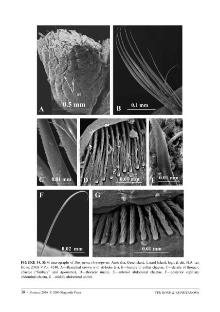
FIGURE 14. SEM micrographs <strong>of</strong> Dasynema chrysogyrus. Australia, Queensland, Lizard Island, legit & det. H.A. ten Hove. ZMA V.Pol. 4540. A—Branchial crown with stylodes (st), B—bundle <strong>of</strong> collar chaetae, C—details <strong>of</strong> thoracic chaetae (“limbate” and Apomatus), D—thoracic uncini, E—anterior abdominal chaetae, F—posterior capillary abdominal chaeta, G—middle abdominal uncini. 38 · <strong>Zootaxa</strong> 2036 © 2009 <strong>Magnolia</strong> <strong>Press</strong> TEN HOVE & KUPRIYANOVA
Remarks. This relatively poorly known monotypic genus is recorded from the Indo-West Pacific (see Imajima & ten Hove 1984, Nishi 1993). Its characteristic feature, outwardly directed stylodes on the radioles, is unique for serpulids. Paired stylodes are found elsewhere only in the sabellid genera Branchiomma and Pseudobranchiomma. See also discussion by Imajima & ten Hove (1984). Dasynema chrysogyrus (Grube, 1876), Japan, Philippine Islands, Ponape, Indonesia. 8. Ditrupa Berkeley, 1835 (Fig. 15) Type-species: Dentalium subulatum Deshayes, 1826 = junior synonym <strong>of</strong> Dentalium arietinum Müller, 1776; designated by Bush 1905: 223. Number <strong>of</strong> species: 2 Tube free, tusk-like, not attached to substratum, circular in cross-section, open at both ends, broadening anteriorly though exterior tapers just prior to tube mouth. Outer layer hyaline or white, inner layer opaque. Granular overlay <strong>of</strong> the tube absent. Operculum inverse conical with chitinous endplate. Peduncle cylindrical, smooth, without wings, gradually merging into operculum, no constriction; it is positioned as first dorsal left radiole. Pseudoperculum absent. Radioles arranged pectinately; up to 15 radioles per lobe. Inter-radiolar membrane, branchial eyes, and stylodes absent. Pair <strong>of</strong> filiform mouth palps present. 6 thoracic chaetigerous segments. Large entire (non-lobed) collar continuous with short thoracic membranes, ending at first chaetiger (second thoracic segment); tonguelets absent. Collar chaetae absent (see Collar segment, p. 22). First thoracic chaetiger biramous (see Collar segment, p. 22) with limbate chaetae (Fig. 15A, B) and with uncini; sometimes with special chaetae (see remarks). Apomatus chaetae absent. Thoracic uncini saw-to-rasp-shaped (dental formula P:2:2:2:1:1:1:1:1:1:1:1:1:1:1:1:1:1:1:1:1:1:1:1:1 or P:3:2:2:1……1) or rasp-shaped; about 25 teeth in pr<strong>of</strong>ile, with 2 or 3 teeth in a row above peg (P); peg blunt, curved upwards and gouged underneath (Fig. 15C). Triangular depression absent. Abdominal chaetae and certainly posterior ones thin, almost capillary, with very faint narrow geniculate tip (to completely capillary in D. gracillima). Abdominal uncini rasp-shaped, with 20–25 in pr<strong>of</strong>ile, up to 8 teeth in a row above peg; anterior peg blunt, almost rectangular (Fig. 15D). Achaetous anterior abdominal zone absent; however, anterior half <strong>of</strong> abdomen with uncini only. Posterior capillary chaetae present. Posterior glandular pad absent. Remarks. The genus is found living unattached (Fig. 1A) in s<strong>of</strong>t sediment marine environments around the world. Like many serpulid genera, Ditrupa has a history <strong>of</strong> taxonomic confusion to the extent that its tubes were included in the Mollusca by some authors. The generic diagnosis was emended by ten Hove & Smith (1990). An unattached free tube similar to that <strong>of</strong> Ditrupa is known only in two (?three) other serpulids, Bathyditrupa hovei and Serpula crenata (possibly incl. S. sinica). Unlike the circular in cross-section tubes <strong>of</strong> Ditrupa, those <strong>of</strong> Bathyditrupa and S. crenata are rectangular to multi-angular in cross-section (see The tube, p. 7). Ten Hove & Smith (1990: 113, 115) describe 2 populations <strong>of</strong> Ditrupa gracillima in which the first thoracic chaetiger shows special chaetae, one almost geniculately terminating in an oblique frayed narrow limbus, the other stoutly acicular. In view <strong>of</strong> the limited distributions <strong>of</strong> these two forms they question whether these populations might be in the process <strong>of</strong> speciation. 1. Ditrupa arietina (Müller, 1776), Northern Norway to Azores and Canary Islands, Mediterranean 2. Ditrupa gracillima Grube, 1878, widely distributed in Indo-West Pacific. TAXONOMY OF SERPULIDS: STATE OF AFFAIRS <strong>Zootaxa</strong> 2036 © 2009 <strong>Magnolia</strong> <strong>Press</strong> · 39
- Page 1 and 2: ZOOTAXA 2036 Taxonomy of Serpulidae
- Page 3 and 4: Zootaxa 2036: 1-126 (2009) www.mapr
- Page 5 and 6: Introduction The family Serpulidae
- Page 7 and 8: Morphology The tube Whereas tubes o
- Page 9 and 10: FIGURE 2. Variability of serpulid t
- Page 11 and 12: large crystals in the structure of
- Page 13 and 14: 30A) most probably too are a series
- Page 15 and 16: Throughout Spiraserpula, there is a
- Page 17 and 18: FIGURE 3. Morphology of serpulid an
- Page 19 and 20: FIGURE 5. General morphology of ser
- Page 21 and 22: FIGURE 7. Serpulid morphology (cont
- Page 23 and 24: occur incidentally in Pomatoceros,
- Page 25 and 26: completely rasp-shaped (Knight-Jone
- Page 27 and 28: Valid genera with diagnoses and lis
- Page 29 and 30: 5. Apomatus globifer Théel, 1878,
- Page 31 and 32: FIGURE 10. SEM micrographs of chaet
- Page 33 and 34: tonguelets between ventral and late
- Page 35 and 36: FIGURE 12. SEM micrographs of chaet
- Page 37: membrane. Radioles with ocellar clu
- Page 41 and 42: 9. Ficopomatus Southern, 1921 (Fig.
- Page 43 and 44: Filograna implexa M. Berkeley, 1835
- Page 45 and 46: with 12-14 teeth in profile, up to
- Page 47 and 48: FIGURE 19. SEM micrographs of chaet
- Page 49 and 50: 14. Galeolaria Lamarck, 1818 (Fig.
- Page 51 and 52: operculate H. cancerum Knight-Jones
- Page 53 and 54: 14. Hydroides bulbosus ten Hove, 19
- Page 55 and 56: 84. Hydroides trilobulus Chen & Wu,
- Page 57 and 58: membrane and stylodes absent. Branc
- Page 59 and 60: Schröder 1971), France (Fauvel 192
- Page 61 and 62: most recent study (Kupriyanova et a
- Page 63 and 64: FIGURE 28. SEM micrographs of chaet
- Page 65 and 66: FIGURE 29. Photos and SEMs of chaet
- Page 67 and 68: FIGURE 30. SEM micrographs of chaet
- Page 69 and 70: absent. Thoracic uncini saw-to-rasp
- Page 71 and 72: Tube white, opaque, circular in cro
- Page 73 and 74: FIGURE 33. Photos (A-E) and a SEM m
- Page 75 and 76: normal radiole and maximally coveri
- Page 77 and 78: 31. Pomatoleios Pixell, 1913 (Fig.
- Page 79 and 80: FIGURE 37. SEM micrographs of chaet
- Page 81 and 82: Jirkov (1997) further extended it t
- Page 83 and 84: Paraprotula was based on the absenc
- Page 85 and 86: limbate chaetae. Apomatus chaetae a
- Page 87 and 88: FIGURE 42. SEM micrographs of chaet
- Page 89 and 90:
FIGURE 43. SEM micrographs of chaet
- Page 91 and 92:
1. Salmacina amphidentata Jones, 19
- Page 93 and 94:
7. Semivermilia torulosa (delle Chi
- Page 95 and 96:
13. Serpula maorica (Benham, 1927),
- Page 97 and 98:
FIGURE 47. SEM micrographs of chaet
- Page 99 and 100:
limbate zone and proximal wing (Fig
- Page 101 and 102:
FIGURE 49. SEM micrographs of chaet
- Page 103 and 104:
46. Vitreotubus Zibrowius, 1979b (F
- Page 105 and 106:
forming ventral apron. Collar chaet
- Page 107 and 108:
** According to Ippolitov (2007: 26
- Page 109 and 110:
38 (37) Thoracic uncini saw shaped
- Page 111 and 112:
chaeta (pl. chaetae): chitinous bri
- Page 113 and 114:
thoracic membranes: thin folds on b
- Page 115 and 116:
Bush, K.J. (1907) Descriptions of t
- Page 117 and 118:
Gravier, C. (1905 (1906?)) Sur deux
- Page 119 and 120:
Hove, H.A. ten & Wolf, P.S. (1984)
- Page 121 and 122:
Marsden, J.R. & Anderson, D.T. (198
- Page 123 and 124:
Radl, E. (1912) Neue Lehre vom Zent
- Page 125 and 126:
Verrill, A.E. (1873) Report upon th