

Identification of important interactions between subchondral bone ...
Identification of important interactions between subchondral bone ...
Identification of important interactions between subchondral bone ...

Create successful ePaper yourself
Turn your PDF publications into a flip-book with our unique Google optimized e-Paper software.
CHAPTER 2: Introduction<br />
Bone is a specialized tissue, which together with cartilage make up the human skeleton. The<br />
<strong>bone</strong>s have three main functions; 1) providing mechanical support for the whole body and acting<br />
as muscle attachment for locomotion, 2) providing protective shields for the vital organs and the<br />
<strong>bone</strong> marrow, and 3) providing a reserve for ions, mainly calcium and phosphate, and for growth<br />
factors and cytokines 15,16 .<br />
There are several subtypes <strong>of</strong> <strong>bone</strong>s; long <strong>bone</strong>, flat <strong>bone</strong>s, short <strong>bone</strong>s and<br />
irregular <strong>bone</strong>s 17 , <strong>of</strong> which only long <strong>bone</strong>s - e.g. femur and tibia, which constitute the knee - will<br />
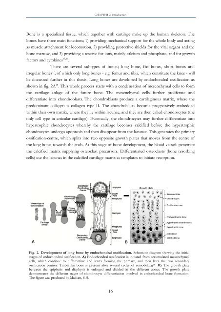
be discussed further in this thesis. Long <strong>bone</strong>s are developed by endochondral ossification as<br />
shown in fig. 2A 18 . This whole process starts with a condensation <strong>of</strong> mesenchymal cells to form<br />
the cartilage anlage <strong>of</strong> the future <strong>bone</strong>. The mesenchymal cells further proliferate and<br />
differentiate into chondroblasts. The chondroblasts produce a cartilaginous matrix, where the<br />
predominant collagen is collagen type II. The chondroblasts become progressively embedded<br />
within their own matrix, where they lie within lacunae, and they are then called chondrocytes (the<br />
only cell type in articular cartilage). Eventually, the chondrocytes may further differentiate into<br />
hypertrophic chondrocytes whereby the cartilage becomes calcified before the hypertrophic<br />
chondrocytes undergo apoptosis and then disappear from the lacunae. This generates the primary<br />
ossification-centre, which splits into two opposite growth plates that moves from the centre <strong>of</strong><br />
the long <strong>bone</strong>, towards the ends. At this stage <strong>of</strong> <strong>bone</strong> development, the blood vessels penetrate<br />
the calcified matrix supplying osteoclast precursors. Differentiated osteoclasts (<strong>bone</strong> resorbing<br />
cells) use the lacunas in the calcified cartilage matrix as templates to initiate resorption.<br />
Fig. 2. Development <strong>of</strong> long <strong>bone</strong> by endochondral ossification. Schematic diagram showing the initial<br />
stages <strong>of</strong> endochondral ossification. A) Endochondral ossification is initiated from accumulated mesenchymal<br />
cells, which continue to differentiate and starts forming the primary, and then later the two secondary<br />
ossification centres. Trabecular <strong>bone</strong> is present after several cycles <strong>of</strong> remodelling 16 . B) The growth plate<br />
<strong>between</strong> the epiphysis and diaphysis is enlarged and divided in the different zones. The growth plate<br />
demonstrates the different stages <strong>of</strong> chondrocyte differentiation involved in endochondral <strong>bone</strong> formation.<br />
The figure was produced by Madsen, S.H.<br />
16


