

Identification of important interactions between subchondral bone ...
Identification of important interactions between subchondral bone ...
Identification of important interactions between subchondral bone ...

You also want an ePaper? Increase the reach of your titles
YUMPU automatically turns print PDFs into web optimized ePapers that Google loves.
CHAPTER 2: Introduction<br />
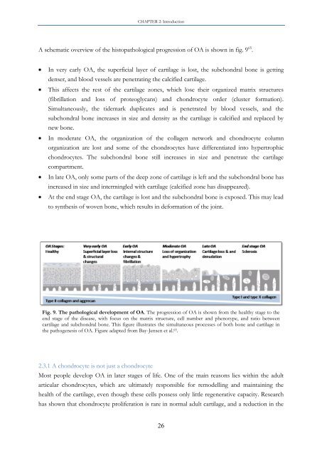
A schematic overview <strong>of</strong> the histopathological progression <strong>of</strong> OA is shown in fig. 9 65 .<br />
In very early OA, the superficial layer <strong>of</strong> cartilage is lost, the <strong>subchondral</strong> <strong>bone</strong> is getting<br />
denser, and blood vessels are penetrating the calcified cartilage.<br />
This affects the rest <strong>of</strong> the cartilage zones, which lose their organized matrix structures<br />
(fibrillation and loss <strong>of</strong> proteoglycans) and chondrocyte order (cluster formation).<br />
Simultaneously, the tidemark duplicates and is penetrated by blood vessels, and the<br />
<strong>subchondral</strong> <strong>bone</strong> increases in size and density as the cartilage is calcified and replaced by<br />
new <strong>bone</strong>.<br />
In moderate OA, the organization <strong>of</strong> the collagen network and chondrocyte column<br />
organization are lost and some <strong>of</strong> the chondrocytes have differentiated into hypertrophic<br />
chondrocytes. The <strong>subchondral</strong> <strong>bone</strong> still increases in size and penetrate the cartilage<br />
compartment.<br />
In late OA, only some parts <strong>of</strong> the deep zone <strong>of</strong> cartilage is left and the <strong>subchondral</strong> <strong>bone</strong> has<br />
increased in size and intermingled with cartilage (calcified zone has disappeared).<br />
At the end stage OA, the cartilage is lost and the <strong>subchondral</strong> <strong>bone</strong> is exposed. This may lead<br />
to synthesis <strong>of</strong> woven <strong>bone</strong>, which results in deformation <strong>of</strong> the joint.<br />
Fig. 9. The pathological development <strong>of</strong> OA. The progression <strong>of</strong> OA is shown from the healthy stage to the<br />
end stage <strong>of</strong> the disease, with focus on the matrix structure, cell number and phenotype, and ratio <strong>between</strong><br />
cartilage and <strong>subchondral</strong> <strong>bone</strong>. This figure illustrates the simultaneous processes <strong>of</strong> both <strong>bone</strong> and cartilage in<br />
the pathogenesis <strong>of</strong> OA. Figure adapted from Bay-Jensen et al. 65 .<br />
2.3.1 A chondrocyte is not just a chondrocyte<br />
Most people develop OA in later stages <strong>of</strong> life. One <strong>of</strong> the main reasons lies within the adult<br />
articular chondrocytes, which are ultimately responsible for remodelling and maintaining the<br />
health <strong>of</strong> the cartilage, even though these cells possess only little regenerative capacity. Research<br />
has shown that chondrocyte proliferation is rare in normal adult cartilage, and a reduction in the<br />
26


