

Identification of important interactions between subchondral bone ...
Identification of important interactions between subchondral bone ...
Identification of important interactions between subchondral bone ...

Create successful ePaper yourself
Turn your PDF publications into a flip-book with our unique Google optimized e-Paper software.
CHAPTER 2: Introduction<br />
18<br />
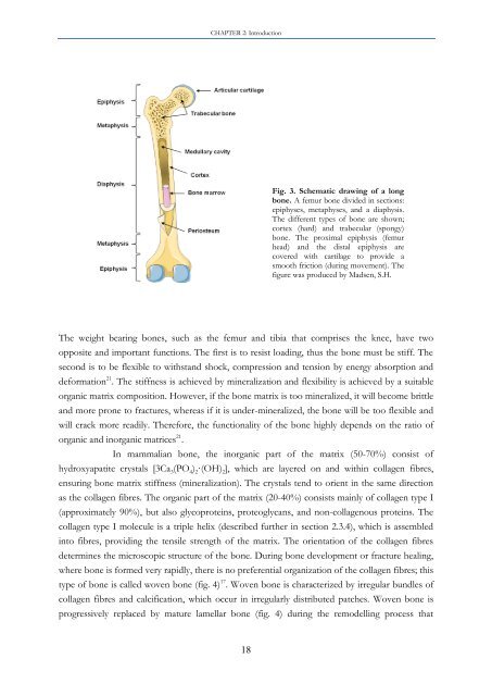
Fig. 3. Schematic drawing <strong>of</strong> a long<br />
<strong>bone</strong>. A femur <strong>bone</strong> divided in sections:<br />
epiphyses, metaphyses, and a diaphysis.<br />
The different types <strong>of</strong> <strong>bone</strong> are shown;<br />
cortex (hard) and trabecular (spongy)<br />
<strong>bone</strong>. The proximal epiphysis (femur<br />
head) and the distal epiphysis are<br />
covered with cartilage to provide a<br />
smooth friction (during movement). The<br />
figure was produced by Madsen, S.H.<br />
The weight bearing <strong>bone</strong>s, such as the femur and tibia that comprises the knee, have two<br />
opposite and <strong>important</strong> functions. The first is to resist loading, thus the <strong>bone</strong> must be stiff. The<br />
second is to be flexible to withstand shock, compression and tension by energy absorption and<br />
deformation 21 . The stiffness is achieved by mineralization and flexibility is achieved by a suitable<br />
organic matrix composition. However, if the <strong>bone</strong> matrix is too mineralized, it will become brittle<br />
and more prone to fractures, whereas if it is under-mineralized, the <strong>bone</strong> will be too flexible and<br />
will crack more readily. Therefore, the functionality <strong>of</strong> the <strong>bone</strong> highly depends on the ratio <strong>of</strong><br />
organic and inorganic matrices 21 .<br />
In mammalian <strong>bone</strong>, the inorganic part <strong>of</strong> the matrix (50-70%) consist <strong>of</strong><br />
hydroxyapatite crystals [3Ca 3(PO 4) 2·(OH) 2], which are layered on and within collagen fibres,<br />
ensuring <strong>bone</strong> matrix stiffness (mineralization). The crystals tend to orient in the same direction<br />
as the collagen fibres. The organic part <strong>of</strong> the matrix (20-40%) consists mainly <strong>of</strong> collagen type I<br />
(approximately 90%), but also glycoproteins, proteoglycans, and non-collagenous proteins. The<br />
collagen type I molecule is a triple helix (described further in section 2.3.4), which is assembled<br />
into fibres, providing the tensile strength <strong>of</strong> the matrix. The orientation <strong>of</strong> the collagen fibres<br />
determines the microscopic structure <strong>of</strong> the <strong>bone</strong>. During <strong>bone</strong> development or fracture healing,<br />
where <strong>bone</strong> is formed very rapidly, there is no preferential organization <strong>of</strong> the collagen fibres; this<br />
type <strong>of</strong> <strong>bone</strong> is called woven <strong>bone</strong> (fig. 4) 17 . Woven <strong>bone</strong> is characterized by irregular bundles <strong>of</strong><br />
collagen fibres and calcification, which occur in irregularly distributed patches. Woven <strong>bone</strong> is<br />
progressively replaced by mature lamellar <strong>bone</strong> (fig. 4) during the remodelling process that


