WOC 6e Guide to Microscopy
WOC 6e Guide to Microscopy
WOC 6e Guide to Microscopy

You also want an ePaper? Increase the reach of your titles
YUMPU automatically turns print PDFs into web optimized ePapers that Google loves.
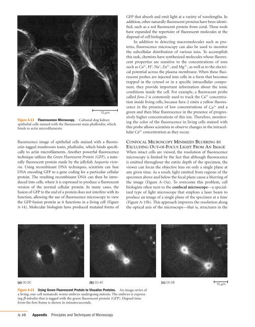
10 m<br />
Figure A-13 Fluorescence <strong>Microscopy</strong>. Cultured dog kidney<br />
epithelial cells stained with the fluorescent stain phalloidin, which<br />
binds <strong>to</strong> actin microfilaments.<br />
fluorescence image of epithelial cells stained with a fluorescein-tagged<br />
mushroom <strong>to</strong>xin, phalloidin, which binds specifically<br />
<strong>to</strong> actin microfilaments. Another powerful fluorescence<br />
technique utilizes the Green Fluorescent Protein (GFP), a naturally<br />
fluorescent protein made by the jellyfish Aequoria vic<strong>to</strong>ria.<br />
Using recombinant DNA techniques, scientists can fuse<br />
DNA encoding GFP <strong>to</strong> a gene coding for a particular cellular<br />
protein. The resulting recombinant DNA can then be introduced<br />
in<strong>to</strong> cells, where it is expressed <strong>to</strong> produce a fluorescent<br />
version of the normal cellular protein. In many cases, the<br />
fusion of GFP <strong>to</strong> the end of a protein does not interfere with its<br />
function, allowing the use of fluorescence microscopy <strong>to</strong> view<br />
the GFP-fusion protein as it functions in a living cell (Figure<br />
A-14). Molecular biologists have produced mutated forms of<br />
(a) 00:00 (b) 03:40 (c) 05:08<br />
Figure A-14 Using Green Fluorescent Protein <strong>to</strong> Visualize Proteins. An image series of<br />
a living, one-cell nema<strong>to</strong>de worm embryo undergoing mi<strong>to</strong>sis. The embryo is expressing<br />
b-tubulin<br />
that is tagged with the green fluorescent protein (GFP). Elapsed time<br />
from the first frame is shown in minutes:seconds.<br />
A-10 Appendix Principles and Techniques of <strong>Microscopy</strong><br />
GFP that absorb and emit light at a variety of wavelengths. In<br />
addition, other naturally fluorescent proteins have been identified,<br />
such as a red fluorescent protein from coral. These <strong>to</strong>ols<br />
have expanded the reper<strong>to</strong>ire of fluorescent molecules at the<br />
disposal of cell biologists.<br />
In addition <strong>to</strong> detecting macromolecules such as proteins,<br />
fluorescence microscopy can also be used <strong>to</strong> moni<strong>to</strong>r<br />
the subcellular distribution of various ions. To accomplish<br />
this task, chemists have synthesized molecules whose fluorescent<br />
properties are sensitive <strong>to</strong> the concentrations of ions<br />
such as and Mg as well as <strong>to</strong> the electri-<br />
2; Ca ,<br />
2; , H ; , Na ; , Zn2; ,<br />
cal potential across the plasma membrane. When these fluorescent<br />
probes are injected in<strong>to</strong> cells in a form that becomes<br />
trapped in the cy<strong>to</strong>sol or in a specific intracellular component,<br />
they provide important information about the ionic<br />
conditions inside the cell. For example, a fluorescent probe<br />
called fura-2 is commonly used <strong>to</strong> track the Ca concentration<br />
inside living cells, because fura-2 emits a yellow fluores-<br />
2;<br />
cence in the presence of low concentrations of and a<br />
Ca 2;<br />
green and then blue fluorescence in the presence of progressively<br />
higher concentrations of this ion. Therefore, moni<strong>to</strong>ring<br />
the color of the fluorescence in living cells stained with<br />
this probe allows scientists <strong>to</strong> observe changes in the intracellular<br />
Ca concentration as they occur.<br />
2;<br />
CONFOCAL MICROSCOPY MINIMIZES BLURRING BY<br />
EXCLUDING OUT-OF-FOCUS LIGHT FROM AN IMAGE<br />
When intact cells are viewed, the resolution of fluorescence<br />
microscopy is limited by the fact that although fluorescence<br />
is emitted throughout the entire depth of the specimen, the<br />
viewer can focus the objective lens on only a single plane at<br />
any given time. As a result, light emitted from regions of the<br />
specimen above and below the focal plane cause a blurring of<br />
the image (Figure A-15a). To overcome this problem, cell<br />
biologists often turn <strong>to</strong> the confocal microscope—a specialized<br />
type of light microscope that employs a laser beam <strong>to</strong><br />
produce an image of a single plane of the specimen at a time<br />
(Figure A-15b). This approach improves the resolution along<br />
the optical axis of the microscope—that is, structures in the<br />
10 m