WOC 6e Guide to Microscopy
WOC 6e Guide to Microscopy
WOC 6e Guide to Microscopy

You also want an ePaper? Increase the reach of your titles
YUMPU automatically turns print PDFs into web optimized ePapers that Google loves.
1<br />
2<br />
3<br />
4<br />
5<br />
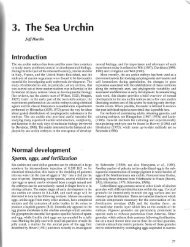
Specimen<br />
Metal a<strong>to</strong>ms<br />
evaporated from<br />
electrode at side<br />
Acid<br />
bath<br />
(a) Shadowing technique<br />
Mica surface<br />
Carbon a<strong>to</strong>ms evaporated<br />
from overhead electrode<br />
Copper grid<br />
Specimen dissolving<br />
away<br />
Metal replica<br />
Metal replica<br />
Metal replica<br />
Metal wire<br />
standard copper specimen grid ( 5 ) for viewing by transmis- hydrophobic interior of membranes, in most cases.<br />
sion electron microscopy.<br />
Platinum/carbon shadowing is then used <strong>to</strong> create a replica<br />
A related procedure is commonly used for visualizing of the fractured surface.<br />
purified molecules such as DNA and RNA. In this technique, Freeze fracturing is illustrated in Figure A-34. It takes<br />
a solution of DNA and/or RNA is spread on an air–water place in a modified vacuum evapora<strong>to</strong>r with an internal<br />
interface, creating a molecular monolayer that is collected on micro<strong>to</strong>me knife for fracturing the frozen specimen. The<br />
a thin film and visualized by uniformly depositing heavy temperature of the specimen support and the micro<strong>to</strong>me<br />
metal on all sides.<br />
arm and knife is precisely controlled. Specimens are generally<br />
fixed prior <strong>to</strong> freeze fracturing, although some living tissues<br />
can be frozen fast enough <strong>to</strong> keep them in almost lifelike con-<br />
FREEZE FRACTURING AND FREEZE<br />
dition. Because cells contain large amounts of water, fixed<br />
ETCHING ARE USEFUL FOR EXAMINING<br />
specimens are usually treated with an antifreeze such as<br />
THE INTERIOR OF MEMBRANES<br />
glycerol <strong>to</strong> provide cryoprotection—that is, <strong>to</strong> reduce the<br />
Freeze fracturing is an approach <strong>to</strong> sample preparation that formation of ice crystals during freezing.<br />
is fundamentally different from the methods described so far. The cryoprotected specimen is mounted on a metal<br />
Instead of cutting uniform slices through a tissue sample (or specimen support (Figure A-34, step 1 ) and immersed<br />
staining unsectioned material), specimens are rapidly frozen rapidly in freon cooled with liquid nitrogen ( 2 ). This proce-<br />
at the temperature of liquid nitrogen or liquid helium, placed dure also reduces the formation of ice crystals in the cells.<br />
in a vacuum, and struck with a sharp knife edge. Samples With the frozen specimen positioned on the specimen table<br />
frozen at such low temperatures are <strong>to</strong>o hard <strong>to</strong> be cut. in the vacuum evapora<strong>to</strong>r ( 3 ), a high vacuum is established,<br />
Instead, they fracture along lines of natural weakness—the the stage temperature is adjusted <strong>to</strong> around :100∞C, and the<br />
A-24 Appendix Principles and Techniques of <strong>Microscopy</strong><br />
Heated filament<br />
Carbon electrode<br />
Specimen<br />
(b) Vacuum evapora<strong>to</strong>r<br />
Metal electrode<br />
To vacuum system<br />
Vacuum evaporate<br />
Figure A-33 The Technique of Shadowing. (a) A stepwise procedure for shadowing.<br />
1<br />
The specimen is spread on a mica surface and dried. 2 The specimen is shadowed<br />
by coating it with a<strong>to</strong>ms of a heavy metal (platinum or gold, shown in blue) that are<br />
evaporated from a heated filament located <strong>to</strong> the side of the specimen in a vacuum<br />
evapora<strong>to</strong>r. This generates a metal replica (orange), the thickness of which reflects the<br />
surface con<strong>to</strong>urs of the specimen. 3 Next, the specimen is coated with carbon a<strong>to</strong>ms<br />
evaporated from an overhead electrode <strong>to</strong> strengthen and stabilize the metal replica.<br />
The replica is then floated on<strong>to</strong> the surface of an acid bath <strong>to</strong> dissolve away the speci-<br />
4<br />
men, leaving a clean metal replica. 5 The replica is washed and picked up on a copper<br />
grid for examination in the TEM. (b) The vacuum evapora<strong>to</strong>r in which shadowing is<br />
done. The carbon electrode is located directly over the specimen, whereas the heavy<br />
metal electrode is off <strong>to</strong> the side.