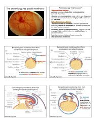
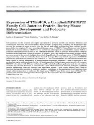
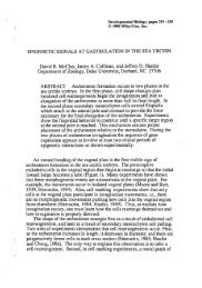
WOC 6e Guide to Microscopy
WOC 6e Guide to Microscopy
WOC 6e Guide to Microscopy

You also want an ePaper? Increase the reach of your titles
YUMPU automatically turns print PDFs into web optimized ePapers that Google loves.
(a) Confocal microscropy (a) Multipho<strong>to</strong>n microscopy<br />
(a) (b)<br />
Figure A-19 Digital Deconvolution <strong>Microscopy</strong>. A fission yeast cell stained with a dye<br />
specific for DNA (red) and a membrane-specific dye (green). The image on the left is<br />
an unprocessed optical section through the center of the cell. The image on the right is<br />
a projection of all the sections following three-dimensional image processing. The ring<br />
of the developing medial septum (red) is forming between the two nuclei (red) that<br />
arose by nuclear division during the previous mi<strong>to</strong>sis.<br />
of the same cell 30 times per second, making it possible <strong>to</strong><br />
moni<strong>to</strong>r rapid changes in the appearance and behavior of<br />
subcellular components. This has allowed scientists <strong>to</strong> obtain<br />
information on the changes in concentration and subcellular<br />
distribution of such cy<strong>to</strong>solic components as second messengers<br />
during cellular signaling, and <strong>to</strong> study the role of<br />
cy<strong>to</strong>skeletal structures in intracellular movements. Thus,<br />
digital video microscopy has greatly expanded our ability <strong>to</strong><br />
moni<strong>to</strong>r events as they occur within living cells.<br />
A-14 Appendix Principles and Techniques of <strong>Microscopy</strong><br />
Figure A-18 Multipho<strong>to</strong>n Excitation<br />
<strong>Microscopy</strong>. (a) In a standard laser<br />
scanning confocal microscope, the<br />
laser results in fluorescence in an<br />
hourglass-shaped path throughout<br />
the specimen. Because a large area<br />
fluoresces, pho<strong>to</strong>damage is much<br />
more likely <strong>to</strong> occur than in<br />
multipho<strong>to</strong>n excitation microscopy.<br />
(b) In a multipho<strong>to</strong>n excitation<br />
microscope, fluorescence is limited<br />
<strong>to</strong> a spot at the focus of the pulsed<br />
infrared laser beam, resulting in<br />
much less damage. The infrared<br />
illumination also penetrates more<br />
deeply in<strong>to</strong> the specimen than<br />
visible light.<br />
4 m<br />
Digital microscopy is not only useful for examining events<br />
in one focal plane. In a variation of this technique, a computer<br />
is used <strong>to</strong> control a focus mo<strong>to</strong>r attached <strong>to</strong> a microscope.<br />
Images are then collected throughout the thickness of a<br />
specimen. When such a series of images is collected at specific<br />
time intervals, such microscopy is called four-dimensional<br />
microscopy (this phrase is borrowed from physics; the four<br />
dimensions are the three dimensions of space plus the additional<br />
dimension of time). Analyzing four-dimensional data