Diploma Thesis - Erich Schmid Institute
Diploma Thesis - Erich Schmid Institute
Diploma Thesis - Erich Schmid Institute

Create successful ePaper yourself
Turn your PDF publications into a flip-book with our unique Google optimized e-Paper software.
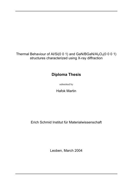
Thermal Behaviour of Al/Si(0 0 1) and GaN/BGaN/Al2O3(0 0 0 1)<br />
structures characterized using X-ray diffraction<br />
<strong>Diploma</strong> <strong>Thesis</strong><br />
submitted by<br />
Hafok Martin<br />
<strong>Erich</strong> <strong>Schmid</strong> Institut für Materialwissenschaft<br />
Leoben, March 2004
Danksagung<br />
Diese Arbeit entstand im Zeitraum September 2003 bis März 2004, wobei sämtliche<br />
Messungen am <strong>Erich</strong> <strong>Schmid</strong> Institut für Materialwissenschaft in Leoben durchgeführt<br />
wurden.<br />
Das Schreiben dieser Arbeit wäre ohne die professionelle Hilfe und Unterstützung von<br />
Herrn Dr. DI. Jozef Keckes nicht möglich gewesen. Ich verdanke ihm, dass er mir die<br />
Methoden der Spannungsmessung mittels Röntgendiffraktion sowie deren<br />
Auswertung und Interpretation näher gebracht hat. Ebenso möchte ich seinen Einsatz<br />
und guten Rat, der mir bei der Lösung vieler Probleme im Rahmen dieser Arbeit<br />
geholfen hat, hervorheben sowie dass er für mich und meine Anliegen immer Zeit<br />
gefunden hat. Aus all diesen Gründen will ich mich herzlich bedanken.<br />
Weiters möchte ich auch Herrn Ao. Univ. Prof. Balder Ortner meinen Dank für seine<br />
Ratschläge und Bemühungen aussprechen.<br />
Ein weiterer großer Dank gilt Herrn DI. Ernst Eiper, der mir bei der schwierigen<br />
Messung der einkristallinen GaN/BGaN/Saphire Probe behilflich war und mir bei der<br />
Korrektur der Arbeit eine wertvolle Hilfestellung gegeben hat. Ebenso möchte ich mich<br />
auch bei Stefan Massl bedanken, der mir bei der Überarbeitung des Textes geholfen<br />
hat.<br />
Zu guter letzt möchte ich meiner Familie meinen Dank aussprechen, die mich in<br />
meinem Bestreben stets unterstützt hat und mir immer einen wertvollen Rückhalt<br />
geboten hat.
Content:<br />
i<br />
Content<br />
1. Motivation.............................................................................................................1<br />
2. Introduction ..........................................................................................................2<br />
3. Basic X-ray diffraction expressions ......................................................................3<br />
3.1. Reciprocal lattice ......................................................................................3<br />
3.2. Bragg equation .........................................................................................4<br />
4. Mechanical properties of crystals .........................................................................6<br />
4.1. Stress, strain and displacement ...............................................................6<br />
4.2. Anisotropic Elasticity.................................................................................7<br />
4.3. Intrinsic and extrinsic stress .....................................................................9<br />
5. Orthogonal tensors and rotation.........................................................................11<br />
5.1. Basic relations ........................................................................................11<br />
5.2. Rotation in a plane..................................................................................12<br />
5.3. Euler angles ...........................................................................................13<br />
6. Basic Physical Properties of layer and substrate materials ................................15<br />
6.1. Polycrystalline aluminium on monocrystalline silicon .............................15<br />
6.1.1. Properties of aluminium layer .................................................................15<br />
6.1.2. Properties of silicon substrate ................................................................16<br />
6.2. Monocrystalline GaN on monocrystalline sapphire.................................17<br />
6.2.1. GaN........................................................................................................17<br />
6.2.2. Boron nitride ...........................................................................................21<br />
6.2.3. Sapphire substrate .................................................................................22<br />
7. Polycrystalline and monocrystalline Model.........................................................24<br />
7.1. Calculating isotropic elastic constants....................................................24<br />
7.1.1. Isotropic material ....................................................................................24<br />
7.1.2. Voigt model ............................................................................................24<br />
7.1.3. Reuss model: .........................................................................................27<br />
7.1.4. Hill model................................................................................................28<br />
7.2. Calculating stresses for a single crystalline material ..............................29<br />
7.3. Strain evaluation.....................................................................................31
ii<br />
Content<br />
8. Deposition of Thin Films.....................................................................................33<br />
8.1. Magnetron sputtering of polycrystalline Al thin films on Si(1 0 0) ...........33<br />
8.2. Molecular beam epitaxy of GaN/BGaN on Al2O3(0 0 0 1).......................34<br />
9. X-ray Diffraction – Measurements and Alignment ..............................................36<br />
9.1. Four Circle Goniometer ..........................................................................36<br />
9.2. DHS 900 Domed Hot Stage ...................................................................37<br />
9.3. The High-Resolution Monochromator.....................................................38<br />
9.4. Alignment of the diffractometer – point focus .........................................41<br />
9.5. Alignment of the diffractometer – line focus............................................42<br />
10. Aluminium on silicon measurement....................................................................45<br />
10.1. Experiment .............................................................................................45<br />
10.2. Shift of diffraction peaks .........................................................................46<br />
10.3. Peak broadening with increasing ψ tilt....................................................48<br />
11. GaN and GaBN on sapphire measurement........................................................50<br />
11.1. Measuring with the high resolution monochromator ...............................50<br />
11.2. Stereographic projections and crystal orientation...................................51<br />
11.3. Phi adjustment........................................................................................55<br />
11.4. Omega adjustment .................................................................................57<br />
11.5. Theta scans:...........................................................................................58<br />
12. Results of aluminium on silicon ..........................................................................61<br />
12.1. Sin(ψ)² vs. a plot.....................................................................................61<br />
12.2. Lattice spacing of aluminium layer .........................................................64<br />
12.3. Thermal expansion coefficient of aluminium...........................................65<br />
12.4. Lattice spacing of silicon substrate.........................................................66<br />
12.5. Thermal expansion coefficient of silicon substrate .................................66<br />
12.6. Stress curve ...........................................................................................67<br />
12.7. Strain curves ..........................................................................................69<br />
12.8. Discussion ..............................................................................................70<br />
13. Results of GaN/GaBN/Al2O3(0 0 0 1) .................................................................74<br />
13.1. Sapphire lattice parameters....................................................................74<br />
13.2. Thermal expansion coefficients of sapphire ...........................................75<br />
13.3. In-plane stress in GaN and GaBN layer: ................................................75<br />
13.4. Discussion: .............................................................................................81
iii<br />
Content<br />
14. Conclusion and Outlook .....................................................................................82<br />
15. Literature ............................................................................................................83<br />
16. Appendix ............................................................................................................87<br />
16.1. Serial port communication ......................................................................87<br />
16.2. Stereographic projection of Silicon (0 0 1)..............................................88<br />
16.3. Stereographic projection of gallium nitride (0 0 1) ..................................89<br />
16.4. Stereographic projection of sapphire (0 0 1)...........................................90<br />
16.5. GaN stress evaluation written in Mathematica: ......................................91
1. Motivation<br />
Motivation<br />
Virtually all types of thin films are expected to contain some amount of residual strain<br />
decisively influencing their mechanical behaviour and, secondary, modifying band-<br />
gaps in semiconductors, transition temperatures in superconductors, magnetic<br />
anisotropy, wear resistance or other important physical parameters. The strains can<br />
be formed unintentionally as an unavoidable consequence of the deposition process<br />
or intentionally in order to control required parameters of devices.<br />
The residual stresses represent very important issue especially in the case of modern<br />
electronics packages representing nowadays complicated composites of<br />
semiconductors, metals, dielectrics and plastics with specific thermal expansion<br />
coefficients, manufacturing temperatures and geometry. For the fabrication as well as<br />
for the practical application of such structures, the control of residual stresses has<br />
turned out to be of utmost importance.<br />
For the production of interconnects in microelectronic chips, aluminium and copper<br />
have been used. When the interconnects are thermally cycled during operation,<br />
various micro-structural effects can occur including grain growth, diffusion, plastic<br />
flow, electromigration etc. All these phenomena are influenced and partly also<br />
controlled by the magnitude of residual stresses in the metals.<br />
On the other hand, residual stresses in semiconductors directly influence important<br />
optical and electronic properties through the deformation of crystal lattice and<br />
subsequently the modification of the band gap parameters. The nitride<br />
semiconductors including the family of refractory materials like indium nitride,<br />
aluminium nitride and especially gallium nitride possess a significant potential for<br />
optoelectronic and piezoelectric applications. The presence of high compressive<br />
residual stresses in nitride-based thin films, however, influence not only the optical<br />
properties but is responsible also for crack formation – a very serious problem in<br />
nitride technology.<br />
The characterization of residual stresses in thin films represent thus a very important<br />
issue for nowadays technology. The main aim of this thesis to perform elevated-<br />
temperature X-ray diffraction characterization of residual stresses in aluminium thin<br />
films and in BGaN/GaN structure focusing micro-structural changes and phenomena<br />
related to intrinsic and extrinsic stresses.<br />
1
2. Introduction<br />
Introduction<br />
Already in ancient times, materials were tested for their reliability. For example the<br />
bending of swords to proof their elasticity and the tapping on ceramic vessels, like<br />
amphora, to detect defects were wide spread testing methods at that time. The first<br />
systematic examinations of materials properties and their quantification have been<br />
reported since the middle age, where this knowledge served for the constructions of<br />
buildings as well as to improve shipbuilding. With the appearance of the first<br />
industrial steam engines in the 19 th century, the demands not only on the material but<br />
also on adequate testing methods raised. Since that time, many important testing<br />
methods have been developed and utilized till nowadays especially for bulk<br />
materials. With the development of microelectronics and with the application of<br />
integrated circuits, new testing methods have been introduced allowing a significant<br />
progress in the miniaturization of the thin film – based structures.<br />
Today’s increasing performance of electronic devices is accompanied by a higher<br />
energy consumption stimulating requirements for the cooling. The heat dissipation<br />
influences not only the electrical, optical and magnetic properties of the devices but<br />
also the magnitude of internal stresses. The presence of residual stresses in thin<br />
films and in sublayers of sandwich structures can not be underestimated due to their<br />
direct influence on all basic physical as well as on mechanical properties of the<br />
devices. Especially in the context of the electromigration in copper or in aluminium<br />
interconnects, the residual stresses represent a very important issue.<br />
Up to now, stresses in thin films have been analyzed predominantly ex situ using<br />
X-ray diffraction, Raman and photoluminescence spectroscopy, the wafer curvature<br />
method, and high resolution transmission electron microscopy. The main practical<br />
advantage of the curvature technique resides in the application for new materials with<br />
unknown elastic constants. The diffraction techniques, on the other hand, are<br />
capable of resolving anisotropic deformation of crystal lattice, thin film behaviour on<br />
anisotropic substrates and stresses even in multilayered structures. Recently, a<br />
significant attention was devoted to the studying of residual stress origins in thin<br />
films. In this case, the elevated-temperature X-ray diffraction provided in important<br />
results leading to the understanding the role of intrinsic-stresses and their formation<br />
in thin films.<br />
Within this thesis structural properties of polycrystalline Al thin film deposited on<br />
Si(1 0 0) substrate and the properties of GaN/BGaN multilayers deposited on c plane<br />
sapphire are studied using elevated-temperature X-ray diffraction technique<br />
implemented recently at <strong>Erich</strong> <strong>Schmid</strong> <strong>Institute</strong> for Materials Science in Leoben. For<br />
the studies of GaN/BGaN/Al2O3(0 0 0 1) structures, a high-resolution monochromator<br />
was used.<br />
2
3. Basic X-ray diffraction expressions<br />
3.1. Reciprocal lattice<br />
Basic X-ray diffraction<br />
A mathematical formalism will be defined to describe scattering phenomena on<br />
crystal structures with translation symmetry [1]. Supposing the crystal periodicity, the<br />
selection of available functions is reduced. A periodical function alone, like sinus or<br />
cosines, is unable to describe the electron density distribution and phase<br />
phenomenon, the basic attributes of X-ray diffraction effect. Fourier series can be<br />
applied to describe the scattering effect.<br />
n<br />
G<br />
= ∫ N(<br />
r)<br />
exp( −i<br />
G . r)<br />
dV<br />
(equ. 3.1)<br />
cell<br />
∑<br />
n ( r ) = nG<br />
exp( i G . r)<br />
(equ. 3.2)<br />
G<br />
The imaginary unit, square root of minus one, is expressed by i. N(r) defines the<br />
electron density within one unit cell while, on the other hand, n(r) denotes the<br />
electron density of the whole crystal. The two vectors r and G in equation 3.1 and 3.2<br />
that appear in the exponential term can be expressed as:<br />
r = x a + y a + z a<br />
(equ. 3.3)<br />
1<br />
1<br />
2<br />
2<br />
3<br />
G = h b + k b + l b<br />
(equ. 3.4)<br />
The summation over G in equation 3.2 should be understood as a summations over<br />
h, k and l from minus infinity to plus infinity. The relation between ai and bi vectors<br />
can be found by calculating the scalar product of G and r. According to the Fourier<br />
series the scalar product must have the following form.<br />
3<br />
G . r = 2π<br />
(h x + k y + l z)<br />
(equ. 3.5)<br />
Considering the equation 3.5, the scalar product of a1 and b2 or a1 and b3 is zero, so<br />
a system of linear equations can be written as:<br />
3
⎛ a1<br />
. b1<br />
⎜<br />
⎜a<br />
2 . b1<br />
⎜<br />
⎝a<br />
3 . b1<br />
The solution of this system is:<br />
a<br />
a<br />
a<br />
1<br />
2<br />
3<br />
b<br />
b<br />
b<br />
. b<br />
. b<br />
2<br />
. b<br />
1<br />
2<br />
3<br />
2<br />
2<br />
a1<br />
. b3<br />
⎞ ⎛1<br />
⎟ ⎜<br />
a2<br />
. b3<br />
⎟ = 2π<br />
⎜0<br />
a ⎟ ⎜<br />
3 . b3<br />
⎠ ⎝0<br />
a2<br />
× a3<br />
= 2π<br />
a . ( a × a )<br />
1<br />
a3<br />
× a1<br />
= 2π<br />
a . ( a × a )<br />
1<br />
a1<br />
× a2<br />
= 2π<br />
a . ( a × a )<br />
1<br />
2<br />
2<br />
2<br />
3<br />
3<br />
3<br />
0<br />
1<br />
0<br />
0⎞<br />
⎟<br />
0⎟<br />
1⎟<br />
⎠<br />
Basic X-ray diffraction<br />
The vectors b1, b2 and b3 of equations 3.7 are called reciprocal lattice vectors.<br />
3.2. Bragg equation<br />
(equ. 3.6)<br />
(equ. 3.7)<br />
The Bragg equation represents a relatively simple way to describe the scattering<br />
phenomenon on a crystal lattice. Consider an incident beam that is reflected by a<br />
family of net planes. It’s worth to mention that the path difference between<br />
neighbouring net planes causes a phase difference between the diffracted beams, so<br />
the reflected radiation shows constructive and destructive interference. The Bragg<br />
equation, formula 3.8, predicts that constructive interference only occurs if the ratio<br />
between the phase difference and wavelength is an integer value of n.<br />
2 d sin( θ)<br />
n =<br />
(equ. 3.8)<br />
λ<br />
The variable θ is the angle between the incident or the reflected beam and the net<br />
plane. Because elastic scattering is assumed, the incident and reflected beam must<br />
have same wavelength λ. The last parameter is the lattice spacing d, the shortest<br />
distance between two neighbouring net planes with same Miller indices (h k l) and the<br />
surface normal G that is inversely proportional to the plane spacing d.<br />
π<br />
=<br />
G<br />
2<br />
d (equ. 3.9)<br />
4
Basic X-ray diffraction<br />
Equation 3.9 is valid for all crystal systems, and the components of G can be<br />
replaced by the points of intersection u1, u2 and u3 of the net plane with the elongated<br />
translation vectors a1, a2 and a3.<br />
Figure 3.1: Crystallographic plane and surface normal<br />
The components of G are for that reason:<br />
1<br />
h =<br />
u1<br />
1<br />
k =<br />
u2<br />
1<br />
l = (equ. 3.10)<br />
u<br />
3<br />
5
4. Mechanical properties of crystals<br />
4.1. Stress, strain and displacement<br />
Mechanical properties of crystals<br />
Internal stresses are produced by external forces acting on a body [2]. These<br />
external forces can be separated into two categories, like the distribution of forces<br />
over the surface, such as hydrostatic pressure, and distributed forces over the<br />
volume, for example gravitational forces or magnetic forces. A motion of the body is<br />
also able to influence the internal stresses. The most important expression to<br />
describe stresses can be derived by assuming the conservation of impulse.<br />
ρ a = div(<br />
σ)<br />
+ f<br />
(equ. 4.1)<br />
Where ρ is the density of the body, a is the acceleration, f denotes the distributed<br />
forces over the volume and σ, a second ranked tensor, represents the internal<br />
stresses.<br />
σ<br />
σ n<br />
n = (equ. 4.2)<br />
The distributed forces or stresses over a surface are written in equation 4.2 that is the<br />
product between the stress tensor and the surface normal vector n, and can be<br />
understood as a boundary condition for equation 4.1. In the case of stress<br />
measurement on thin films, which is a static problem, the acceleration is zero and the<br />
volume forces are neglected.<br />
div ( σ ) = 0<br />
(equ. 4.3)<br />
A body under stresses will deform, this means that a point P of the unstressed body<br />
moves to the position P’. The vector connecting P and P’ is called displacement u.<br />
We consider a short and a long rod with same cross section and made of the same<br />
material. Let us say that the change of length is the displacement, than one can see<br />
that the displacement of the longer rod is greater than the displacement of the small<br />
rod, if the same stress is applied at the ends of both rods. An assessment based on<br />
strains to characterize the deformation will show same results for both rods. This<br />
means that the influence of geometry does not play a role.<br />
6
ε<br />
ij<br />
=<br />
1 ⎛<br />
⎜<br />
∂ ui<br />
2 ⎜<br />
⎝ ∂x<br />
j<br />
∂ u j ⎞<br />
+ ⎟<br />
∂x<br />
⎟<br />
i ⎠<br />
Mechanical properties of crystals<br />
(equ. 4.4)<br />
Equation 4.4 defines the strain tensor for small displacements. A closer look on the<br />
strain tensor reveals the symmetric property. It was not mentioned above but the<br />
stress tensor is also symmetric.<br />
4.2. Anisotropic Elasticity<br />
ε = ε<br />
(equ. 4.5)<br />
ij<br />
ij<br />
ji<br />
σ = σ (equ. 4.6)<br />
Further equations are needed to solve a general mechanical problem, because if all<br />
unknown quantities of the previous chapter are count together the total sum will be<br />
15, in detail there are six stresses, just as much strains and additionally three<br />
displacements. On the other side we have three expressions to describe stresses<br />
(equation 4.1 or 4.3) and six formulas of strain/displacement relations (equation 4.4),<br />
so remains a lack of six equations.<br />
The missing equations are based on the mechanical behaviour of a material that is<br />
assumed to be elastic. In literature a material is often treated in an isotropic way. It<br />
means that a property like the electrical resistant or thermal expansion coefficient is<br />
not depending on the materials direction. However, a crystal is anisotropic [3], and<br />
therefore a general expression will define the elastic stresses/strains relations.<br />
ij<br />
ijkl<br />
ji<br />
σ = c ε<br />
(equ. 4.7)<br />
ij<br />
ijkl<br />
kl<br />
ε = s σ<br />
(equ. 4.8)<br />
The stiffness tensor cijkl of equation 4.7 is the relation between stress and strain<br />
tensor, both of second rank including nine components, thus the stiffness tensor must<br />
consist of 81 elements. Very similar to the stiffness is the compliance tensor sijkl in<br />
equation 4.8. It is simple to find a relation between both expressions.<br />
−1<br />
c = s<br />
kl<br />
−1<br />
s = c<br />
(equ. 4.9)<br />
7
Mechanical properties of crystals<br />
Because of the anisotropic material behaviour, with unequal properties in different<br />
directions, the position of the crystal system with reference to the sample is of<br />
interest. Generally the crystal and sample system do not coincide due to angular<br />
differences. In order to express the anisotropic behaviour in a sample system, the<br />
compliance or stiffness tensors must be rotated.<br />
A ' = O O A<br />
(equ. 4.10)<br />
For a fourth ranked tensor the expression has the following form:<br />
ijkl<br />
ij<br />
im<br />
ik<br />
jn<br />
jl<br />
ko<br />
kl<br />
A ' = O O O O A<br />
(equ. 4.11)<br />
In literature a second preferred way to express the relation between stresses and<br />
strains according to W. Voigt [4] is often used. By applying the matrix notation, the<br />
stress and strain tensors are reduced to vectors with six components, thus the<br />
compliance and stiffness matrices have 36 components. A comparison between<br />
stresses and strains written in matrix notation and tensor notation shows:<br />
⎛ ε<br />
⎜<br />
⎜ε<br />
⎜<br />
⎝ ε<br />
⎛ σ<br />
⎜<br />
⎜σ<br />
⎜<br />
⎝ σ<br />
11<br />
12<br />
13<br />
11<br />
12<br />
13<br />
ε<br />
ε<br />
ε<br />
12<br />
22<br />
23<br />
σ<br />
σ<br />
σ<br />
12<br />
22<br />
23<br />
ε<br />
ε<br />
ε<br />
13<br />
23<br />
33<br />
σ<br />
σ<br />
σ<br />
13<br />
23<br />
33<br />
⎞ ⎛ σ<br />
⎟ ⎜<br />
⎟ = ⎜σ<br />
⎟ ⎜<br />
⎠ ⎝ σ<br />
⎛<br />
⎜ ε1<br />
⎞<br />
⎟<br />
⎜<br />
⎜ 1<br />
⎟ = ε<br />
⎟<br />
⎜ 2<br />
⎠ ⎜ 1<br />
⎜ ε<br />
⎝ 2<br />
6<br />
5<br />
1<br />
6<br />
5<br />
lp<br />
1<br />
2<br />
σ<br />
σ<br />
σ<br />
ε<br />
1<br />
2<br />
ε<br />
2<br />
ε<br />
6<br />
2<br />
4<br />
mnop<br />
6<br />
4<br />
σ5<br />
⎞<br />
⎟<br />
σ4<br />
⎟<br />
σ ⎟<br />
3 ⎠<br />
1<br />
ε<br />
2<br />
1<br />
ε<br />
2<br />
ε<br />
3<br />
5<br />
4<br />
⎞<br />
⎟<br />
⎟<br />
⎟<br />
⎟<br />
⎟<br />
⎟<br />
⎠<br />
(equ. 4.12)<br />
(equ. 4.13)<br />
The first two and last two suffixes of the tensor notation are merged to a single<br />
number running from one to six, according to the scheme:<br />
Tensor<br />
notation<br />
Matrix<br />
notation<br />
11 22 33 23, 32 31, 13 12, 21<br />
1 2 3 4 5 6<br />
8
The relations between the compliance tensor and matrix are:<br />
sijkl = smn when m and n are 1, 2 or 3.<br />
2 sijkl = smn when either m or n are 4, 5 or 6.<br />
4 sijkl = smn when both m and n are 4, 5 or 6.<br />
Mechanical properties of crystals<br />
The advantage of working with matrix notation is the compacter formalism, though<br />
the rotation of the compliance and the stiffness matrix is more complicated than the<br />
rotation in the tensor notation. For example an isotropic material can be<br />
characterized by the following stiffness matrix:<br />
⎛ c11<br />
c12<br />
c12<br />
0 0 0 ⎞<br />
⎜<br />
⎟<br />
⎜c12<br />
c11<br />
c12<br />
0 0 0 ⎟<br />
⎜c<br />
⎟<br />
12 c12<br />
c11<br />
0 0 0<br />
c = ⎜<br />
⎟<br />
(equ. 4.14)<br />
⎜ 0 0 0 c44<br />
0 0 ⎟<br />
⎜ 0 0 0 0 c ⎟<br />
44 0<br />
⎜<br />
⎟<br />
⎜<br />
⎟<br />
⎝ 0 0 0 0 0 c44<br />
⎠<br />
Isotropic materials have only two independent components, so the third one can be<br />
calculated:<br />
4.3. Intrinsic and extrinsic stress<br />
1<br />
c44 = ( c11<br />
− c12<br />
)<br />
2<br />
The residual stresses in thin films results from a growth procedure and from a cooling<br />
down to the operation temperature after the deposition with specific intrinsic and<br />
extrinsic stress contributions, respectively. The intrinsic stresses originate from a<br />
specific microstructure development and the film densification during the growth [5].<br />
An atomic disorder caused by foreign atoms integrated into the lattice can serve as<br />
an example of the intrinsic stress origin. An impurity atom can substitute a lattice<br />
atom or it can be found in lattice gaps as an interstitial atom. The compressive/tensile<br />
stress is getting higher if the atomic radius of the impurity atoms is larger/smaller than<br />
the atomic radius of the original lattice atoms. Intrinsic stresses can be reduced by<br />
applying high substrate temperatures during deposition, because of the high mobility<br />
of the atoms reduces the disorder. An additional part of intrinsic stresses occur in<br />
sputtered layers. The incoming sputtered atoms are hitting the layer and this is like<br />
9
Mechanical properties of crystals<br />
shot peening, setting the layer under compressive stress. This effect is known as<br />
atomic peening.<br />
Extrinsic stresses originate from the cooling down procedure and basically depend on<br />
the different constants of thermal expansion of layer and substrate. After cooling or<br />
heating, starting from the deposition temperature TS, the layer material is elastically<br />
or plastically deformed. In the elastic region the extrinsic stress is:<br />
hkl<br />
σ = ( α − α ) ( T − T )<br />
(equ. 4.15)<br />
ex<br />
M l s<br />
S<br />
The thermal expansion coefficient αl belongs to the layer, the other one αs to the<br />
substrate and the quantity M hkl is the biaxial modulus of the layer [6] that is<br />
depending on the crystallographic orientation. The final stresses are the sum of<br />
intrinsic and extrinsic stresses.<br />
Residual stresses are reduced through crack formation under tensile stress,<br />
delamination of the layer or plastic deformation forming hills under compressive<br />
stress.<br />
10
5. Orthogonal tensors and rotation<br />
5.1. Basic relations<br />
Orthogonal tensors and rotation<br />
Crystals are anisotropic bodies, which means that the mechanical behaviour depends<br />
on the crystal orientation. For this reason, it is important to exactly define the<br />
orientation of the crystal in the sample coordinate system. Generally the sample and<br />
the crystal coordinate systems can be related by a rotation (Chapter 4.2) expressed<br />
by an orthogonal tensor O.<br />
We consider two right angled coordinate systems with the unit vectors ei and ei’<br />
(figure 5.1). A unit vector belonging to one coordinate system, for example ei, is<br />
projected on the axes of the other system so that the position of this vector can be<br />
obtained in the new coordinate system ei’ [7]. The length of the projected unit vector<br />
on one axis is:<br />
Figure 5.1: Relation between two tilted coordinate systems<br />
e i ' . e j = ei<br />
' e j cos( ei<br />
',<br />
e j)<br />
e i ' = 1<br />
j 1 = e<br />
e . e = cos( e ',<br />
e )<br />
(equ. 5.1)<br />
i ' j<br />
i j<br />
By applying equation 5.1 on all unit vectors of one coordinate system, an orthogonal<br />
tensor is received that contains only cosine functions. Such a expression is also<br />
known as direction cosine. For example let us use e1 to express e1‘, where the<br />
components of e1‘ and e1 are x1’, y1’, z1’ and x1, y1, z1.<br />
11
⎛x<br />
1'⎞<br />
⎛ cos( e1'<br />
, e1)<br />
⎜ ⎟ ⎜<br />
⎜ y1'⎟<br />
= ⎜cos(<br />
e 2'<br />
, e1)<br />
⎜ ⎟ ⎜<br />
⎝ z1'<br />
⎠ ⎝cos(<br />
e3'<br />
, e1)<br />
5.2. Rotation in a plane<br />
cos( e ',<br />
e )<br />
1<br />
2<br />
3<br />
2<br />
cos( e ',<br />
e )<br />
2<br />
cos( e ',<br />
e )<br />
1<br />
1<br />
2<br />
Orthogonal tensors and rotation<br />
cos( e1'<br />
, e3<br />
) ⎞ ⎛ x1<br />
⎞<br />
⎟ ⎜ ⎟<br />
cos( e 2'<br />
, e3<br />
) ⎟ ⎜ y1<br />
⎟<br />
cos( e ⎟ ⎜ ⎟<br />
3'<br />
, e3<br />
) ⎠ ⎝ z1<br />
⎠<br />
(equ. 5.2)<br />
e ' = O e<br />
(equ. 5.3)<br />
To learn more about how a rotation around a certain axis will affect the position of the<br />
sample, it is necessary to consider a body on which points the vector r, like in figure<br />
5.2. A positive rotation of the body is synonymous with the rotation of the vector r<br />
around the e3 axis and at the end of the operation the vector r coincides with r’.<br />
The new point r’ with the components x’, y’ and z’ can be expressed by the starting<br />
point r:<br />
r ' = O<br />
3<br />
r<br />
⎛cos(<br />
ϕ)<br />
− sin( ϕ)<br />
0⎞<br />
⎜<br />
⎟<br />
3<br />
O = ⎜ sin( ϕ)<br />
cos( ϕ)<br />
0⎟<br />
(equ. 5.4)<br />
⎜<br />
⎟<br />
⎝ 0 0 1⎠<br />
The orthogonal tensors, that are describing a rotation around the two other axes, are<br />
shown in equation 5.5 and 5.6.<br />
Figure 5.2: Rotation of an object<br />
⎛1<br />
0 0 ⎞<br />
⎜<br />
⎟<br />
1<br />
O = ⎜0<br />
cos( ψ)<br />
− sin( ψ)<br />
⎟<br />
(equ. 5.5)<br />
⎜<br />
⎟<br />
⎝0<br />
sin( ψ)<br />
cos( ψ)<br />
⎠<br />
12
Orthogonal tensors and rotation<br />
⎛ cos( ω)<br />
0 sin( ω)<br />
⎞<br />
⎜<br />
⎟<br />
2<br />
O = ⎜ 0 1 0 ⎟<br />
(equ. 5.6)<br />
⎜<br />
⎟<br />
⎝−<br />
sin( ω)<br />
0 cos( ω)<br />
⎠<br />
The body in position r’ can be turned back by a rotation around e3 axis in the<br />
negative direction. Thus the angle’s sign is negative and the body’s starting position<br />
is supposed to be r’.<br />
⎛x<br />
⎞ ⎛ cos( ϕ)<br />
⎜ ⎟ ⎜<br />
⎜ y⎟<br />
= ⎜ − sin( ϕ)<br />
⎜ ⎟ ⎜<br />
⎝ z ⎠ ⎝ 0<br />
sin( ϕ)<br />
cos( ϕ)<br />
0<br />
0⎞<br />
⎛ x'⎞<br />
⎟ ⎜ ⎟<br />
0⎟<br />
⎜ y'⎟<br />
1⎟<br />
⎜ ⎟<br />
⎠ ⎝ z'<br />
⎠<br />
A closer look shows that the orthogonal tensor is the transposed or the inverse<br />
version of O 3 .<br />
5.3. Euler angles<br />
3 T<br />
( O ) r'<br />
r = (equ. 5.7)<br />
In the previous chapter we only thought about a rotation in a plane. In practice more<br />
possibilities are needed to express the tilt between two coordinate systems. One<br />
possibility is the concept of Euler angles [8], that can be understood as three<br />
separated rotations around certain axes, similar like before. The first step is the<br />
rotation around the e3 axis so e1 and e2 will change their position. After that the new<br />
e1 is supposed to be the next rotation axis, which will lead to two new e2 and e3 axes,<br />
and the last step is like the first one.<br />
After the first rotation the new axis is e1.<br />
⎛cos(<br />
ϕ1)<br />
− sin( ϕ1)<br />
0⎞<br />
⎜<br />
⎟<br />
ei<br />
' = ⎜ sin( ϕ1)<br />
cos( ϕ1)<br />
0⎟<br />
e<br />
⎜ 0 0 1⎟<br />
⎝<br />
⎠<br />
⎛1<br />
0 0 ⎞<br />
⎜<br />
⎟<br />
ei<br />
'' =<br />
⎜0<br />
cos( φ)<br />
sin( φ)<br />
⎟ ei<br />
'<br />
⎜0<br />
sin( ) cos( ) ⎟<br />
⎝ − φ φ ⎠<br />
i<br />
13
The last step is the rotation around the new e3 axis.<br />
All equations written in full are:<br />
⎛cos(<br />
ϕ2<br />
) − sin( ϕ2<br />
) 0⎞<br />
⎜<br />
⎟<br />
ei<br />
'' ' = ⎜ sin( ϕ2<br />
) cos( ϕ2<br />
) 0⎟<br />
ei<br />
''<br />
⎜ 0 0 1⎟<br />
⎝<br />
⎠<br />
Orthogonal tensors and rotation<br />
⎛cos(<br />
ϕ2<br />
) − sin( ϕ2<br />
) 0⎞<br />
⎛1<br />
0 0 ⎞ ⎛cos(<br />
ϕ1)<br />
− sin( ϕ1)<br />
0⎞<br />
⎜<br />
⎟ ⎜<br />
⎟ ⎜<br />
⎟<br />
ei<br />
' ' ' = ⎜ sin( ϕ2<br />
) cos( ϕ2<br />
) 0⎟<br />
⎜0<br />
cos( φ)<br />
sin( φ)<br />
⎟ ⎜ sin( ϕ1)<br />
cos( ϕ1)<br />
0⎟<br />
e<br />
⎜ 0 0 1⎟<br />
⎜0<br />
sin( ) cos( ) ⎟ ⎜ 0 0 1⎟<br />
⎝<br />
⎠ ⎝ − φ φ ⎠ ⎝<br />
⎠<br />
After the multiplications the orthogonal Euler tensor has the following formula:<br />
e<br />
O<br />
⎛ cos( ϕ1)<br />
cos( ϕ2<br />
) − cos( φ)<br />
sin( ϕ1)<br />
sin( ϕ2<br />
)<br />
⎜<br />
= ⎜−<br />
cos( ϕ1)<br />
sin( ϕ2<br />
) − cos( φ)<br />
sin( ϕ1)<br />
cos( ϕ2<br />
)<br />
⎜<br />
⎝<br />
sin( φ)<br />
sin( ϕ1)<br />
sin( ϕ ) cos( ϕ ) + cos( φ)<br />
cos( ϕ ) sin( ϕ )<br />
1<br />
− sin( ϕ ) sin( ϕ ) + cos( φ)<br />
cos( ϕ ) cos( ϕ )<br />
1<br />
2<br />
2<br />
− sin( φ)<br />
cos( ϕ )<br />
1<br />
1<br />
1<br />
2<br />
2<br />
sin( φ)<br />
sin( ϕ2<br />
) ⎞<br />
⎟<br />
sin( φ)<br />
cos( ϕ2<br />
) ⎟<br />
cos( φ)<br />
⎟<br />
⎠<br />
i<br />
(equ. 5.8)<br />
14
Basic Physical Properties<br />
6. Basic Physical Properties of layer and substrate materials<br />
6.1. Polycrystalline aluminium on monocrystalline silicon<br />
6.1.1. Properties of aluminium layer<br />
Aluminium ore, most commonly bauxite, occurs mainly in tropical and sub-tropical<br />
areas. The raw material bauxite is converted into alumina in the Bayer process, which<br />
is reduced to aluminium metal in electrolytic cells known as pots by adding cryolite [9].<br />
Pure aluminium shows no phase transformation in the solid state and will crystallise at<br />
an equilibrium temperature of about 660°C in a closed packed face centred lattice [10]<br />
with a mono-atomar basis (figure 6.1).<br />
As basis for the stress evaluation the anisotropic elastic behaviour of a cubic material<br />
is represented in equation 6.1, where the low number of independent elastic constants<br />
corresponds to the high symmetry of the cubic system [3, 4].<br />
⎛s11<br />
⎜<br />
⎜s12<br />
⎜s12<br />
s = ⎜<br />
⎜ 0<br />
⎜ 0<br />
⎜<br />
⎝ 0<br />
s<br />
s<br />
s<br />
12<br />
11<br />
12<br />
0<br />
0<br />
0<br />
s<br />
s<br />
s<br />
12<br />
12<br />
11<br />
0<br />
0<br />
0<br />
s<br />
0<br />
0<br />
0<br />
44<br />
0<br />
0<br />
Figure 6.1: Cubic face centred unit cell<br />
s<br />
0<br />
0<br />
0<br />
0<br />
44<br />
0<br />
0 ⎞<br />
⎟<br />
0 ⎟<br />
0 ⎟<br />
⎟<br />
0 ⎟<br />
0 ⎟<br />
⎟<br />
s ⎟<br />
44 ⎠<br />
⎛ c<br />
⎜<br />
⎜c<br />
⎜c<br />
c = ⎜<br />
⎜ 0<br />
⎜ 0<br />
⎜<br />
⎝ 0<br />
11<br />
12<br />
12<br />
c<br />
c<br />
c<br />
12<br />
11<br />
12<br />
0<br />
0<br />
0<br />
c<br />
c<br />
c<br />
12<br />
12<br />
11<br />
0<br />
0<br />
0<br />
c<br />
0<br />
0<br />
0<br />
44<br />
0<br />
0<br />
c<br />
0<br />
0<br />
0<br />
0<br />
44<br />
0<br />
0 ⎞<br />
⎟<br />
0 ⎟<br />
0 ⎟<br />
⎟<br />
0 ⎟<br />
0 ⎟<br />
⎟<br />
c ⎟<br />
44 ⎠<br />
(equ 6.1)<br />
15
Basic Physical Properties<br />
The components of the compliance and stiffness matrix or tensor are not real constant<br />
values due to their temperature dependence. In figure 6.2 the three independent<br />
compliance matrix components of aluminium were plotted [11]. Another characteristic<br />
parameter associated with the temperature influence is the coefficient of thermal<br />
expansion that has a value of 23,8 10 -6 K -1 for aluminium [12].<br />
s 12 / [10 -3 GPa -1 ]<br />
-5<br />
-6<br />
-7<br />
-8<br />
-9<br />
-10<br />
s 11 / [10 -3 GPa -1 ]<br />
24<br />
22<br />
20<br />
18<br />
16<br />
14<br />
34<br />
0 100 200 300 400 500<br />
6.1.2. Properties of silicon substrate<br />
The Czochralski technique is a widespread production method capable of providing<br />
silicon single crystals with a relatively large size. After cutting the silicon single crystals<br />
into thin wafers, followed by surface treatments like lapping, etching and polishing,<br />
they are ready to serve as substrate for thin film deposition.<br />
Silicon has a diamond-like structure [1, 10], which is based on a face centred cubic<br />
lattice with the primitive basis consisting of two atoms at position [0 0 0] and [¼¼¼].<br />
This basis reduces therefore the symmetry but it is not changing the general elastic<br />
mechanical behaviour [13] of the cubic lattice (figure 6.3) described by the compliance<br />
or stiffness matrix (at room-temperature).<br />
T / °C<br />
Figure 6.2: Temperature dependent compliance constants of aluminium<br />
c11 = 165,64 GPa ; c12 = 63,94 GPa ; c44 = 79,51 GPa;<br />
s 11<br />
s 12<br />
s 44<br />
50<br />
48<br />
46<br />
44<br />
42<br />
40<br />
38<br />
36<br />
s 44 / [10 -3 GPa -1 ]<br />
16
Basic Physical Properties<br />
During cooling, starting from the melting point at 1410°C, silicon crystallises in the<br />
diamond structure without showing allotropic transformation in the solid state. The<br />
cooling process is associated with the thermal contraction of the silicon material that<br />
has a thermal expansion coefficient of 2,616 10 -6 K -1 at room temperature [13].<br />
6.2. Monocrystalline GaN on monocrystalline sapphire<br />
6.2.1. GaN<br />
Unfortunately, bulk crystals of nitrides cannot be obtained by conventional methods of<br />
liquid phase epitaxy, because of extremely high melting temperatures and very high<br />
decomposition pressures at the melting point.<br />
Several production techniques are available for growing thin gallium nitride (GaN)<br />
films. These methods can be divided in two groups, one where chemical reactions play<br />
an important role and the other one where only physical process are responsible for<br />
the layer formation. For example a representative of the first group would be<br />
metalorganic vapour deposition (MOCVD) technique or reactive sputtering and for<br />
instance a production method belonging to the second group would be molecular<br />
beam epitaxy (MBE).<br />
Figure 6.3: Diamond structure<br />
17
Basic Physical Properties<br />
Group III nitrides like AlN, GaN and InN can crystallize in wurtzite, zinc-blende and<br />
rock-salt crystal structures. At ambient conditions the thermodynamically stable phase<br />
is the wurtzite structure and only at higher pressures an allotropic transformation<br />
changes the crystal to rock salt structure. The zinc-blende structure is metastable and<br />
may be stabilized by heteroepitaxial growth on substrates reflecting topological<br />
compatibility.<br />
In the preparation phase of the experiment several Bragg reflections of the thin layer<br />
were measured, leading to the result that the thin GaN film is based on wurtzite<br />
structure (figure 6.4), where the anions form a closed packed hexagonal structure and<br />
the cations with smaller atomic radius will occupy the tetrahedral gaps [10]. Owing to<br />
the stoichiometry only the half of all available tetrahedral positions can be filled.<br />
GaN crystallized in wurtzite structure and therefore it has a lower symmetry than<br />
native hexagonal structures. All hexagonal based lattices have same compliance and<br />
stiffness matrix [3] that are expressed in equation 6.2.<br />
⎛s11<br />
⎜<br />
⎜s12<br />
⎜s13<br />
s = ⎜<br />
⎜ 0<br />
⎜ 0<br />
⎜<br />
⎝ 0<br />
s<br />
s<br />
s<br />
12<br />
11<br />
13<br />
0<br />
0<br />
0<br />
s<br />
s<br />
s<br />
13<br />
13<br />
33<br />
0<br />
0<br />
0<br />
s<br />
0<br />
0<br />
0<br />
44<br />
0<br />
0<br />
s<br />
0<br />
0<br />
0<br />
0<br />
44<br />
0<br />
2 ( s<br />
Figure 6.4: Wurtzite structure<br />
11<br />
0<br />
0<br />
0<br />
0<br />
0<br />
− s<br />
11<br />
⎞<br />
⎟<br />
⎟<br />
⎟<br />
⎟<br />
⎟<br />
⎟<br />
⎟<br />
) ⎟<br />
⎠<br />
⎛ c11<br />
⎜<br />
⎜c12<br />
⎜c13<br />
⎜<br />
c =<br />
⎜ 0<br />
⎜<br />
0<br />
⎜<br />
⎜ 0<br />
⎝<br />
c<br />
c<br />
c<br />
12<br />
11<br />
13<br />
0<br />
0<br />
0<br />
c<br />
c<br />
c<br />
13<br />
13<br />
33<br />
0<br />
0<br />
0<br />
c<br />
0<br />
0<br />
0<br />
44<br />
0<br />
0<br />
c<br />
0<br />
0<br />
0<br />
0<br />
44<br />
0<br />
1<br />
( c<br />
2<br />
11<br />
0<br />
0<br />
0<br />
0<br />
0<br />
− c<br />
11<br />
⎞<br />
⎟<br />
⎟<br />
⎟<br />
⎟<br />
⎟<br />
⎟<br />
⎟<br />
) ⎟<br />
⎠<br />
(equ 6.2)<br />
18
Basic Physical Properties<br />
During the measurement the temperature is increased or decreased, and it must be<br />
taken into account that the elastic stiffness tensor has no longer constant values. In<br />
the evaluation of the data, the temperature dependence of the elastic constants,<br />
measured by R. R. Reeber and K. Wang [14], will be taken into consideration.<br />
c 13 / GPa<br />
100<br />
98<br />
96<br />
94<br />
92<br />
c11 / GPa<br />
c33 / GPa<br />
390<br />
385<br />
380<br />
375<br />
370<br />
365<br />
360<br />
134<br />
200 300 400 500 600 700 800 900 1000<br />
T / °C<br />
Figure 6.5: Temperature dependent stiffness constants of wurtzite GaN<br />
c 11<br />
c 33<br />
c 12<br />
c 13<br />
c 44<br />
146<br />
144<br />
142<br />
140<br />
138<br />
136<br />
c 12 / GPa<br />
99,0<br />
98,5<br />
98,0<br />
97,5<br />
97,0<br />
96,5<br />
c 44 / GPa<br />
19
Basic Physical Properties<br />
The plane spacing of hexagonal lattices is expressed through h, k, l, a0 and c0. Both<br />
unknown unstressed lattice parameters a0 and c0 must be considered when refining<br />
the structural parameters. R.R. Reeber and K. Wang [15] examined the lattice<br />
parameters of 99,99% pure and annealed GaN powder using neutron scattering. The<br />
results of their experiment can be seen in figure 6.6. With this data a relation between<br />
a0 and c0 is found by expressing c0 through the lattice parameter a0 and the ratio c0/ a0<br />
given by the neutron scattering measurement.<br />
-1<br />
c 0 a 0<br />
1,6263<br />
1,6262<br />
1,6261<br />
1,6260<br />
1,6259<br />
1,6258<br />
1,6257<br />
1,6256<br />
a 0 / A<br />
3,200<br />
3,198<br />
3,196<br />
3,194<br />
3,192<br />
3,190<br />
3,188<br />
3,186<br />
200 300 400 500 600 700 800 900 1000<br />
T / K<br />
a 0 spacing<br />
c 0 spacing<br />
-1<br />
c0 a0 Figure 6.6: Lattice parameters of wurtzite GaN<br />
5,195<br />
5,190<br />
5,185<br />
5,180<br />
c 0 / A<br />
20
6.2.2. Boron nitride<br />
Basic Physical Properties<br />
Boron nitride (BN) has different properties than other group III nitride members [16].<br />
The crystal structures and related physical properties are analogous to modifications<br />
of carbon, thus boron nitride exists in graphite like hexagonal structure. The former<br />
includes the stable zinc-blende and metastable wurtzite crystal structure. In contrast to<br />
the other group III nitrides no transition to the rock-salt structure at high pressures has<br />
been observed. The hexagonal boron nitride has outstanding mechanical properties,<br />
but it is less interesting for electronic applications.<br />
Figure 6.7: Modifications of boron nitride [17]<br />
gBN … hexagonal BN, zBN … zinc-blende BN, wBN … wurtzite BN<br />
The buffer layer contains only a small fraction of boron nitride, thus it is supposed that<br />
the boron will substitute gallium, so the buffer layer will also crystallise in wurtzite<br />
structure. The different lattice constants between boron nitride (a0=2,553A and<br />
c0=4,228A) and gallium nitride (a0=3,188A and c0=5,185A) [16] will cause tension in<br />
the buffer layer because of the smaller atomic radius of boron.<br />
21
Basic Physical Properties<br />
The elastic stiffness constants for wurtzite boron nitride at 300K, according to K. Kim,<br />
W.R.L. Lambrecht and B. Segall [18], are:<br />
c11 = 987 GPa; c12 = 143 GPa; c13= 70 GPa; c33 = 1020GPa; c44= 369GPa;<br />
6.2.3. Sapphire substrate<br />
Sapphire is the most used substrate for the growth of group III nitrides, which can be<br />
produced by the Czrochalski technique with high crystal quality and at low cost.<br />
Based on the 2:3 stoichiometry aluminium cations that take an octahedral position<br />
must fill two third of available sites [19]. To see how this occur a cation layer between<br />
two layers of close packed oxygen ions is drawn in figure 6.8.<br />
Figure 6.8: Shifting of aluminium cation layer<br />
These octahedral sites occupied by aluminium ions form a hexagonal array with the<br />
same spacing as the oxygen layer. The next cation layer has the same honeycomb<br />
configuration but is shifted by one atomic spacing in the direction of the vector 1. After<br />
another close packed oxygen layer, a third cation layer is placed, that is shifted by the<br />
vector 2. If a vertical slice as indicated by the dashed line is taken than the<br />
arrangement of cathions is drawn like in figure 6.9. The columns of octahedral sited<br />
perpendicular to the (0 0 0 1) plane alternate in having every two sides occupied and<br />
one empty. Only by considering the stacking of the closed packed oxygen ion layers<br />
must follow that the sapphire crystal is based on a hexagonal lattice. The anisotropic<br />
22
Basic Physical Properties<br />
elastic behaviour is for that reason expressed by equation 6.2. The following stiffness<br />
constants for sapphire are valid for room temperature [20].<br />
c11 = 496 GPa; c12 = 164 GPa; c13= 115 GPa; c33 = 498GPa; c44= 148GPa;<br />
A thermal property influencing the origin of extrinsic stress is the thermal expansion<br />
coefficient that is distinguished between expansion parallel and perpendicular to<br />
c-axis. According to Landolt and Börnstein [21] the thermal expansion coefficients for<br />
sapphire are 7,5 10 -6 K -1 perpendicular to c-axis and 8,5 10 -6 K -1 parallel to c-axis.<br />
Figure 6.9: (1 0 1 0) vertical slice according to dashed line in figure 6.8<br />
23
7. Polycrystalline and monocrystalline Model<br />
7.1. Calculating isotropic elastic constants<br />
7.1.1. Isotropic material<br />
Polycrystalline and monocrystalline Model<br />
An untextured material will behave in an isotropic way due to the randomly oriented<br />
grains, though a grain can be seen as single crystal with anisotropic elastic<br />
properties. For calculating stresses in isotropic materials, the Hill model [22] can be<br />
used which is based on Voigt and Reuss approach.<br />
7.1.2. Voigt model<br />
W. Voigt [4] assumed an untextured polycrystalline material where all grains are<br />
under the same strain. Starting with the matrix notation of the stiffness matrix for a<br />
cubic material (equation 6.1) and converting the expression into tensor notation<br />
(chapter 4.2) a rotation can be performed using Euler angles (equation 5.8) to rotate<br />
the crystal in any possible position. A mean value of all random orientated crystals is<br />
taken to express an isotropic behaviour (equation 4.14) of an untextured cubic<br />
material.<br />
2π<br />
π 2π<br />
V 1<br />
e e e e<br />
c ijkl = ∫ ∫ ∫ Oim<br />
O jn Oko<br />
Olp<br />
cmnop<br />
sin( φ)<br />
dϕ1<br />
dφ<br />
dϕ2<br />
(equ 7.1)<br />
8π<br />
0 0<br />
c<br />
c<br />
c<br />
0<br />
V<br />
1111<br />
V<br />
1122<br />
V<br />
2323<br />
= c<br />
= c<br />
= c<br />
V<br />
11<br />
V<br />
12<br />
V<br />
44<br />
1<br />
= ( 3c<br />
5<br />
1<br />
= ( c11<br />
5<br />
1<br />
= ( c11<br />
5<br />
11<br />
+ 2 c<br />
+ 4 c<br />
− c<br />
12<br />
12<br />
12<br />
+<br />
−<br />
+<br />
3 c<br />
4 c<br />
2 c<br />
44<br />
44<br />
)<br />
44<br />
)<br />
)<br />
(equ 7.2)<br />
The stiffness form is unusable for calculating strains, thus the elastic constants must<br />
be converted into compliance form by applying equation 4.9. First the stiffness<br />
components of the cubic system must be replaced on the right side, and afterwards<br />
the isotropic stiffness constants of the Voigt average must be expressed by<br />
compliance components of an isotropic material, that has only two independent<br />
parameters.<br />
s<br />
V<br />
44<br />
=<br />
2 ( s<br />
V<br />
11<br />
− s<br />
V<br />
12<br />
)<br />
24
Polycrystalline and monocrystalline Model<br />
Therefore the compliance components of the Voigt average defined by compliance<br />
constants of a cubic material are:<br />
s<br />
V<br />
12<br />
s<br />
V<br />
11<br />
=<br />
= 2 s<br />
11<br />
1 ⎛<br />
⎜<br />
⎜−<br />
s<br />
2 ⎝<br />
s<br />
11<br />
− s<br />
12<br />
+ 3s<br />
2<br />
5 ( s11<br />
− s12<br />
)<br />
−<br />
3s<br />
− 3s<br />
+ s<br />
12<br />
5 ( s<br />
11<br />
12<br />
44<br />
2<br />
5 ( s11<br />
− s12<br />
)<br />
+<br />
3s<br />
− 3s<br />
+ s<br />
− s<br />
11<br />
) s<br />
12<br />
44<br />
⎟ ⎞<br />
⎠<br />
V<br />
11 12 44<br />
44 = (equ 7.3)<br />
3s11<br />
− 3s12<br />
+ s44<br />
The sample can be rotated around three different angles ω, ψ and ϕ, but the rotation<br />
cannot be performed in any order of these angles. In figure 7.1 the sample and two<br />
coordinate systems are shown. The first one is the laboratory coordinate system eL<br />
which will be fixed and independent of the sample rotation. In this system the plane<br />
spacing is measured, because G will always be parallel to eL3. The second one is the<br />
sample coordinate system eS that is associated with the sample and will change<br />
position during rotation. The problem is, that the strains are measured in the<br />
laboratory system, but the stresses must be expressed in the sample system, so a<br />
rotation order must be performed to describe the sample’s tilt during the<br />
measurement. Let us start at a position where both systems coincide. The first<br />
rotation performed by the diffractometer is around eS1 direction by an angle ω,<br />
marked with the double arrow. In this position the new rotation is done around the<br />
new eS2 axis and the last one around the new eS3 axis. This is similar to the Euler<br />
angles procedure.<br />
a.) starting position b.) ψ-rotation c.) ϕ-rotation d.) end position<br />
and ω-rotation<br />
Figure 7.1: Rotation of the sample with respect to the laboratory system<br />
According to chapter 4.2 the laboratory system can be transformed by rotation into<br />
the sample system.<br />
25
e =<br />
The transformation matrix written in full is:<br />
Polycrystalline and monocrystalline Model<br />
3 1 2<br />
D<br />
S = O O O e L O e L<br />
(equ 7.4)<br />
⎛cos(<br />
ϕ)<br />
cos( ω)<br />
− sin( ϕ)<br />
sin( ψ)<br />
sin( ω)<br />
− sin( ϕ)<br />
cos( ψ)<br />
cos( ϕ)<br />
sin( ω)<br />
+ sin( ϕ)<br />
sin( ψ)<br />
cos( ω)<br />
⎞<br />
⎜<br />
⎟<br />
D<br />
O = ⎜sin(<br />
ϕ)<br />
cos( ω)<br />
+ cos( ϕ)<br />
sin( ψ)<br />
sin( ω)<br />
cos( ϕ)<br />
cos( ψ)<br />
sin( ϕ)<br />
sin( ω)<br />
− cos( ϕ)<br />
sin( ψ)<br />
cos( ω)<br />
(equ 7.5)<br />
⎟<br />
⎜<br />
⎟<br />
⎝ − cos( ψ)<br />
sin( ω)<br />
sin( ψ)<br />
cos( ψ)<br />
cos( ω)<br />
⎠<br />
Equation 7.4 will transform the laboratory system into the sample system, but for the<br />
evaluation of stresses the opposite way is demanded. For that reason the inverse<br />
orthogonal tensor O D is used.<br />
D D<br />
σ = O O σ<br />
ϕψ (equ 7.6)<br />
ij<br />
ki<br />
The change of the orthogonal tensor’s suffix, in equation 7.6, represents the<br />
transposed or inverted version of O D . The thin film is considered to be under plane<br />
stress, which means that every component of the stress tensor on the right side that<br />
has at least one three as suffix is zero.<br />
ε<br />
ϕψ,<br />
V<br />
33<br />
=<br />
lj<br />
kl<br />
V ϕψ<br />
s33kl σkl<br />
In the experiment the ω−angle has no significance and it is set to zero. After<br />
simplifying the expression for the Voigt model is:<br />
= ( σ<br />
) V<br />
ϕψ,<br />
V<br />
ε33 11 + σ22<br />
1 + σϕ<br />
2 ( s<br />
V =<br />
1<br />
σ ϕ<br />
11<br />
− s<br />
V<br />
2<br />
12<br />
V<br />
) ( s11<br />
+ 2 s12<br />
) − ( s<br />
2 (3s<br />
− 3s<br />
+ s<br />
11<br />
12<br />
2<br />
11<br />
44<br />
5 ( s11<br />
− s12<br />
) s44<br />
=<br />
2 (3s<br />
− 3s<br />
+ s<br />
11<br />
12<br />
44<br />
sin<br />
2<br />
− 3s<br />
)<br />
)<br />
( ψ)<br />
2<br />
2<br />
= σ11<br />
sin ( ϕ)<br />
− σ12<br />
sin( 2 ϕ)<br />
+ σ22<br />
cos ( ϕ)<br />
(equ 7.7)<br />
12<br />
) s<br />
44<br />
26
7.1.3. Reuss model:<br />
Polycrystalline and monocrystalline Model<br />
In contrast to Voigt supposes Reuss that all grains of an untextured material are<br />
under same stresses, so only grains with a net plane normal parallel to eL3 will diffract<br />
[23]. Therefore the crystal must be rotated in a position where the net plane is<br />
perpendicular to the measuring direction eL3.<br />
v<br />
3<br />
h b1<br />
+ k b2<br />
+ l b<br />
=<br />
h b + k b + l b<br />
v<br />
2<br />
1<br />
l a<br />
=<br />
l a<br />
1<br />
2<br />
2<br />
2<br />
− k a<br />
− k a<br />
v = v × v<br />
The wanted net plane normal can be rotated into a parallel position to eL3 by using<br />
the direction cosine.<br />
⎛ ec1<br />
. v1<br />
ec2<br />
. v1<br />
ec3<br />
. v1<br />
⎞<br />
⎜<br />
⎟<br />
e L = ⎜e<br />
c1 . v 2 ec2<br />
. v2<br />
ec3<br />
. v2<br />
⎟ eC<br />
= O<br />
⎜e<br />
c1 . v3<br />
ec2<br />
. v3<br />
ec3<br />
. v ⎟<br />
⎝<br />
3 ⎠<br />
The following othogonal tensor O hkl is only valid for cubic systems.<br />
O<br />
hkl<br />
⎛<br />
⎜<br />
⎜<br />
⎜<br />
= ⎜<br />
⎜<br />
⎜<br />
⎜<br />
⎝<br />
h<br />
h<br />
2<br />
2<br />
k<br />
2<br />
+ k<br />
+ l<br />
0<br />
h<br />
2<br />
+ k<br />
2<br />
2<br />
+ l<br />
2<br />
+ l<br />
2<br />
−<br />
k<br />
2<br />
+ l<br />
h<br />
+ k<br />
2<br />
2<br />
k + l<br />
k<br />
2<br />
2<br />
h k<br />
h<br />
l<br />
2<br />
2<br />
+ k<br />
2<br />
+ l<br />
2<br />
3<br />
2<br />
3<br />
3<br />
+ l<br />
2<br />
3<br />
3<br />
−<br />
k<br />
hkl<br />
2<br />
e<br />
−<br />
C<br />
+ l<br />
h<br />
2<br />
2<br />
h l<br />
h<br />
k<br />
+ k<br />
2<br />
2<br />
k + l<br />
l<br />
An untextured material will have lots of grains with G parallel to eL3 so in every<br />
position around the net plane normal the same amount of crystals is present.<br />
O<br />
λ<br />
⎛cos(<br />
λ)<br />
⎜<br />
= ⎜ sin( λ)<br />
⎜<br />
⎝ 0<br />
− sin( λ)<br />
cos( λ)<br />
0<br />
0⎞<br />
⎟<br />
0⎟<br />
1⎟<br />
⎠<br />
2<br />
+ k<br />
2<br />
+ l<br />
2<br />
2<br />
+ l<br />
2<br />
⎞<br />
⎟<br />
⎟<br />
⎟<br />
⎟<br />
⎟<br />
⎟<br />
⎟<br />
⎠<br />
27
2π<br />
hkl,<br />
R 1 λ λ λ λ λ λ λ λ<br />
sijkl =<br />
2π<br />
∫ Oim<br />
O jn Oko<br />
Olp<br />
Omq<br />
Onr<br />
Oos<br />
Opt<br />
sqrst<br />
0<br />
Polycrystalline and monocrystalline Model<br />
After calculating the Reuss average the mean elastic constants are dependent of the<br />
miller indices h, k and l. The strain is written in the following way, by using the<br />
stresses in the laboratory system from before:<br />
After simplifying:<br />
ϕψ,<br />
hkl,<br />
R hkl,<br />
R ϕψ<br />
ε33 = s33 kl σkl<br />
ϕψ , hkl,<br />
R<br />
hkl<br />
ε33 = ( σ11<br />
+ σ22<br />
) R1<br />
+ σϕ<br />
R<br />
R<br />
7.1.4. Hill model<br />
R<br />
hkl<br />
2<br />
hkl<br />
1<br />
= s<br />
= s<br />
11<br />
12<br />
− s<br />
+ Γ<br />
12<br />
⎛<br />
⎜s<br />
⎝<br />
11<br />
− s<br />
⎛<br />
+ 3 Γ ⎜s<br />
⎝<br />
11<br />
12<br />
hkl<br />
2<br />
s<br />
−<br />
2<br />
− s<br />
12<br />
44<br />
2<br />
sin ( ψ)<br />
⎞<br />
⎟<br />
⎠<br />
s<br />
−<br />
2<br />
44<br />
⎞<br />
⎟<br />
⎠<br />
dλ<br />
2 2 2 2 2 2<br />
h k + k l + h l<br />
Γ =<br />
(equ 7.8)<br />
2 2 2 2<br />
( h + k + l )<br />
The Reuss and Voigt model are both limits of elastic stresses. In the hill model, that<br />
shows good correspondence with practical results, simply the average value of both<br />
limits is taken by allocating x in equation 7.9 to be a half.<br />
ε = x ε + ( 1 − x)<br />
ε<br />
(equ 7.9)<br />
hill<br />
33<br />
ϕψ,<br />
hkl,<br />
R<br />
33<br />
The strain in direction of the net plane normal is:<br />
After rewriting equation 7.10 follows:<br />
ε<br />
hill<br />
33<br />
ϕψ<br />
d − d<br />
=<br />
d<br />
0<br />
0<br />
ϕψ,<br />
V<br />
33<br />
(equ 7.10)<br />
28
d<br />
ϕψ<br />
ϕψ,<br />
hkl,<br />
R<br />
= d (1+<br />
( x ε + ( 1−<br />
x)<br />
ε<br />
0<br />
33<br />
Polycrystalline and monocrystalline Model<br />
ϕψ,<br />
V<br />
33<br />
Equation 7.11 will be used for nonlinear regression and d0 is substituted by:<br />
d<br />
ϕψ<br />
a 0 ϕψ,<br />
hkl,<br />
R<br />
ϕψ,<br />
V<br />
= (1+<br />
( x ε33<br />
+ ( 1−<br />
x)<br />
ε33<br />
)) (equ 7.11)<br />
2 2 2<br />
h + k + l<br />
7.2. Calculating stresses for a single crystalline material<br />
The examination of isotropic polycrystalline material differs from anisotropic single<br />
crystalline material in such a way that an untextured polycrystalline material fulfils the<br />
Bragg condition at every ψ-ϕ tilt because there are always crystals with net plane<br />
normals parallel to the measuring direction in the laboratory system. However Bragg<br />
reflections of single crystals can only be detected at strict defined tilts [24].<br />
ε = O O O O s σ<br />
(equ 7.12)<br />
ij<br />
e<br />
mi<br />
e<br />
nj<br />
e<br />
ok<br />
Only strains can be measured, hence the stresses must be expressed with a linear<br />
elastic anisotropic material model. The strains in the sample system are depending<br />
on the crystal position therefore the orthogonal tensor O e , which is described by Euler<br />
angles, is introduced to express the relation between the sample and the crystal<br />
system.<br />
ij<br />
D<br />
ki<br />
e<br />
pl<br />
D<br />
lj<br />
kl<br />
mnop<br />
kl<br />
))<br />
ε = O O ε<br />
ϕω (equ 7.13)<br />
The strain measurement is done in the laboratory system, so the sample’s tilt is<br />
defined by equation 7.13 which is similar to equation 7.6 except that the strain tensor<br />
is rotated and ω−angle is used instead of ψ.<br />
In the case of gallium nitride and gallium boron nitride on sapphire the basal plane of<br />
the GaN crystal and BGaN crystal is perpendicular to the surface normal, so further<br />
calculations are simplified in such a way that no orthogonal tensor O e is needed,<br />
because of the transversal isotropy of the hexagonal lattice.<br />
ε = O O s σ<br />
ϕω (equ 7.14)<br />
33<br />
D<br />
i3<br />
D<br />
j3<br />
ijkl<br />
kl<br />
29
Polycrystalline and monocrystalline Model<br />
If we are assuming a biaxial stress state than all stresses that have at least one three<br />
as suffix will be zero.<br />
ε ϕ<br />
= ( s<br />
11<br />
σ<br />
11<br />
+ s<br />
12<br />
σ<br />
22<br />
2<br />
) cos ( ϕ)<br />
+ ( s<br />
12<br />
ε<br />
= ε<br />
+ ε<br />
sin ( ω)<br />
ϕω<br />
33<br />
C ϕ 2<br />
ε<br />
σ<br />
c<br />
11<br />
= s13 ( σ11<br />
+ σ22<br />
)<br />
+ s<br />
11<br />
σ<br />
22<br />
2<br />
) sin ( ϕ)<br />
+ ( s<br />
11<br />
− s<br />
12<br />
) σ<br />
12<br />
sin(2 ϕ)<br />
− s13<br />
( σ11<br />
+ σ<br />
(equ 7.15)<br />
The plane spacing is calculated by applying the Bragg equation and can be<br />
expressed by the two lattice parameters a and c of the hexagonal lattice.<br />
The strain parallel to G is:<br />
2<br />
2 2<br />
1 4 (h + h k + k ) l<br />
= +<br />
(equ 7.16)<br />
2<br />
2<br />
2<br />
d 3a<br />
c<br />
ε<br />
ϕω<br />
33<br />
ϕω<br />
d − d<br />
=<br />
d<br />
The final regression formula has the following form:<br />
d<br />
0<br />
0<br />
ϕω<br />
C ϕ<br />
= d0<br />
(1+<br />
ε ) = d0<br />
(1+<br />
ε + ε sin ( ω)<br />
)<br />
(equ 7.17)<br />
ϕω 2<br />
33<br />
It is possible to replace one lattice parameter by using the ratio of cN0 and aN0<br />
measured by R. R. Reeber and K. Wang [15].<br />
c = a<br />
0<br />
The unstressed lattice parameter a0 is found using equation 7.16 and by replacing d0<br />
in the regression formula.<br />
0<br />
c<br />
a<br />
N0<br />
N0<br />
30<br />
22<br />
)
d<br />
2<br />
2<br />
N0<br />
Polycrystalline and monocrystalline Model<br />
2<br />
N0<br />
0 = a 0<br />
(equ 7.18)<br />
2<br />
l<br />
4<br />
+ (h<br />
3<br />
⎛ c<br />
⎜<br />
⎝ a<br />
⎞<br />
⎟<br />
⎠<br />
2 ⎛ c<br />
+ h k + k ) ⎜<br />
⎝ a<br />
The elastic behaviour of the buffer layer that is containing a small fraction of boron<br />
nitride, about 3%, can be estimated by supposing a linear change of elastic constants<br />
with increasing boron nitride content.<br />
s<br />
BGaN<br />
ij<br />
= s<br />
GaN<br />
ij<br />
+ ( s<br />
It is assumed that the compliance constants of the BGaN show the same thermal<br />
behaviour like the GaN layer constants.<br />
s ( T)<br />
=<br />
BGaN<br />
ij<br />
s(<br />
T<br />
)<br />
GaN<br />
ij<br />
BN<br />
ij<br />
− s<br />
s(<br />
300<br />
GaN<br />
ij<br />
s(<br />
300 K)<br />
The approximation of the temperature dependence was done by assuming that the<br />
compliance constants will increase in the same way like the compliance constants of<br />
gallium nitride. To estimate the new lattice parameters of BGaN the same procedure<br />
) x<br />
K)<br />
N0<br />
N0<br />
BGaN<br />
ij<br />
GaN<br />
ij<br />
was applied like before, hence the value of x is again 3%.<br />
And the new c0/a0 ratio is:<br />
c<br />
a<br />
0<br />
0<br />
a<br />
c<br />
( T)<br />
( T)<br />
7.3. Strain evaluation<br />
BGaN<br />
0<br />
BGaN<br />
0<br />
BGaN<br />
BGaN<br />
= a<br />
= c<br />
GaN<br />
0<br />
GaN<br />
0<br />
c0<br />
=<br />
a<br />
0<br />
+<br />
+<br />
( T)<br />
( T)<br />
( a<br />
( c<br />
GaN<br />
GaN<br />
BN<br />
0<br />
BN<br />
0<br />
− a<br />
− c<br />
0<br />
GaN<br />
0<br />
GaN<br />
0<br />
) x<br />
) x<br />
c0<br />
( 300 K)<br />
a 0(<br />
300 K)<br />
c0<br />
( 300 K)<br />
a ( 300 K)<br />
The strains are related to the sample system like in the stress evaluation. The only<br />
strain that can be measured is expressed in the laboratory system. This strain is<br />
parallel to G and so to eL3 vector of the laboratory system, thus the sample<br />
coordinates must be expressed by the laboratory coordinates. The opposite way is<br />
GaN<br />
⎞<br />
⎟<br />
⎠<br />
BGaN<br />
BGaN<br />
GaN<br />
31
Polycrystalline and monocrystalline Model<br />
expressed in equation 7.7, where the laboratory system is transformed into sample<br />
system. For that reason the inverse tensor O D will be used to transform the strains<br />
from the sample into the laboratory system.<br />
ε = O O ε<br />
ϕψ (equ 7.19)<br />
ij<br />
D<br />
ki<br />
The transposed matrix of O D is obtained by changing the suffixes. Like before the<br />
measured strain in the laboratory system is written as:<br />
ε<br />
ϕψ<br />
33<br />
D<br />
lj<br />
ϕψ<br />
d − d<br />
=<br />
d<br />
The equation used in the nonlinear regression has the following form:<br />
d<br />
ϕψ<br />
= d ( 1+<br />
( ε<br />
0<br />
33<br />
+ ( ε<br />
22<br />
+ ( ε<br />
13<br />
2<br />
cos ( ϕ)<br />
− ε<br />
sin( ϕ)<br />
− ε<br />
12<br />
23<br />
0<br />
0<br />
kl<br />
sin( 2 ϕ)<br />
+ ε<br />
11<br />
cos( ϕ))<br />
sin( 2 ψ)))<br />
2<br />
sin ( ϕ)<br />
− ε<br />
If the ω−axis is involved in the sample rotation the plane spacing is:<br />
d<br />
ϕω<br />
= d ( 1+<br />
( ε<br />
0<br />
33<br />
+ ( ε<br />
11<br />
+ ( ε<br />
2<br />
cos ( ϕ)<br />
+ ε<br />
13<br />
cos( ϕ)<br />
+ ε<br />
12<br />
23<br />
sin( 2 ϕ)<br />
+ ε<br />
22<br />
sin( ϕ))<br />
sin( 2 ω)))<br />
33<br />
2<br />
sin ( ϕ)<br />
− ε<br />
33<br />
2<br />
) sin ( ψ)<br />
2<br />
) sin ( ω)<br />
(equ 7.20)<br />
(equ 7.21)<br />
32
8. Deposition of Thin Films<br />
Deposition of Thin Films<br />
8.1. Magnetron sputtering of polycrystalline Al thin films on Si(1 0 0)<br />
Magnetron sputtering of polycrystalline thin films is a widely used deposition<br />
technique. Thin layers of chromium, gold, titanium, aluminium and many other<br />
materials can be produced using the technique in a deposition chamber<br />
schematically depicted in Fig. 8.1. In the evacuated chamber, ionized gas atoms<br />
(usually argon ions) are accelerated under influence of an electrical field towards a<br />
target. The high kinetic energy enables the ions to strike out surface bound atoms<br />
which are leaving, with rather low kinetic energy (some keV), the target’s surface in<br />
all possible directions. A substrate, fixed usually on the opposited side of the<br />
chamber, is covered by a thin film of the sputtered atoms. The advantages of the<br />
magnetron sputtering process are a good control of process parameters, high purity<br />
of the films and a possibility to use low process temperatures.<br />
For the production of polycrystalline Al thin films on monocrystalline Si(1 0 0) wafers,<br />
magnetron sputtering device installed at the “<strong>Institute</strong> of Physics, Technical University<br />
Wien” was used. As a substrate for the specimen preparation, a 1 mm thick Si(1 0 0)<br />
wafer cleaned using isopropanol and acetone was used. On this native-oxidized<br />
silicon wafer, a polycrystalline aluminium thin film with a thickness of 2 µm was<br />
deposited using magnetron sputtering. The deposition was performed at 150 °C<br />
applying a pressure of 4 x 10 -3 mbar.<br />
+<br />
Figure 8.1: A schematic view of a sputtering reactor<br />
1 Vacuum chamber, 2 upper electrode, 3 plasma,<br />
4 lower electrode, 5 gas inlet, 6 vacuum pump connection<br />
7 substrate, 8 high frequency voltage, 9 target<br />
10 plasma ion, 11 target atom<br />
33
8.2. Molecular beam epitaxy of GaN/BGaN on Al2O3(0 0 0 1)<br />
Deposition of Thin Films<br />
The molecular beam epitaxy (MBE) is a thin film deposition process in which thermal<br />
beams of atoms or molecules react on a single crystalline substrate surface that is<br />
held under ultra high vacuum at elevated temperatures [25, 26].<br />
Such devices usually consist of at least three chambers (Figure 8.2). The first<br />
chamber serves as a sample container at medium high vacuum. The second<br />
chamber is used for sample preparations such as outgassing or sputter etching and<br />
for an accommodation of surface analytical facilities under ultra high vacuum. The<br />
last chamber or growth chamber is also under ultrahigh vacuum and equipped with<br />
facilities capable of forming and monitoring the vacuum level, heating and monitoring<br />
the temperature of the substrate, generating and determining the molecular or atomic<br />
beam’s intensity and controlling of the composition profiles.<br />
The composition of the grown film and its doping level depend on the relative<br />
deposition rates of the constituent elements, which is controlled by the evaporation<br />
rate and the geometrical configuration between source and substrate. The<br />
evaporation rate of particular elements is set by temperature of the effusion cells,<br />
containing the element in the liquid phase.<br />
MBE has in comparison with other epitaxial growth methods some unique<br />
advantages. The growth rate is generally low allowing a compositional and doping<br />
profile changes within atomic dimensions leading also to an atomic smooth surface.<br />
The growth temperature is relatively low and thus diffusion between layers of different<br />
composition is negligible. A disadvantage is due to low growth rates the small<br />
throughput having an effect on long depositions times, about one hour for a one<br />
micron high layer.<br />
Figure 8.2: A schematic drawing of the growth chamber<br />
For the purposes of this diploma thesis, the GaN/B0.03Ga0.97N multilayer structure<br />
(with the individual thickness of 1 µm in the case of both sublayers) was prepared in<br />
34
Deposition of Thin Films<br />
the development laboratories of “Infineon AG, München”. In the preparation phase<br />
the substrate is degreased and etched for the removal of surface contaminates and<br />
mechanical damage due to polishing, and finally rinsed in deionised water. The<br />
deposition of both sublayers on monocrystalline Al2O3(0 0 0 1) was performed at<br />
700 °C in ultra-high vacuum gradually - at first BGaN sublayer was prepared and<br />
then the deposition process was concluded with the deposition of GaN sublayer. The<br />
original goal was to understand an influence of the BGaN buffer layer on the stress<br />
state in the GaN top layer.<br />
35
X-ray Diffraction – Measurements and Alignment<br />
9. X-ray Diffraction – Measurements and Alignment<br />
9.1. Four Circle Goniometer<br />
The basic principle of characterising residual stress in thin layers with the help of a<br />
diffractometer is to measure lattice spacing by using X-ray diffraction at different<br />
sample tilts. The deviation from the unstressed lattice parameters in combination with<br />
a material model enables the evaluation of the residual stresses. All present<br />
measurements were carried out on a Seifert 3000 PTS four circle diffractometer<br />
equipped with the DHS 900 heating stage.<br />
The X-ray radiation is produced by rapid deceleration of electrons and can be<br />
separated in continuous and characteristic radiation. In a diffraction experiment it is<br />
desirable to have monochromatic Kα radiation. This is achieved by placing a filter<br />
material, which has its K absorption edge between the Kα and Kβ wavelengths, in<br />
the beam path. A common filter material for copper radiation is nickel [27].<br />
Figure 9.1: Diffraction geometry<br />
36
X-ray Diffraction – Measurements and Alignment<br />
In order to obtain a convergent and parallel beam, a collimator is mounted on the X-<br />
ray tube housing. The collimated beam irradiates the sample surface, where the<br />
biggest part is absorbed and only a small part is diffracted, which can be detected by<br />
a scintillation counter at certain θ-angles.<br />
The collimator is replaced if the high resolution monchromator will be used. To<br />
achieve a sufficient high intensity the disturbing filter material must be removed, so<br />
the restriction of wavelengths is now guaranteed by the high resolution<br />
monochromator, because only beams of a certain wavelength undergo two times<br />
double diffraction.<br />
The geometry of a diffraction experiment is sketched in figure 9.1, where the θ-angle<br />
concerns the detector rotation and the three other angles ψ, ϕ and ω characterise the<br />
sample tilt.<br />
9.2. DHS 900 Domed Hot Stage<br />
A stress measurement at elevated temperatures requires a devise capable of a<br />
controlled heating of various thin film samples. In cooperation with the company<br />
“Anton Paar”, the “<strong>Erich</strong> <strong>Schmid</strong> Institut für Materialwissenschaften“ and the<br />
“Technische Universität Graz” the heating chamber DHS 900, Domed Hot stage, was<br />
developed to fulfil important demands on elevated-temperature stress analysis.<br />
The DHS 900 operates in a temperature range from 25°C up to 900°C controlled by<br />
the TCU TEX temperature controller, where temperatures can be entered manually<br />
or by a simple C-program. The advantage of setting the temperature via the serial<br />
port is an automatic controlled temperature dependent stress measurements. The C-<br />
Program can be seen in the appendix.<br />
During the heating the reactivity of a sample increases with temperature. To protect<br />
the layer from oxidation an inert gas or nitrogen is kept under a small dome. This<br />
circumstance means that the primary beam and the scattered beams must go<br />
through the dome, and no additional diffraction should be produced by the dome<br />
itself. Thus organic material like PEEK is an excellent option, because of its small<br />
absorption and scattering length, but the disadvantage of plastic is the low melting<br />
temperature and therefore it is necessary to cool the dome with air. So the supply by<br />
gas, air and electricity causes a limitation in rotation, thus it is recommended to stay<br />
in a ϕ range from -100° to 50°.<br />
37
9.3. The High-Resolution Monochromator<br />
X-ray Diffraction – Measurements and Alignment<br />
To measure residual stresses in a multilayered structure with very small differences<br />
in lattice spacing, it is necessary to use a monochromatic radiation with a very narrow<br />
wavelength (or energy) distribution. In this case, it is usual to use a high-resolution<br />
monochomator<br />
Figure 9.2: Monolithic monochromator with housing<br />
Figure 9.2 shows a picture of the high resolution monochromator, produced by the<br />
“Instituto dei Materiali per l’Elettronica ed il Magnetismo, Parma, Italy”. The design is<br />
based on the Bartel’s monochromator, shown in figure 9.3, which is including two<br />
channel-cut crystals cut out of a piece single crystalline germanium. In each crystal<br />
two separate symmetric {2 2 0} diffractions occur, so the final beam is in the same<br />
direction as the incident beam from the X-ray source.<br />
Figure 9.3: Bartels monochromator with possible rotations for alignment<br />
38
X-ray Diffraction – Measurements and Alignment<br />
In the present case, the high resolution monochromator was manufactured out of a<br />
piece of germanium single crystal, where the two channel cut crystals are joined by a<br />
spring. This significantly simplifies the usually complicated alignment procedure.<br />
The cut through the two blocks was done by taking into account two different<br />
scattering planes. In the first block (1 1 1) and ( 1 1 1 ) and in the second block<br />
( 2 2 0) and (2 2 0) planes are involved. The basic idea is that two couples of<br />
reflection with non parallel scattering vectors are being obtained in the same crystal<br />
for some specific geometry and by inducing a small bending δ of the crystal, as can<br />
be seen in figure 9.4. The monolithic design makes it not necessary to tilt the<br />
alignment like it is done with Bartels monochromator, since the crystal cut is designed<br />
to guarantee that the scattering plane in the first and the second block coincide within<br />
a good accuracy.<br />
Figure 9.4: Monolithic monochromator under small bending<br />
The final wavelength, dispersion and divergence are of interest for the measurement.<br />
A way to qualitatively evaluate these parameters can be found by using DuMond<br />
diagrams for multiple diffractions. According to the dynamical theory of X-ray<br />
diffraction, the reflectivity of a perfect crystal for a fixed wavelength is close to 100%<br />
in a finite ∆α range of several seconds of arc. Thus the DuMond diagram of a perfect<br />
crystal is a band of width ∆α in the λ−α space. In the DuMond diagram, shown in<br />
figure 9.5, two bands of wavelengths for several diffractions are plotted, one for the<br />
first block with asymmetric (1 1 1) reflection and one for the second block with<br />
symmetric (2 2 0) reflection. The cross section of the DuMond diagram for two<br />
crystals gives the wavelength dispersion and the divergence of the final X-ray beam.<br />
For the high resolution monochromator a wavelength dispersion ∆λ / λ = 2·10 -4 and a<br />
divergence of ∆α = 15,4 arcsec is obtained, by using CuKα X-ray radiation.<br />
39
X-ray Diffraction – Measurements and Alignment<br />
Figure 9.5: DuMond diagram for of the the Ge(1 1 1, 1 1 1 )( 2 2 0, 2 2 0) setting<br />
This device produces a diffracted beam with the minimum divergence in the<br />
scattering plane but with the maximum divergence of several degrees in the<br />
perpendicular direction, which does not affect the width of the diffraction profiles to be<br />
measured if the scattering plane of the sample is aligned to the scattering plane of<br />
the monochromator.<br />
The final intensity of the high resolution monochromator is more than 50% higher<br />
than that of the Bartels setting, thus the high resolution monochromator is a powerful<br />
device for high resolution X-Ray diffraction.<br />
40
9.4. Alignment of the diffractometer – point focus<br />
X-ray Diffraction – Measurements and Alignment<br />
To obtain reliable and reproducible results from X-ray diffraction stress<br />
characterization in ψ-geometry, the beam must be directed to the intersection of<br />
three rotation axes ω, ϕ and ψ, so that the beam spot is not allowed to move on the<br />
sample surface during rotation or tilting. The alignment procedure is carried out for<br />
the collimator with a point focus.<br />
At the beginning the detector is set to 90° position an d the ω, ψ and ϕ angles are set<br />
to 0°. Afterwards a laser is mounted on the shade hol der. If the laser is turned on, a<br />
little red spot will appear on the diffractometer plate. Now a small fluorescent plate is<br />
fixed on the diffractometer plate and the grid’s origin printed on the fluorescent plate<br />
must be in the middle of the laser spot.<br />
A dial gauge is fixed in front of the diffractometer plate to adjust the height until the<br />
distance measured from the small fluorescent plate surface is 5,75 mm. After that the<br />
dial gauge is removed.<br />
The ϕ angle is set to 180° and the laser spot must stay in the origin of the grid. If not<br />
the shade holder must be oriented, in such a way that the laser spot is back in the<br />
middle. After turning back to ϕ = 0° the spot should stay in the origin.<br />
The laser spot is now parallel to the ϕ-axis and, as next, the ψ angle is set to 85°.<br />
The spot is distorted into a line, but the middle of the line must stay in the origin of the<br />
grid. One more verification is made by setting ψ back to 0° and going with θ to 5°.<br />
Again a line appears while its midpoint must coincide with the grid’s origin on the<br />
fluorescent plate. After this procedure, the laser beam is directed to the intersection<br />
of ω, ϕ and ψ axes (table 9.1).<br />
θ ψ ω φ Note<br />
90° 0° 0° 0° / 180° The laser spot must stay in the origin.<br />
90° ± 85° 0° 0° The midpoint must stay in the origin.<br />
5° 0° 0° 0° The midpoint must stay in the origin.<br />
Table 9.1: Detector alignment<br />
The next alignment procedure will concern the collimator (table 9.2). First the ψ angle<br />
is set to 0°, the detector is driven to 155° and after wards the ω−angle must be in<br />
90° position. If the shutter is opened a weak spot will appear on the fluorescent plate<br />
surface. Like in the detector system alignment the middle of the spot must coincide<br />
with the grid’s origin. If not, the shutter must be closed at first and then the collimator<br />
will be adjusted. For that reason the whole procedure must be repeated several times<br />
until the middle of the X-ray spot coincides with the grid’s origin.<br />
41
X-ray Diffraction – Measurements and Alignment<br />
The previous step was only a rough alignment. To be sure that the spot is in the<br />
origin, ψ angle is set to 85°. The beam spot will appear as a li ne, and if the midpoint<br />
does not match with the origin, the collimator has to be adjusted in vertical direction.<br />
The alignment is nearly finished only the adjustment in the horizontal direction must<br />
be done, thus ψ is changed to 0° and ω is set to 3°. Afterwards the procedure is done<br />
in a similar way like in the vertical alignment.<br />
At the end the small fluorescent plate is replaced by an annealed gold film. A θ-scan,<br />
to measure the (4 2 0) peak with Cu-Kα radiation at a ψ-tilt of 0° and 60°, is<br />
performed after the height adjustment, where the dial gauge must show a value of<br />
5,75mm. The difference of the peak’s maxima at different ψ-tilts must not be more<br />
than 0,015° and the small angular misalignment betwe en measurement value and<br />
(4 2 0) peak position will be electronically corrected.<br />
θ ψ ω φ Note<br />
155° 0° 90° 0° The x-ray spot must be in the origin.<br />
155° 85° 90° 0° The midpoint must be in the origin.<br />
155° 0° 3° 0° The midpoint must be in the origin.<br />
Table 9.2 Collimator alignment<br />
9.5. Alignment of the diffractometer – line focus<br />
In the case of the point focus alligment, the main aim was to find the intersection<br />
point of all three rotation axes and focus the centre of the X-ray beam into this point.<br />
Performing measurements with the high resolution monochromator in line focus<br />
mode, the beam must just intersect the ω−rotation axis.<br />
Before the X-ray beam alignment is done it is important to ensure that the Bragg<br />
condition is fulfilled by the high resolution monochromator. Therefore all angles are<br />
set to 0° and the secondary slit is removed so the whole detector area is used to<br />
collect incoming photons. The tube’s voltage and current must be less than 20kV and<br />
20mA to avoid a damage of the detector. By inducing a small bending on the<br />
germanium crystal, the channel cut parts of the crystal are tilted a little bit, turning the<br />
net planes into diffraction position. A maximum intensity of 80.000 to 90.000 counts<br />
per second is a good standard value.<br />
Then the shade holder with the laser is mounted and the θ-angle is set to 90°. A<br />
cuboid shape like metallic block with parallel faces is fixed on the diffractometer plate,<br />
42
X-ray Diffraction – Measurements and Alignment<br />
so that the centre of the top face is in the middle of the laser spot. Afterwards the<br />
shade holder together with the laser is removed and the detector is turned back to<br />
0° position. Afterwards a dial gauge is mounted in fro nt of the diffractometer plate to<br />
adjust the height to 5,75 mm.<br />
The top surface of the metallic block intersects with the ω−axis. If no radiation is<br />
detected by opened shutter the height of the monochromator is too low and all<br />
radiation will be adsorbed by the metallic block (figure 9.6a). Otherwise if it is too high<br />
the detector will measure the full intensity (figure 9.6b). To find the correct height the<br />
monochromator is moved in such a way that half of the maximum intensity is<br />
measured, so it is guaranteed that the X-ray beam intersects with the ω−axis<br />
(figure 9.6c).<br />
a.) Monochromator is too low b.) Monochromator is too high c.) X-ray beam intersects with<br />
rotation axe<br />
The fine adjustment is done by rotating the metal block around ω−axis. A scan shows<br />
that the intensity has a maximum at a certain ω−angle (figure 9.7). It is supposed that<br />
at the maximum intensity the top surface of the metallic block is parallel to the<br />
incident beam, so the deviation from the zero ω−angle can be electronically<br />
corrected. The last step is to measure gold standard like it is done in the point focus<br />
alignment.<br />
Figure 9.6: Height adjustment of the monochromator<br />
43
I / counts s -1<br />
83000<br />
82000<br />
81000<br />
80000<br />
79000<br />
78000<br />
77000<br />
76000<br />
75000<br />
X-ray Diffraction – Measurements and Alignment<br />
-0,4 -0,2 0,0 0,2 0,4<br />
ω / °<br />
Figure 9.7: Fine adjustment<br />
44
10. Aluminium on silicon measurement<br />
10.1. Experiment<br />
Aluminium on silicon measurement<br />
The aluminium layer, deposited by magnetron sputtering on silicon substrate, was<br />
examined in a temperature controlled stress analysis performed on a four circle<br />
diffractometer operating in point focus mode. The thermal load of the sample, which<br />
included two thermal subcycles at elevated temperatures, was in a range from 25°C<br />
to 450°C.<br />
The temperature remained unaltered during the X-ray diffraction analysis and the<br />
production of new measurement files by copying and by rewriting the θ-range. All<br />
these processes took about 27 minutes together with the setting of the new<br />
temperature followed by the beginning of the next X-ray diffraction analysis. The<br />
order of temperature steps that were set in the experiment can be seen in figure 10.1.<br />
At the beginning the specimen was heated up from room temperature to 275°C by a<br />
step width of 25°C. The last temperature coincided wit h the beginning of the first<br />
thermal subcycle, where the temperature was decreased step by step to a lower limit<br />
of 150°C followed by an increase up to 375°C. During this heating the first thermal<br />
subcycle ended at a temperature of 275°C.<br />
T / °C<br />
500<br />
400<br />
300<br />
200<br />
100<br />
0<br />
First thermal<br />
subcycle<br />
0 200 400 600 800 1000 1200<br />
t / min<br />
Second thermal<br />
subcycle<br />
Figure 10.1: Time table of the cycle experiment<br />
45
Aluminium on silicon measurement<br />
The last temperature was also the beginning of the second thermal subcycle that<br />
caused the sample’s temperature to decrease by a step width of 25°C to the lowest<br />
value of 250°C. Like at the lowest temperature in th e first subcycle, the sample was<br />
heated up again to the maximum temperature of 450°C . At the end, the cooling of the<br />
specimen was shortened by an increase of the step width to a value of 100°C.<br />
10.2. Shift of diffraction peaks<br />
The peak positions were changing during heating, caused by three effects, without<br />
considering delamination, crack formation and precipitation or solution of phases at<br />
elevated temperatures. The first effect is based on thermal or extrinsic stresses that<br />
are strongly depending on the difference of thermal expansion coefficients between<br />
film and substrate and/or between matrix and precipitations. The second effect is the<br />
thermal expansion of the sample and the last effect is due to the changes in thin film<br />
architecture by diffuse processes, like grain growth and recrystallisation. In figure<br />
10.2 the change of peak position is shown for the (6 2 0) substrate reflection at a<br />
ψ-angle of 0°.<br />
I / counts s -1<br />
60000<br />
50000<br />
40000<br />
30000<br />
20000<br />
10000<br />
0<br />
127,0 127,2 127,4 127,6 127,8 128,0 128,2 128,4 128,6 128,8<br />
2 θ / °<br />
Figure 10.2: Peak shift of (6 2 0) substrate peak at ψ = 0°<br />
300<br />
250<br />
200<br />
150<br />
100<br />
50<br />
0<br />
T / °C<br />
46
Aluminium on silicon measurement<br />
The two peaks originated from identical Bragg reflection and were traced back to<br />
characteristical copper Kα1 and Kα2 radiation. The peak located at lower θ-angles<br />
was referred to copper Kα1 and the other one to copper Kα2 radiation.<br />
The shown silicon peaks in figure 10.2 were measured in the first heating phase from<br />
room temperature to 275°C. With increasing temperatur e the sample expanded, so<br />
that the size of the lattice enlarged and therefore the reciprocal lattice spacing was<br />
reduced.<br />
I / counts s -1<br />
In figure 10.3 the peak shift of the (3 3 1) aluminium reflection at ψ = 0° is shown in<br />
the same temperature range as mentioned before, where the peaks shifted to lower<br />
θ-angles due to the thermal expansion of the lattice in combination with residual<br />
stresses.<br />
300<br />
250<br />
200<br />
150<br />
100<br />
110<br />
111<br />
112<br />
2 θ / °<br />
It is obvious, that the intensity of the layer peaks in comparison with the substrate<br />
peaks was much lower because of the structural differences. The substrate is single<br />
113<br />
300<br />
Figure 10.3: Peak shift of (3 3 1) aluminium peak at ψ = 0°<br />
250<br />
200<br />
150<br />
100<br />
50<br />
0<br />
T / °C<br />
47
Aluminium on silicon measurement<br />
crystalline, so at a certain θ, ϕ and ψ position all net planes within the irradiated<br />
volume were involved in the scattering process, in contrast to the polycrystalline<br />
layer, where only grains in particular orientations were diffracting.<br />
Another difference between layer and substrate is the peak shift which is reflected by<br />
the thermal expansion coefficients of the silicon substrate, 2,6 10 -6 K -1 , and the thin<br />
aluminium layer, 23,8 10 -6 K -1 . The mean peak shift obtained from the measurement<br />
data was for the aluminium layer 0,159°K -1 and that of the substrate had a value of<br />
0,0187°K -1 , so the ratio of thermal expansion coefficients and the ratio of peak shifts<br />
are similar.<br />
10.3. Peak broadening with increasing ψ tilt<br />
During the stress analysis it was essential to tilt the sample by combination of two<br />
angles, like it was done in the experiment by using ψ and ϕ-angle.<br />
I / counts s -1<br />
150<br />
100<br />
50<br />
0<br />
(3 3 1); ψ = 0°<br />
(3 3 1); ψ = 30°<br />
(3 3 1); ψ = 45°<br />
(3 3 1); ψ = 60°<br />
111 112 113 114<br />
2 θ / °<br />
Figure 10.4: Aluminium peaks at 25°C<br />
Figure 10.4 shows (3 3 1) aluminium reflections of the first X-ray diffraction<br />
measurement at room temperature for ϕ = 0° and at different ψ-tilts. Consider the<br />
black curve, where a separation between Kα1 and Kα2 peak can be easily done. At a<br />
48
Aluminium on silicon measurement<br />
tilt of ψ = 30° the peak shape is becoming broader (red), theref ore both copper Kα<br />
peaks start to merge, but one can still separate between Kα1 and Kα2 peak. The<br />
peak profile at ψ = 45° (purple) is asymmetric and broader than the last two curves.<br />
At the highest ψ-tilt, the peak profile (blue) seems to be symmetric, although it<br />
consists of Kα1 and Kα2 radiation.<br />
One reason for the peak broadening was the elongation of the beam spot caused by<br />
sample tilt. Figure 10.5 shows the change of an originally circular beam spot at<br />
different ψ and θ-angles. The region from where X-rays were diffracted enlarged with<br />
increasing sample and decreasing θ-tilt. A rotation around the ψ-axis had the effect<br />
that half of the sample surface was behind and half of it was in front of its original<br />
plane. In these regions the beam was not longer exactly focused on the sample<br />
surfaces, which led to a decreasing intensity simultaneously with a broadening of the<br />
reflection profile.<br />
Usually the integral intensity of the peaks remains constant, but the detector<br />
equipped with a system of receiving slits, recorded only radiation diffracted from a<br />
small, constant area of the sample surface, therefore the intensity decreased with<br />
increasing sample tilt. It is worth to mention that such a beam spot distortion has no<br />
effect on the θ-position of the peaks.<br />
Figure 10.5: Distortion of beam spot<br />
49
11. GaN and GaBN on sapphire measurement<br />
GaN and GaBN on sapphire measurement<br />
11.1. Measuring with the high resolution monochromator<br />
For this measurement it should be noted that the GaN-layer, the BGaN buffer-layer<br />
as well as the substrate are single crystalline, so a Bragg reflection can only be<br />
obtained by rotating the sample in a certain position depending on the<br />
crystallographic orientation. Therefore a couple of certain angles has to be used to<br />
measure lattice spacing for each (h k l) reflection.<br />
To differentiate between BGaN and GaN reflections, a high resolution X-ray<br />
diffraction experiment was performed to resolve the small difference in-plane spacing<br />
between GaN layer and BGaN buffer layer. For that reason the collimator was<br />
replaced by the high resolution monochromator that operates in line focus mode.<br />
In figure 11.1a the combination of ψ and ϕ-axes would distort the primary beam’s<br />
projection and would cause a radiation of different specimen regions by varying the<br />
sample tilt. On the other hand the ω - ϕ couple is more favourable, because there is<br />
no influence on the spot shape, as can be seen in figure 11.1b.<br />
a.) Psi – ϕ tilting b.) Omega - ϕ tilting<br />
Figure 11.1: Choice of rotation axes and distortion of the primary beam’s projection<br />
50
GaN and GaBN on sapphire measurement<br />
It has to be noted that the ω−axis will always be parallel to the θ-axis if the<br />
ω - ϕ couple is chosen. Thus the measured ω value is not the true tilt of the crystal<br />
plane with respect to the sample system.<br />
Figure 11.2: Definition of omega tilt<br />
Figure 11.2 illustrates a small volume cut out of the sample. The thick black line<br />
symbolizes a net plane where only a small part of incident beam is diffracted, by<br />
showing an angular difference of 2θ between primary and scattered beam.<br />
The measured ω−angle ωm is defined as angle between the sample surface and the<br />
primary beam. However, a true ω value, which is demanded in the evaluation, is<br />
represented as angle between net plane and surface.<br />
2 θ<br />
ω = − ωm<br />
(equ 11.1)<br />
2<br />
In figure 11.2 it is obvious that the true ω can be calculated in equation 11.1 by<br />
calculating the difference between θ and the measured ω−angle.<br />
11.2. Stereographic projections and crystal orientation<br />
Because there exists no simple procedure to estimate the find reflections in the<br />
orientation space, the whole ϕ - ω space must be scanned to detect a certain Bragg<br />
reflection. After two peaks were found, a calculation can be performed by using<br />
numerical solvers to estimate the reflection that is perpendicular to the layer surface.<br />
In the case of GaN and BGaN the (0 0 0 1) direction is parallel to the surface normal.<br />
This information was used to generate a stereographic projection by the program<br />
JWulf [28], that helped to find additional Bragg reflections by converting the true<br />
51
GaN and GaBN on sapphire measurement<br />
ω−angle, that is obtained by the stereographic projection, into the ω−value, that was<br />
set in the measurement (equation 11.1). Due to the monochromatic wavelength and<br />
the loss of intensity by the monochromator, it was necessary to perform scans<br />
without shade, because the expected peaks had a low intensity and a low peak<br />
width.<br />
ω / °<br />
22,0<br />
21,5<br />
21,0<br />
20,5<br />
20,0<br />
19,5<br />
19,0<br />
18,5<br />
The peak detection of a single crystalline material in a high resolution X-ray<br />
diffraction measurement is a very sensitive process, where a small change of the tilt<br />
angle ω can cause a peak to vanish as can be seen in figure 11.3. In the scan the<br />
GaN peaks in ϕ−direction had a peak broadening of about 3,5° and the broadening in<br />
ω−direction was about 0,5°.<br />
-80 -60 -40 -20 0 20 40<br />
A scan of the sapphire substrate can be seen in figure 11.4. The X-ray beam that<br />
penetrates into the substrate was diffracted in a bigger volume than in the layer, so a<br />
higher amount of atoms were taking part in the scattering process, which led to a<br />
ϕ / °<br />
Figure 11.3: {2 1 3 4} GaN Peaks<br />
smaller peak width in comparison to the layer peaks.<br />
50 counts s -1<br />
100 counts s -1<br />
150 counts s -1<br />
200 counts s -1<br />
250 counts s -1<br />
52
GaN and GaBN on sapphire measurement<br />
The substrate peak was of about 1° in ϕ-direction, and in ω−direction the broadening<br />
was only about 0,2°. Since the peaks shifted with increasi ng temperature, their<br />
position could sometimes be only detected by performing a scan without slit a small<br />
ϕ - ω range.<br />
ω / °<br />
All Bragg peaks that were used to examine stresses in the thin film are plotted in the<br />
stereographic projection, shown in figure 11.5. The surface normal of the sample<br />
coincides with the (0 0 0 1) direction and the ( 1 0 1 0) direction is parallel to eS2<br />
vector of the sample system if the misalignment of the sample in the DHS 900 is<br />
neglected.<br />
25,5<br />
25,0<br />
24,5<br />
24,0<br />
23,5<br />
23,0<br />
22,5<br />
22,0<br />
-80 -60 -40 -20 0 20 40<br />
ϕ /°<br />
0 counts s -1<br />
20 counts s -1<br />
40 counts s -1<br />
60 counts s -1<br />
80 counts s -1<br />
Figure 11.4: {3 1 4 11} Sapphire peak<br />
100 counts s -1<br />
120 counts s -1<br />
140 counts s -1<br />
53
GaN and GaBN on sapphire measurement<br />
eS2<br />
Figure 11.5: Stereographic projection of GaN peaks<br />
54<br />
eS1
11.3. Phi adjustment<br />
GaN and GaBN on sapphire measurement<br />
Figure 11.3 and 11.4 show that the reflections were stretched in the ω - ϕ space, so<br />
the main task was to find the peak maximum by varying ϕ and ω−angle. Before the<br />
first ϕ-scan was carried out by setting an estimated ω and θ-angle, the shade holder<br />
was removed to ensure that the whole detector area could be used to detect a peak<br />
maxima. The ϕ-scan at room temperature is plotted in figure 11.6 where three peaks<br />
belonging to the same family of net planes are shown.<br />
I / counts s -1<br />
300<br />
200<br />
100<br />
0<br />
ϕ (2 2 0 5) / °<br />
18 20 22 24 26 28 30 32 34<br />
400<br />
-105 -100 -95 -90<br />
ϕ (2 0 2 5) / °<br />
-42 -40 -38 -36 -34 -32 -30 -28<br />
ϕ (0 2 2 5) / °<br />
Figure 11.6: Phi scans of {2 0 2 5} peaks<br />
ϕ (2 0 2 5)<br />
ϕ (0 2 2 5)<br />
ϕ (2 2 0 5)<br />
55
GaN and GaBN on sapphire measurement<br />
The peak profiles in figure 11.6 were not fitted by a Gaussian peak profile. An<br />
explanation of such a peak shape is given by the wide detector range measuring<br />
without a shade. Figure 11.7 shows a peak that is going to be scanned where the<br />
detector is assumed to be the reference system. For that reason the environment and<br />
the peaks are moving. A peak that will come into the measuring range of the detector<br />
increases the measured intensity until the whole peak is in the detector range, thus<br />
the peak profile will show a plateau region.<br />
So the peaks were not fitted by a simple Gaussian peak function that is unable to<br />
describe the plateau region of the measured ϕ-peaks, but the evaluation can be done<br />
by introducing the regression parameter c instead of the quadratic function in the<br />
exponential term.<br />
Figure 11.7: Peak shape distortion<br />
I ( θ)<br />
= I<br />
0<br />
⎛<br />
⎜ ⎛ θ - θ<br />
+ a exp - ⎜<br />
⎜ ⎜<br />
⎝ ⎝ b<br />
0<br />
c<br />
⎞ ⎞<br />
⎟ ⎟<br />
⎟<br />
⎠<br />
⎟<br />
⎠<br />
56
11.4. Omega adjustment<br />
GaN and GaBN on sapphire measurement<br />
Like in chapter 11.3 all scans were carried out without a slit. In the ω−scans the new<br />
ϕ-angle at maximum intensity was set and the former estimated θ-angle was kept.<br />
Such scans, performed at room temperature, of {2 0 2 5} net planes are shown in<br />
figure 11.8. The second peak cannot be traced back to copper Kα2 radiation<br />
because of the restricted wavelength spectrum. Therefore it is supposed that the<br />
peak at higher ω−angles refers to BGaN due to lower lattice constant caused by<br />
substitution of gallium by boron atoms.<br />
The ω−position of GaN and BGaN is assumed to be hardly influenced by the<br />
ϕ-position because of the large peak width in ϕ-direction. Therefore ω−scans for both<br />
layers were performed at same ϕ-tilt.<br />
I / counts s -1<br />
800<br />
600<br />
400<br />
200<br />
0<br />
31,0 31,2 31,4 31,6 31,8 32,0<br />
ω / °<br />
Figure 11.8: Omega scans of {2 0 2 5} peaks<br />
ω (2 0 2 5)<br />
ω (0 2 2 5)<br />
ω (2 2 0 5)<br />
57
11.5. Theta scans:<br />
GaN and GaBN on sapphire measurement<br />
The θ-angle is required to calculate the plane spacing, the crucial parameter for<br />
stress and strain evaluation. At first, the sample was tilted into the ω and ϕ-orientation<br />
determined by the procedures from Sec. 11.3 and 11.4. It was necessary to<br />
distinguish between GaN and BGaN θ-peaks with respect to their ω−positions. These<br />
scans were performed using a 1mm slit shade. The results are presented in figure<br />
11.9 for GaN and in figure 11.10 for BGaN.<br />
I / counts s -1<br />
600<br />
500<br />
400<br />
300<br />
200<br />
100<br />
0<br />
135,8 136,0 136,2 136,4 136,6 136,8 137,0 137,2<br />
2 θ / °<br />
2θ (2 0 2 5)<br />
2θ (0 2 2 5)<br />
2θ (2 2 0 5)<br />
Figure 11.9: Theta scans of {2 0 2 5} GaN peaks<br />
58
I / counts s -1<br />
800<br />
600<br />
400<br />
200<br />
GaN and GaBN on sapphire measurement<br />
0<br />
136,4 136,6 136,8 137,0 137,2 137,4 137,6 137,8<br />
The same procedure was used to gain information about sapphire peaks as well. The<br />
substrate peaks, like (0 0 0 12), showed much higher intensity but smaller peak width<br />
than the layer peaks, so the detection was very difficult even the shade holder was<br />
removed. The substrate peak shape had no Gaussian profile, as can be seen in<br />
figure 11.11, because of the same reasons mentioned in chapter 11.3 or maybe<br />
because of the dynamic theory of X-ray diffraction, where this peak could be<br />
interpreted as Darwin curve.<br />
2 θ / °<br />
2θ (2 0 2 5)<br />
2θ (0 2 2 5)<br />
2θ (2 2 0 5)<br />
Figure 11.10: Omega scans of {2 0 2 5} GaBN peaks<br />
59
I (2 2 0 5) / counts s -1<br />
500<br />
400<br />
300<br />
200<br />
100<br />
2 θ (0 0 0 12) / °<br />
2 θ (2 2 0 5) / °<br />
GaN and GaBN on sapphire measurement<br />
90,3 90,4 90,5 90,6 90,7 90,8 90,9<br />
600<br />
7000<br />
2θ (0 0 0 12)<br />
2θ (2 2 0 5)<br />
0<br />
0<br />
135,6 135,8 136,0 136,2 136,4 136,6 136,8 137,0 137,2 137,4<br />
6000<br />
5000<br />
4000<br />
3000<br />
2000<br />
1000<br />
Figure 11.11: Comparison between GaN layer and sapphire substrate peak<br />
I (0 0 0 12) / counts s -1<br />
60
12. Results of aluminium on silicon<br />
12.1. Sin(ψ)² vs. a plot<br />
Results of aluminium on silicon<br />
The principle behind the stress evaluation is the projection of stresses defined in the<br />
sample coordinate system on the measuring direction that is usually the eL3 axis of<br />
the laboratory system. If the sample is not tilted around the ψ-axis the eS3 direction<br />
will be parallel to the eL3 direction. For that reason no stresses can be evaluated if the<br />
sample is in a biaxial stress state that has no components in eS3 direction.<br />
σ33 ϕψ<br />
= ( σ<br />
11<br />
2<br />
sin ( ϕ)<br />
− σ<br />
12<br />
sin( 2<br />
ϕ)<br />
+ σ<br />
22<br />
2<br />
2<br />
cos ( ϕ))<br />
sin ( ψ)<br />
Because of the isotropic behaviour of the thin Aluminium film, it is supposed that the<br />
in-plane stresses (σ11, σ22) are equal, which leads to the non-diagonal elements<br />
σ12 = 0.<br />
a / A<br />
4,080<br />
4,075<br />
4,070<br />
4,065<br />
4,060<br />
4,055<br />
4,050<br />
4,045<br />
4,040<br />
275°C<br />
250°C<br />
225°C<br />
200°C<br />
175°C<br />
150°C<br />
125°C<br />
100°C<br />
75°C<br />
25°C<br />
0,0 0,2 0,4 0,6 0,8<br />
sin 2 (ψ)<br />
Figure 12.1: Lattice spacing dependence on sin² (ψ)<br />
61
Results of aluminium on silicon<br />
Such a simplified regression is mainly depending on the ψ-tilt and implies a linear<br />
behaviour of a vs sin²(ψ). The measurement points, in figure 12.1, show the lattice<br />
spacing at ψ = 0°, 30°, 45° and 60° evaluated from the 2- θ positions of the peaks,<br />
starting from room temperature to 275°C.<br />
If the sample is tilted the peak position will be influenced by residual stresses that are<br />
responsible for the change of lattice spacing and the shift of peaks at constant<br />
temperature. The slope value of the linear smoothing function provides information<br />
about the stress state, so a positive sign indicates that the layer is under tensile<br />
stress and if a negative sign is obtained then the opposite stress state will be present.<br />
Furthermore the absolute value of the slope is proportional to the magnitude of<br />
stress.<br />
In figure 12.1 can be seen that the layer was in a tensile stress state at room<br />
temperature. With raising temperature the slope is slightly decreasing and is nearly<br />
zero at 125°C. A further heating was increasing the com pressive stress until 225°C<br />
where the layer started to relax to lower compressive stress values.<br />
a / A<br />
4,080<br />
4,075<br />
4,070<br />
4,065<br />
4,060<br />
4,055<br />
4,050<br />
275°C<br />
250°C<br />
225°C<br />
200°C<br />
175°C<br />
275°C<br />
250°C<br />
225°C<br />
200°C<br />
175°C<br />
150°C<br />
0,0 0,2 0,4 0,6 0,8<br />
sin 2 (ψ)<br />
Figure 12.2: First thermal subcycle<br />
Heating<br />
Cooling<br />
62
Results of aluminium on silicon<br />
In the first thermal subcycle the temperature was changed in steps of 25°C from<br />
275°C to 150°C, followed by an increase of temperatur e up to 275°C. In figure 12.2<br />
the stresses increased from compression to tension during the cooling, which is<br />
expressed by the blue lines. The approximate border between the two stress states<br />
was found at 225°C.<br />
The heating, symbolised by the black lines, reduced the tension and turned the layer<br />
back into a compressive stress state at a temperature of about 225°C. It can be seen<br />
that there is nearly no difference in stress behaviour between heating and cooling<br />
operation because the linear smoothing functions are nearly parallel.<br />
The dependence of the lattice spacing on the sample tilt in the second thermal<br />
subcycle, which is shown in figure 12.3, was measured in a temperature range from<br />
250°C to 375°C with a step width of again 25°C. In t he first X-ray analysis of the<br />
second subcycle at a temperature of 375°C the specimen was under compressive<br />
stresses. The followed cooling increased the stress value until 325°C, where a further<br />
reduction of heat did not seem to affect the stress of the following temperature steps.<br />
a / A<br />
4,090<br />
4,085<br />
4,080<br />
4,075<br />
4,070<br />
4,065<br />
375°C<br />
350°C<br />
325°C<br />
300°C<br />
275°C<br />
250°C<br />
375°C<br />
350°C<br />
325°C<br />
300°C<br />
275°C<br />
0,0 0,2 0,4 0,6 0,8<br />
sin 2 (ψ)<br />
Figure 12.3: Second thermal subcycle<br />
Heating<br />
Cooling<br />
63
Results of aluminium on silicon<br />
Tension was dominating at the lowest temperature of 250°C. A followed heating<br />
enlarged the lattice spacing and decreases the slope of the smoothing function, so<br />
the stress changed back to compression at 300°C.<br />
In the cooling operation the border between tension and compression was detected<br />
at about 350°C and in the heating operation it was f ound about 50°C lower than<br />
before. At the end of the second thermal subcycle the black and blue linear<br />
smoothing functions are nearly at same position.<br />
12.2. Lattice spacing of aluminium layer<br />
In equation 7.11 not only the in-plane stress but also the mean unstressed lattice<br />
parameter, of all cubic face centred aluminium crystals that are present in the<br />
irradiated sample volume, were evaluated.<br />
a 0 / A<br />
4,10<br />
4,09<br />
4,08<br />
4,07<br />
4,06<br />
4,05<br />
4,04<br />
0 100 200 300 400 500<br />
T / °C<br />
a 0 = a + b T + c T 2<br />
a = 4,0488<br />
b = 6,8827 10 -5<br />
c = 7,4653 10 -8<br />
Figure 12.4: Thermal expansion of aluminium lattice spacing<br />
The thermal expansion of the lattice spacing a0 of the thin aluminium layer is plotted<br />
in figure 12.4 and shows only weak scattering of the data. An exponential function<br />
proved to be favourable to fit the temperature dependent lattice spacing.<br />
64
12.3. Thermal expansion coefficient of aluminium<br />
Results of aluminium on silicon<br />
The thermal expansion coefficient of the aluminium layer can be calculated [29] by<br />
entering the temperature dependent fit function, which expresses the unstressed<br />
lattice spacing of the previous paragraph, in equation 12.1.<br />
1 ∂ a 0(<br />
T)<br />
α ( T)<br />
=<br />
(equ. 12.1)<br />
a ( T)<br />
∂T<br />
0<br />
The result of this application is plotted in figure 12.5. A comparison between values<br />
from literature (chapter 6.1.1) and the calculated expansion shows a difference of<br />
20% at room temperature.<br />
α Al / 10 -6 K -1<br />
34<br />
32<br />
30<br />
28<br />
26<br />
24<br />
22<br />
20<br />
18<br />
16<br />
0 100 200 300 400 500<br />
T / °C<br />
Figure 12.5: Thermal expansion coefficient of aluminium<br />
65
12.4. Lattice spacing of silicon substrate<br />
Results of aluminium on silicon<br />
The lattice spacing of silicon substrate was evaluated by making use of the (6 2 0)<br />
reflection. It is assumed that the thermal stresses of the substrate are negligible with<br />
respect to relatively deep penetration depth of the X-ray beam, so the measurements<br />
can provide an information about the thermal expansion of the substrate.<br />
a 0 / A<br />
5,440<br />
5,438<br />
5,436<br />
5,434<br />
5,432<br />
5,430<br />
12.5. Thermal expansion coefficient of silicon substrate<br />
The procedure of calculating the thermal expansion coefficient of the silicon substrate<br />
is done in the same manner as it was mentioned in chapter 12.3. The result can be<br />
seen in figure 12.7, where the thermal expansion coefficient at room temperature is<br />
2,43 10 -6 K -1 .<br />
0 100 200 300 400 500<br />
T / °C<br />
a 0 = a + b T + c T 2<br />
a = 5,4308<br />
b = 1,2380 10 -5<br />
c = 1,2058 10 -8<br />
Figure 12.6: Thermal expansion of silicon lattice spacing<br />
66
α Si / 10 -6 K -1<br />
4,5<br />
4,0<br />
3,5<br />
3,0<br />
2,5<br />
2,0<br />
12.6. Stress curve<br />
Results of aluminium on silicon<br />
0 100 200 300 400 500<br />
Owing to the heating and cooling operations, it is necessary to differ between these<br />
two temperature gradients. Consequently the stress dependence of the heating is<br />
drawn using black lines and the blue lines symbolize the stress dependence during<br />
the cooling in figure 12.8.<br />
T / °C<br />
Figure 12.7: Thermal expansion coefficient of silicon substrate<br />
At 25°C the layer was in a tensile stress state of 185 M Pa, but with raising<br />
temperature the stress decreased linearly by 1,702 MPa·K -1 (12.8-1) until the<br />
temperature reached 200°C where the linearity was lost (12.8-2) and the curve<br />
shows a minimum at 225°C. At a temperature of 275°C the first thermal subcycle<br />
was commenced. The linear stress-temperature dependences of the heating and<br />
cooling are very similar which is reflected by the slope values of 1,957 MPa·K -1 of the<br />
cooling operation and 1,995 MPa·K -1 of the heating operation (12.8-3 and 12.8-4).<br />
67
Results of aluminium on silicon<br />
After the first subcycle, the residual stress was in range between -70MPa and -<br />
40MPa that was measured in a temperature interval from 275°C to 375°C.<br />
The specimen temperature of 375°C was also the beginnin g of the second thermal<br />
subcycle that started with a cooling operation (12.8-5). From 375°C to 325°C the<br />
stress increased linearly with a slope of 1,581 MPa·K -1 , afterwards the slope changed<br />
to 0,663 MPa·K -1 thus the stress increased less than before, but the dependence was<br />
still linear. At the lower limit of the second thermal subcycle at 250°C, the sample<br />
was heated again. During the heating the stress decreased with 1,876 MPa·K -1 until a<br />
temperature of 325°C was reached (12.8-6). Afterwards the stress remained in a<br />
range between -55MPa and -40MPa. At the end the sample was cooled to room<br />
temperature with a step width of 100°C between the X -ray diffraction measurements.<br />
It is supposed that the temperature dependent stress of the last cooling operation<br />
(12.8-8) can be described by a linear behaviour with a slope value of 0,815 MPa·K -1 .<br />
σ 11 / MPa<br />
400<br />
300<br />
200<br />
100<br />
0<br />
-100<br />
-200<br />
1<br />
4<br />
3<br />
0 100 200 300 400 500<br />
T / °C<br />
Heating<br />
Cooling<br />
Figure 12.8: In-plane stress in aluminium layer<br />
1 First heating phase, 2 deviation from linear behaviour,<br />
3 cooling operation of first thermal subcycle, 4 heating operation of first thermal subcycle,<br />
5 cooling operation of second thermal subcycle, 6 heating operation of second thermal subcycle,<br />
7 decrease of compressive yield stress, 8 last cooling operation<br />
2<br />
6<br />
8<br />
5<br />
7<br />
68
12.7. Strain curves<br />
Results of aluminium on silicon<br />
The evaluated stress from chapter 12.6 is based on the strain measurement, since<br />
the shift of Bragg reflections is referred to the change of plane spacing, which is<br />
proportional to the change of strain ε33 in laboratory system.<br />
In the chapter 12.1, the in-plane stresses σ11 and σ22 were assumed to be equal, and<br />
the shear stress was neglected, so the strain evaluation must be based on this<br />
assumptions. In the evaluation of the strain data use has been made of the<br />
unstressed lattice spacing a0 received from the previous stress evaluation. The<br />
reason why not calculating a0 in the strain evaluation is that it was done before, and<br />
therefore more measurement points per regression parameter can be used in the<br />
nonlinear regression. The results are shown in figure 12.9 and 12.10 for the in-plane<br />
and out of plane strain-temperature dependence.<br />
ε 11<br />
0,004<br />
0,003<br />
0,002<br />
0,001<br />
0,000<br />
-0,001<br />
-0,002<br />
0 100 200 300 400 500<br />
T / °C<br />
Figure 12.9: In-plane strain of aluminium layer<br />
Heating<br />
Cooling<br />
69
ε 33<br />
0,002<br />
0,000<br />
-0,002<br />
-0,004<br />
12.8. Discussion<br />
Results of aluminium on silicon<br />
0 100 200 300 400 500<br />
T / °C<br />
Figure 12.10: Out of plane strain of aluminium layer<br />
During the deposition of the thin film, target atoms were adsorbed on the layer<br />
surface transferring a part of their kinetic energy to the layer and the substrate, where<br />
this additional energy is converted into heat and plastic deformation of the layer. The<br />
rather low deposition temperature of 150°C, that was kept constant by a temperature<br />
control devise during the film formation, led to a high supercooling accompanied by a<br />
high density of nuclei. Owing to the low temperature diffusion was limited, and so<br />
grain growth was negligible, resulting in a fine-grained film structure.<br />
The deposition process was followed by a cooling operation that caused a<br />
contraction of the layer as well as the substrate. The prevention of free contraction by<br />
the substrate during cooling down was responsible for extrinsic in-plane tensile<br />
stresses in the aluminium layer, because of the ten times higher thermal expansion<br />
coefficient of aluminium. For that reason tensile in-plane stresses were obtained in<br />
the first X-ray diffraction experiment at room temperature.<br />
Heating<br />
Cooling<br />
70
Results of aluminium on silicon<br />
The measured temperature dependent stress behaviour is shown in figure 12.8,<br />
where the stress/temperature dependence of the first heating operation from room<br />
temperature to 200°C is elastic. Only the extrinsic in-p lane stresses, but no additional<br />
plastic deformation, are superimposed on the intrinsic in-plane stress state (12.8-1).<br />
Therefore, the intrinsic in-plane stress value of -39MPa [30, 31] was derived at<br />
150°C, which corresponds to the deposition temperature where no extrinsic stresses<br />
were assumed to be present. The compressive intrinsic in-plane stress state was<br />
traced back to atomic peening caused by the fabrication technique.<br />
In the present case diffuse processes were activated at elevated temperatures<br />
[6, 32], where at 200°C a deviation from the linear elastic behaviour was observed<br />
(12.8-2). This nonlinearity is related to plastic deformation, grain growth and<br />
recovery. According to F. Vollertsen and S. Vogler [32] dynamic recrystallisation of<br />
aluminium can be excluded, because a high dislocation density is rapidly reduced by<br />
recovery processes at elevated temperatures.<br />
As mentioned before, the deposited layer formed as fine-grained structure, so the<br />
reduction of interface boundary that is synonymous with a reduction of the integral<br />
surface energy is the driving force behind the coarsening process [32]. The grain<br />
growth will be inhibited if the grains reach a dimension which is in the order of the<br />
layer thickness, and so it is assumed that the grain growth process ended at a<br />
temperature of 275°C [6]. Such a coarsened structure m ay be regarded as bamboo<br />
structure which represents an obstacle for electro migration.<br />
The material responded in an elastic manner during the first thermal subcycle (12.8-3<br />
and 12.8-4). A completely different behaviour was measured in the second thermal<br />
subcycle which showed a hysteresis. The stress elastically increased starting at<br />
375°C and -40,3MPa until 325°C, where the sample sta rted to yield again at a tensile<br />
stress value of 38,5MPa (12.8-5). At 250°C the sample w as heated again and an<br />
elastic behaviour was obtained up to 325°C (12.8-6). It can be seen that the<br />
compressive yield stress was decreasing in the temperature range from 275°C to<br />
450°C (12.8-7). This is a well known phenomenon. Plasti c deformation is described<br />
through dislocation movement and diffusion driven processes. Those processes are<br />
temperature dependent and cause a material to yield at lower stress values than at<br />
room temperature.<br />
Regarding to figure 12.8, it is obvious that the elastic behaviour of the cooling<br />
operation in the second thermal subcycle (12.8-5) was measured within a<br />
temperature interval of 325°C to 375°C, which is small er than the elastic response of<br />
the heating operation (12.8-6) that lay in a temperature interval of 250°C to 325°C. If<br />
the plastic yield stress of the cooling operation in the second thermal subcycle is<br />
extrapolated to higher temperatures values, like it is done in figure 12.11, than the<br />
extrapolated yield stress will intersect with the compressive yield stress of the cycle<br />
(12.8-7) at a temperature lower than 450°C. Conseque ntly it is questionable if an<br />
71
Results of aluminium on silicon<br />
elastic behaviour is obtained during the last cooling operation (12.8-8) which cannot<br />
be shown by the experimental data because of the rough step width of 100°C.<br />
However, an elastic behaviour would have only little influence on the in-plane<br />
stresses of the last cooling operation starting from 450° to room temperature that is<br />
dominated by plastic deformation (12.8-8).<br />
σ 1 / MPa<br />
200<br />
100<br />
0<br />
-100<br />
-200<br />
In figure 12.12 the decrease of intensity as a function of temperature is shown. The<br />
data is scattered but an exponential decay is obvious. Of interest are the points<br />
belonging to the last cooling steps (350°C, 250°C, 150 °C and 25°C), which lie close<br />
to the dotted line.<br />
250 300 350 400 450<br />
T / °C<br />
Figure 12.11: Extrapolation of plastic yield stress<br />
Heating<br />
Cooling<br />
An explanation of the deviation of the exponential decay could be given by<br />
considering the elongation of grains, caused by plastic deformation accompanied by<br />
a preferred orientation of crystallographic planes. For that reason the lower intensity<br />
values may be traced back to a change of texture during the last cooling operation.<br />
72
I / counts s -1<br />
180<br />
160<br />
140<br />
120<br />
100<br />
80<br />
60<br />
40<br />
T / °C<br />
Results of aluminium on silicon<br />
0 100 200 300 400 500<br />
Figure 12.12: Decrease of intensity<br />
(3 3 1); ψ = 0°<br />
73
13. Results of GaN/GaBN/Al2O3(0 0 0 1)<br />
13.1. Sapphire lattice parameters<br />
Results of GaN/GaBN/Al2O3(0 0 0 1)<br />
Temperature dependencies of sapphire lattice parameters were evaluated by<br />
measuring (0 0 0 12) and (3 1 4 11) reflections of the substrate. Since the<br />
penetration depth of the X-ray beam is much higher than the GaN/BGaN film<br />
thickness, it can be supposed that the lattice spacings evaluated from the measured<br />
data represent unstressed parameters.<br />
The lattice parameters can be calculated using equation 7.16 and by entering the<br />
calculated spacing from the Bragg equation 3.8. The temperature dependent lattice<br />
parameters are plotted in Figure 13.1 and were fitted by a quadratic function.<br />
c 0 / A<br />
13,06<br />
13,04<br />
13,02<br />
13,00<br />
12,98<br />
12,96<br />
c 0<br />
a 0<br />
a 0 = a + b T + c T 2<br />
a = 4,7581<br />
b = 2,9193 10 -5<br />
c = 7,5746 10 -9<br />
100 200 300 400 500 600<br />
T / °C<br />
c 0 = a + b T + c T 2<br />
a = 12,9849<br />
b = 9,6315 10 -5<br />
c = 1,5527 10 -8<br />
Figure 13.1: Temperature dependencies of sapphire lattice parameters.<br />
4,790<br />
4,785<br />
4,780<br />
4,775<br />
4,770<br />
4,765<br />
4,760<br />
4,755<br />
74<br />
a 0 / A
13.2. Thermal expansion coefficients of sapphire<br />
Results of GaN/GaBN/Al2O3(0 0 0 1)<br />
The thermal expansion coefficients of the sapphire were evaluated from the functions<br />
describing temperature dependencies of the lattice spacings using equation 12.1.<br />
The results are plotted in figure 13.2, where the evaluated thermal expansion<br />
coefficients show good correlation to literature values.<br />
α / 10 -6 K -1<br />
9,0<br />
8,5<br />
8,0<br />
7,5<br />
7,0<br />
6,5<br />
6,0<br />
100 200 300 400 500 600<br />
T / °C<br />
Figure 13.2: Thermal expansion coefficient of sapphire<br />
13.3. In-plane stress in GaN and GaBN layer:<br />
The stress evaluation in GaB and in BGaN is based on the assumption that single<br />
crystalline hexagonal thin films are oriented with (0 0 0 1) parallel to the sample’s<br />
surface normal as described in chapter 7.2. Considering these specific geometrical<br />
conditions, it can be supposed that the in-plane stress is isotropic (σ11 = σ22) while<br />
surface normal stress components σi3 = 0. As a consequence of the assumed stress<br />
state follows that the non-diagonal components σ12 = 0.<br />
75
Results of GaN/GaBN/Al2O3(0 0 0 1)<br />
The unstressed lattice spacing a0 and c0 of the hexagonal crystals were evaluated<br />
using the equation 7.18. It is assumed that the ratio of the unstressed lattice<br />
parameters a0/c0 is corresponding to the literature values measured by K. Wang and<br />
R.R. Reeber [15]. The evaluated unstressed lattice parameter a0 is plotted in<br />
figure 13.3.<br />
a 0 / A<br />
3,200<br />
3,198<br />
3,196<br />
3,194<br />
3,192<br />
3,190<br />
3,188<br />
3,186<br />
The in-plane stresses were measured in a temperature range from 50°C to 600°C<br />
with a step width of 50°C. They behave in a nearly l inear way in the lower<br />
temperature region, as can be seen in figure 13.4. In the beginning the in-plane<br />
stress was in a compressive state of 440 MPa that is further reduced during the<br />
heating cycle. By approaching the end of the heating procedure the thin film was in<br />
an unstressed state.<br />
0 100 200 300 400 500 600<br />
T / °C<br />
Figure 13.3: Measured lattice parameter a0 of GaN<br />
76
σ 11 / GPa<br />
0,0<br />
-0,1<br />
-0,2<br />
-0,3<br />
-0,4<br />
-0,5<br />
T / °C<br />
Results of GaN/GaBN/Al2O3(0 0 0 1)<br />
0 100 200 300 400 500 600<br />
Figure 13.4: Inplane stress of GaN layer<br />
The properties of BGaN sublayer were evaluated using the same approach as those<br />
of the GaN top layer. Both have wurtzite structure and same orientation with respect<br />
to the substrate. For the BGaN sublayer, the unstressed lattice parameter as well as<br />
the in-plane stresses were evaluated. The unstressed lattice spacing ao is slightly<br />
smaller than the ao spacing of the GaN layer, as can be seen in figure 13.5.<br />
77
a 0 / A<br />
3,194<br />
3,192<br />
3,190<br />
3,188<br />
3,186<br />
3,184<br />
3,182<br />
3,180<br />
Results of GaN/GaBN/Al2O3(0 0 0 1)<br />
0 100 200 300 400 500 600<br />
Figure 13.6 shows the in-plane stress of the buffer layer containing about 3% boron<br />
nitride that is at 50°C under tensile stress of 350MPa. An increase of temperature<br />
was attended by an increase of in-plane stress, for that reason the in-plane stress at<br />
600°C has a value of 710MPa.<br />
T / °C<br />
Figure 13.5: Unstressed lattice parameter a0 of the GaBN buffer layer<br />
78
σ 11 / GPa<br />
0,8<br />
0,7<br />
0,6<br />
0,5<br />
0,4<br />
0,3<br />
Results of GaN/GaBN/Al2O3(0 0 0 1)<br />
0 100 200 300 400 500 600<br />
In figure 13.7 both in-plane and out of plane strain of the GaN top layer are<br />
approaching an unstrained state with raising sample temperature. The in-plane strain<br />
of the buffer layer in figure 13.8 is in a tensile state forcing the out of plane stress into<br />
compressive strain regions.<br />
T / °C<br />
Figure 13.6: Inplane stress of GaBN buffer layer<br />
79
ε 11<br />
ε 11<br />
0,0000<br />
-0,0002<br />
-0,0004<br />
-0,0006<br />
-0,0008<br />
-0,0010<br />
0,0016<br />
0,0014<br />
0,0012<br />
0,0010<br />
0,0008<br />
0,0006<br />
0,0004<br />
T / °C<br />
Figure 13.7: In and out of plane strains of GaN layer<br />
Results of GaN/GaBN/Al2O3(0 0 0 1)<br />
0 100 200 300 400 500 600<br />
0 100 200 300 400 500 600<br />
T / °C<br />
Figure 13.8: In and out of plane strains of BGaN buffer layer<br />
ε 11<br />
ε 33<br />
ε 11<br />
ε 33<br />
5e-4<br />
4e-4<br />
3e-4<br />
2e-4<br />
1e-4<br />
0<br />
-0,0002<br />
-0,0003<br />
-0,0004<br />
-0,0005<br />
-0,0006<br />
-0,0007<br />
ε 33<br />
ε 33<br />
80
13.4. Discussion:<br />
Results of GaN/GaBN/Al2O3(0 0 0 1)<br />
Sapphire is no perfectly suitable substrate for the growth of GaN layer due to the<br />
enormous lattice mismatch. For that reason, attempts are made to reduce the<br />
mismatch by introducing a buffer layer to minimize the defect density which would<br />
degrade the GaN layer’s quality.<br />
In the present case a GaN buffer layer containing about 3% boron nitride was<br />
deposited on the sapphire substrate. If the anion sublattices of the sapphire and the<br />
BGaN are continuous than the deposited buffer layer should be under compressive<br />
stress [33], which cannot be shown by the experimental data.<br />
The simple approach, by only considering geometrical aspects, is not sufficient to<br />
explain the transition region between sapphire and GaN buffer layer that is<br />
influencing the intrinsic stresses. Not only the structural difference between GaN and<br />
sapphire that seems to be responsible for a tensile stress state, caused by island<br />
coalescence [34] of the growing GaN layer and surface tension, but also the<br />
substitution of GaN by BN, which has a smaller unit cell than GaN, contributes to<br />
tensile stress in the buffer layer. An extrapolation of buffer layer’s in-plane<br />
stresses/temperature dependence shows a value of 766MPa at the deposition<br />
temperature at 700°C. The tensile stresses of the buffe r layer affects the GaN layer<br />
that is, if the smoothing function is extrapolated, in a tensile stress state of 128MPa at<br />
the deposition temperature. Therefore the intrinsic stresses of the buffer and GaN<br />
layer are supposed to have a value of 766MPa and 128MPa, respectively.<br />
Extrinsic stresses do not exist at a temperature of 700°C, because they can only<br />
originate from a heating or cooling after the thin film formation. A cooling procedure<br />
starting from the deposition temperature will cause a higher contraction of the<br />
substrate than of the layer materials. For that reason the substrate will induce<br />
compressive in-plane stresses in the BGaN and GaN layer, as is observed in the<br />
experiment where the stresses at room temperature were much lower than at<br />
elevated temperatures.<br />
High resolution X-ray diffraction stress measurements were performed during the<br />
cooling to provide information about an eventually plastic flow of the material.<br />
However, the results showed a good correspondence between heating and cooling of<br />
the sample, so it is assumed that the sample is only elastically deformed.<br />
81
14. Conclusion and Outlook<br />
Conclusion and Outlook<br />
Elevated-temperature X-ray diffraction has been applied to evaluate temperature<br />
dependencies of residual stresses in aluminium thin film on silicon (0 0 1) and in<br />
sublayers of BGaN/GaN structure on sapphire. The results indicate that the method<br />
can provide useful information with respect to the elastic and plastic response of the<br />
(sub)layers, resolve residual stresses in sublayers with very small differences in<br />
composition, extrapolate intrinsic and extrinsic stresses, observe phenomena related<br />
to the changes of thin film architecture etc. The part of the data has been published<br />
and submitted [31, 35]<br />
Further studies on this topic should focus the stress relaxation in transition region<br />
between plastic and elastic deformation that is attributed to grain growth of the<br />
polycrystalline aluminium. A peak profile analysis coupled with scanning electron<br />
microscopy measurements should provide an information about the coarsening<br />
process of the aluminium layer in future examinations.<br />
The weak textured layer was treated in an isotropic way during the evaluation. This<br />
assumption cannot be applied on all polycrystalline thin films due to the circumstance<br />
that most layers show a strong texture. The isotropic Hill model in chapter 7.1 can be<br />
seen as groundwork for a future based ODF dependent formula capable of serving<br />
as basis for a nonlinear stress evaluation of textured layer materials.<br />
The sign and magnitude of the GaN layer in-plane stresses is an important issue in<br />
the manufacturing process. For that reason a buffer layer is usually deposited on the<br />
substrate, in order to provide a suitable lattice for the GaN film. The qualitiy of the<br />
buffer lattice is influenced by residual stresses that are supporting defect formation<br />
and finally leading to a degradation of the BGaN and GaN layer.<br />
It was demonstrated that a separation of Bragg reflections belonging to different<br />
materials with nearly same lattice parameters can be done by a restriction of the<br />
wavelength spectrum using the high resolution monochromator. Further examinations<br />
of GaN/BGaN/Sapphire multilayered structures should be performed to characterise<br />
the in-plane stresses/temperature behaviour depending on the boron nitride content<br />
of the buffer layer, so that the boron nitride influence on the temperature dependent<br />
stresses can be resolved. Such results can serve in a future development of GaN<br />
based applications.<br />
82
15. Literature<br />
[1] C. Kittel<br />
Einführung in die Festkörperphysik<br />
R. Oldenbourg Verlag, 1999<br />
[2] P. Chadwick<br />
Continuum mechanics, concise theory and problems<br />
Dover publications, 1999<br />
[3] J. F. Nye<br />
Properties of crystals, their representation by tensor and matrices<br />
Oxford university press, 2000<br />
[4] W. Voigt<br />
Lehrbuch der Kristallphysik<br />
B. G. Teubners Lehrbücher, 1910<br />
[5] K. O’Donell, J. Kostetsky, R. S. Post<br />
Controlling stress in thin films<br />
IMAPS Flipchips, Wilmington, 2002<br />
[6] W. D. Nix<br />
Mechanical properties of thin films<br />
Metallurgical transactions A, Vol 20A., pp 2217, November 1989<br />
[7] Bronstein, Semendjajew, Musiol, Mühlig<br />
Taschenbuch der Mathematik<br />
Verlag Harri Deutsch, 1995<br />
[8] V. Randle, O. Engler<br />
Introduction to texture analysis, macrotexture, microtexture & Orientation<br />
Mapping<br />
Gordon and Breach Science Publishers, 2000<br />
[9] Hermann Schumann<br />
Metallographie, 13. neu bearbeitete Auflage<br />
Deutscher Verlag für Grundstoffindustrie Stuttgart, 1991<br />
Literature<br />
83
[10] G. Harsch, R. <strong>Schmid</strong>t<br />
Kristallgeometrie, Packungen und Symmetrie in Stereodarstellungen<br />
Diesterweg/Salle, 1993<br />
[11] Landolt Börnstein<br />
Literature<br />
Elastische, piezoelektrische, piezooptische und elektrooptische Konstanten<br />
von Kristallen<br />
Springer Verlag Berlin – Heidelberg – New York, 1966<br />
[12] Horst Kuchling<br />
Taschenbuch der Physik<br />
Fachbuchverlag Leipzig, 1996<br />
[13] Micheal H. Jones, Stephan H. Jones<br />
Basic Mechanical and Thermal Properties of Silicon<br />
www.virginiasemi.com<br />
[14] R. R. Reeber, K. Wang<br />
High Temperature Elastic Constant Prediction of Some Group III-Nitrides<br />
MRS Internet Journal Nitride Semiconductor Research Vol. 6, 2001<br />
[15] K. Wang, R.R. Reeber<br />
Thermal Expansion of GaN and AlN<br />
Nitride Semiconductor Symposium 863-8, 1998<br />
[16] J. I. Pankove, T. D. Moustakas<br />
Gallium nitride (GaN) I<br />
Semiconductors and semimetals, volume 50, 1998<br />
[17] Anna E. McHale<br />
Phase Equilibria Diagrams / Phase diagrams for ceramicists<br />
Volume X, Figures 8665-9114<br />
The American Ceramic Society, Inc. 1994<br />
[18] K. Kim, W.R.L. Lambrecht, B. Segall<br />
Elastic constants and related properties of tetrahedrally bonded BN, AlN, GaN,<br />
and InN.<br />
Physical Review B 53, S. 16310, 1996<br />
84
[19] Y. M. Chiang, W. D. Kingery, D. Birnie III<br />
Physical ceramics, principles for ceramic science and engineering<br />
Wiley, 1997<br />
[20] MarkeTech International<br />
Sapphire Table of General Properties<br />
www.mkt-intl.com<br />
[21] Landolt-Börnstein<br />
Numerical Data and Functional Relationship in Science and Technology<br />
Crystal and solid Physics, Vol. 17, Semiconductors, Springer New York.<br />
[22] P. van Houtte, L. de Buyser<br />
The influence of crystallographic texture on diffraction measurement of<br />
residual stress<br />
Acta Metallurgica et Materialia, Vol. 41, No. 2, pp 323, 1991<br />
[23] T. Uchida, N. Funamori, T. Yagi<br />
Lattice strains in crystals under uniaxial stress field<br />
Journal of applied physics, Vol 80, No. 2, pp 739, 1996<br />
[24] J. Keckes, J. W. Gerlach, B. Rauschenbach<br />
Literature<br />
Residual stresses in cubic and hexagonal GaN grown on sapphire using ion<br />
beam-assisted deposition<br />
Journal of crystal growth 219 1-9, 2000<br />
[25] J. I. Pankove, T. D. Moustakas<br />
Gallium nitride (GaN) II<br />
Semiconductors and semimetals, volume 57, 1999<br />
[26] G. Koblmüller<br />
Molecular beam epitaxy of group III-nitrides on silicon carbide<br />
<strong>Diploma</strong> thesis TU Wien, 2001<br />
[27] I. C. Noyan, J. B. Cohen<br />
Residual stress, measurement by diffraction and interpretation<br />
Springer Verlag, 1986<br />
85
[28] Steffen Weber<br />
JWulf<br />
http://jcrystal.com/steffenweber/java.html<br />
[29] P. A. Tipler<br />
Physik<br />
Spektrum Akademischer Verlag, Heidelberg – Berlin – Oxford, 1995<br />
[30] E. Eiper<br />
Literature<br />
Einfluss der Temperatur auf mechanische Spannungen in dünnen, auf Silizium<br />
gewachsenen Al-Filmen<br />
<strong>Diploma</strong>rbeit TU Graz, 2003<br />
[31] E. Eiper, R. Resel, et al.<br />
Thermally-induced stresses in thin aluminium layers grown on silicon<br />
Powder Diffraction 19(1), March 2004<br />
[32] F. Vollertsen, S. Vogler<br />
Werkstoffeigenschaft und Mikrostruktur<br />
Hanser Verlag, 1989<br />
[33] E. V. Etzkorn, D. R. Clarke<br />
Cracking of GaN films<br />
Journal of Applied Physics, Vol. 89, Nr. 2, pp 1025, 2000<br />
[34] K. A. Dunn, S.E Babcock, D. S. Stone, et al.<br />
Dislocation Arrangement in a thick LEO GaN Film on Sapphire<br />
MRS Internet Journal Nitride Semiconductor Research 5S1, W2.11, 2000<br />
[35] J. Keckes, M. Hafok, E. Eiper, et al.<br />
Evaluation of experimental stress-strain dependence from thermally cycled Al<br />
thin film on Si<br />
Acta Materialica<br />
86
16. Appendix<br />
16.1. Serial port communication<br />
#include <br />
#include <br />
#include <br />
#include <br />
#include <br />
void main(int argc, char *argv[])<br />
{<br />
FILE *fschr;<br />
unsigned int i, bcc, zeit;<br />
char temp[6],zeich[0];<br />
}<br />
strcpy(temp,argv[1]);<br />
bcc= 'S'^'L';<br />
for (i=0;i
16.2. Stereographic projection of Silicon (0 0 1)<br />
Appendix<br />
88
16.3. Stereographic projection of gallium nitride (0 0 1)<br />
Appendix<br />
89
16.4. Stereographic projection of sapphire (0 0 1)<br />
Appendix<br />
90