

View PDF Version - RePub - Erasmus Universiteit Rotterdam
View PDF Version - RePub - Erasmus Universiteit Rotterdam
View PDF Version - RePub - Erasmus Universiteit Rotterdam

Create successful ePaper yourself
Turn your PDF publications into a flip-book with our unique Google optimized e-Paper software.
osteosclerosis, carly depigmentation of hair, sebaceous gland hyperplasia, reduced<br />
female fertility and starvation. TTD mice are a valuable experimental model to<br />
study a possible link between compromised genome functioning (transcription)<br />
induced by DNA damage and the process of aging.<br />
RESULTS<br />
Tluough regular observation of a large group of aging TTD mice and wild-type<br />
littelTI13tes, we noticed that TTD mice develop an overall aged appearance (see<br />
Figure 1) at a young age. This, together with the reduced lifespan and characteristic<br />
stagnation of development tTiggered us to conduct a more systematic analysis of<br />
specific parameters indicative of premature aging. For proper comparison all mice<br />
were in a 50% C57BLl6 and 50% 129 background.<br />
Cutaneous features of premature aging<br />
As reported previously, both skin and hair keratinocytes display abnormalities at<br />
late stages of the telTI1inal differentiation process [20]. A more prominent granular<br />
and cornified layer in the skin is associated with reduced expression of the SPRR2<br />
gene which is considered a marker for late stages of terminal differentiation of skin<br />
keratinocytes [21]. Similarly, TID hairs are brittle due to a deficiency in the group<br />
of cystein-rich matrix proteins. These proteins crosslink hair keratin filaments and<br />
are expressed at the end of the differentiation program [22]. Another hair<br />
abnormality we observed frequently in aging TTD mice was depigmentation, for<br />
which the onset and manifestation was heterogeneous but which was significantly<br />
earlier and more frequent than the sporadic cases of graying we noticed in wild-type<br />
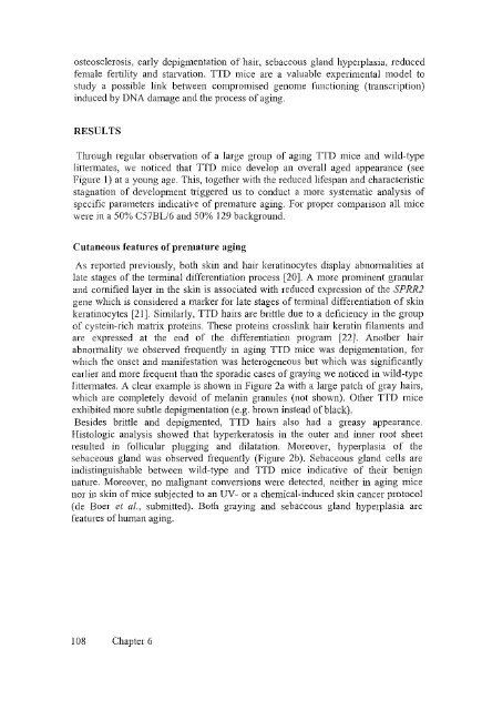
littem13tes. A clear example is shown in Figure 2a with a large patch of gray hairs,<br />
which are completely devoid of melanin granules (not shown). Other TTD mice<br />
exhibited more subtle depigmentation (e.g. brown instead of black).<br />
Besides brittle and depigmented, TrD hairs also had a greasy appearance.<br />
Histologic analysis showed that hyperkeratosis in the outer and inner root sheet<br />
resulted in follicular plugging and dilatation. Moreover, hyperplasia of the<br />
sebaceous gland was observed frequently (Figure 2b). Sebaceous gland cells are<br />
indistinguishable between wild-type and TTD mice indicative of their benign<br />
nature. Moreover, no malignant conversions were detected, neither in aging mice<br />
nor in skin of mice subjected to an UV- or a chemical-induced skin cancer protocol<br />
(de Boer et aI., submitted). Both graying and sebaceous gland hyperplasia are<br />
features of human aging.<br />
108 Chapter 6


