

View PDF Version - RePub - Erasmus Universiteit Rotterdam
View PDF Version - RePub - Erasmus Universiteit Rotterdam
View PDF Version - RePub - Erasmus Universiteit Rotterdam

Create successful ePaper yourself
Turn your PDF publications into a flip-book with our unique Google optimized e-Paper software.
cells involved in bone homeostasis underlie demineralization. Similarly, the<br />
heterogeneous picture of infertility in TID females may be a result of starvationinduced<br />
honnonal changes. Fertility in mammals requires adequate nuh'ition and<br />
reserves of metabolic fuel. Failure of the hypothalamus-pituitary-gonadal axis<br />
occurs in both sexes after starvation, although the female appears to be more<br />
sensitive to the effect of undernourishment [33]. Body weight and, more<br />
specifically, body fat mass has been implicated to influence the timing of menarche<br />
[34] and low body fat mass can cause secondary amenorrhea [35]. It has become<br />
clear that the cytokine leplin is produced by adipocytes and is required for fertility<br />
in the mouse [36] as it affects luteinizing hormone (LH) secretion [37]. Therefore,<br />
the broad range of ovarian dysfunction in the TID mice may be caused by a<br />
decrease in the level of LH and/or misliming of the ovulatory LH peak resulting<br />
from an effect of malnutrition on the hypothalamus or pituitary. The fact that<br />
starvation in TID mice cannot be appointed to abnonnalities in a certain target<br />
organ suggests that the defect in TID is a general cellular defect. Unfortnnately the<br />
phenotype leaves but little clues about the molecular defect underlying this very<br />
pleiotTOpic and heterogeneous syndrome.<br />
Molecular basis of aging<br />
Genetic evidence from yeast to mouse suggests that the rate of cellular metabolism<br />
and the oxidative damage load in pa11icular underlie at least part of the pathogenic<br />
mechanism of aging [I]. Two TID mouse phenotypes i.e. early graying (also<br />
observed in Werner syndrome) and sebaceous gland hyperplasia are conelated to<br />
oxidative damage. First, melanogenesis generates free radicals and melanocytes<br />
may be vulnerable to free radical attack by melanin products because they have<br />
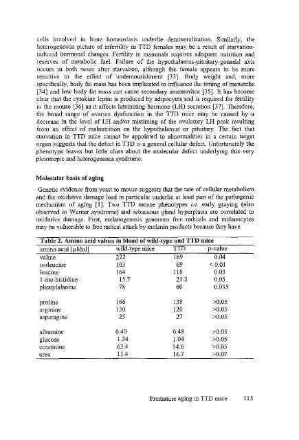
Table 2. Amino acid values in blood of wild-type and TTD mice<br />
amino acid [flMol] wild-type mice TTD p-value<br />
valine 222 169 0.04<br />
isoleucine 103 69 < 0.01<br />
leucine 164 118 0.03<br />
I-me-histidine 15.7 21.2 0.05<br />
phenylalanine 78 60 0.035<br />
proline 166 139 >0.05<br />
arginine 130 120 >0.05<br />
asparagme 25 27 >0.05<br />
albumine 0040 0048 >0.05<br />
glucose 1.34 1.04 >0.05<br />
creatinine 63.4 54.6 >0.'05<br />
urea 11.4 14.7 >0.07<br />
Premature aging in TTD mice 115


