- Page 2:
PRESENTED BY PUBLISHER
- Page 6 and 7:
CONTENTS OF VOLUME THREE. NEW SERIE
- Page 8 and 9:
F. W. Aston, D.Sc, F.R.S. Atoms and
- Page 10 and 11:
2 Radium P. M. the apparatus is app
- Page 12 and 13:
Radium value of the metal container
- Page 14 and 15:
Radium Table I—Continued Classifi
- Page 16 and 17:
8 RADIUM The affections of the thyr
- Page 18 and 19:
10 R a d i u m The treatment should
- Page 20 and 21:
12 Rapitjm ooo per cubic millimetre
- Page 22 and 23:
14 Radium tines of tubercle bacilli
- Page 24 and 25:
16 R A D I U M 4. Lymphatic Leukaem
- Page 26 and 27:
18 RADIUM than in anv other organs
- Page 28 and 29:
20 R a d i u m In a previous paper
- Page 30 and 31:
99 Radium 2 mm. thick; the whole ap
- Page 32 and 33:
2-1 Radium inum tubes in use. These
- Page 34 and 35:
26 R a d i u m 5-gr. capsule and sw
- Page 36 and 37:
28 R A D I U M 1 tared, as in the f
- Page 38 and 39:
80 R a d i u m (6) The greatly less
- Page 40 and 41:
32 R a d i u m and at times three,
- Page 42 and 43:
04 R a d i u m capsule is placed un
- Page 44 and 45:
36 R a d i u m 1 have observed one
- Page 46 and 47:
38 R A D I U M Frequency and Distri
- Page 48 and 49:
10 R a d i u m holder with comparat
- Page 50 and 51:
42 HADITJ M gestion of the eve whic
- Page 52 and 53:
44 R a d i u m The low polymorphonu
- Page 54 and 55:
46 Radium examined, it is believed
- Page 56 and 57:
48 I^ADITJM Positive results of exp
- Page 58 and 59:
50 R a d i u m 1 Is e III , -m If "
- Page 60 and 61:
»2 R a d i u m The summary of phys
- Page 63 and 64:
Radium 55
- Page 65 and 66:
R a d i u m 57 s V. — •7 CO N
- Page 67 and 68:
R a d i u m 59 Measuring radium by
- Page 69 and 70:
R a d i u m 6i Bibliography I. Gudz
- Page 71 and 72:
R a d i u m 68 Ited cells, SUBJECT
- Page 73 and 74:
R a d i u m TECHNIQUE OF RADIUM TRE
- Page 75 and 76:
R a d i u m B7 INDEX OF ARTICLES RE
- Page 77 and 78:
R a d i u m 69 Bower, J. O. and Cla
- Page 79 and 80:
R a d i u m 71 —Treatment of mali
- Page 81 and 82:
R a d i u m 73 —Carcinoma of oeso
- Page 83 and 84:
R a d i u m 75 —Uses of radium in
- Page 85 and 86:
R a d i u m 77 Kimbrough. J. B. and
- Page 87 and 88:
R a d i u m 7^ Lymph Nodes—Histol
- Page 89 and 90:
R a d i u m si Nicolich—Carcinoma
- Page 91 and 92:
R a d i u m 83 Radium— —Large d
- Page 93 and 94:
a d i u m 85 -Physical conditions o
- Page 95 and 96:
R a d i u m ST Arch. f. Gynak. 118:
- Page 97 and 98:
R a d i u m 89 Scholten, G. C. J.
- Page 99 and 100:
R a d i u m 91 (A. J. Larkin) Am. J
- Page 101 and 102:
R a d i u m 93 —Xecessity for com
- Page 103 and 104:
RADIUM 95 Wallerstein, E. U.—X-ra
- Page 105 and 106:
A QUARTERLY JOURNAL DEVOTED TO THE
- Page 107 and 108:
R a d i u m 99 casionally a carcino
- Page 109 and 110:
R a d i u m 101 would be detected w
- Page 111 and 112:
R a d i u m 103 cially likely if th
- Page 113 and 114:
R A D I U M 105 radium irradiations
- Page 115 and 116:
R a d i u m 107 that in nearly ever
- Page 117 and 118:
R a d i u m 109 liver and lung meta
- Page 119 and 120:
Radium in Appleton Co., 1922. (2) P
- Page 121 and 122:
R a d i u m 113 BLOCKING LYMPHATICS
- Page 123 and 124:
R a d i u m 115 removed, and the ra
- Page 125 and 126:
R a d i u m in PATHOLOGY Pathologic
- Page 127 and 128:
4. Cauhape: Thesis. Paris. 1911. R
- Page 129 and 130:
R a d i u m 121 is necessary by thi
- Page 131 and 132:
Radium 123 is injected into the tis
- Page 133 and 134:
R a d i u m 125 (3) Polonaise Numbe
- Page 135 and 136:
R a d i u m 127 modern laboratories
- Page 137 and 138:
R a d i u m 129 NINTH ANNUAL MEETIN
- Page 139 and 140:
R a d i u m 131 Several distinguish
- Page 141 and 142:
R a d i u m 133 which X-rays or rad
- Page 143 and 144:
R A D I U M 135 In the autobiograph
- Page 145 and 146:
R A D I U M 137 of radical operatio
- Page 147 and 148:
R a d i u m 139 reports 26 cases, 8
- Page 149 and 150:
R a d i u m 141 George W. Crile, M.
- Page 151 and 152:
R a d i u m 143 "In carcinoma of th
- Page 153 and 154:
R a d i u m 145 " 1 he principal ob
- Page 155 and 156:
IUM 147 have found no means of obvi
- Page 157 and 158:
R a d i u m 149 and six hours. Now
- Page 159 and 160:
R a d i u m 151 thoroughly transfix
- Page 161 and 162:
R a d i u m 153 weakness, palpitati
- Page 163 and 164:
R a d i u m 155 had all been benefi
- Page 165 and 166:
R a d i u m i57 tation of the kidne
- Page 167 and 168:
Radium 159 Can walk only with a can
- Page 169 and 170:
A QUARTERLY JOURNAL DEVOTED TO THE
- Page 171 and 172:
R a d i u m 163 in operative clinic
- Page 173 and 174:
a d i u m 165 In Table II we have d
- Page 175 and 176:
Hajmu.m 167 tery was used in combin
- Page 177 and 178:
R a d i u m 169 TABLE IX.—RESULTS
- Page 179 and 180:
R a d i u m m Radium alone Class i.
- Page 181 and 182:
R a d i u m 173 and papillary carci
- Page 183 and 184:
R a d i u m 175 It grows rapidly, i
- Page 185 and 186:
R a d i u m 177 emanation tubes. Th
- Page 187 and 188:
R a d i u m 179 never at any time f
- Page 189 and 190:
R a d i u m 181 THE TREATMENT OF MA
- Page 191 and 192:
R a d i u m 183 Cast 3. Carcinoma a
- Page 193 and 194: R a d i u m 185 Chm 7. Showing cond
- Page 195 and 196: R a d i u m 187 courting disaster b
- Page 197 and 198: R a d i u m 189 radium in the treat
- Page 199 and 200: R a d i u m 191 Histologic changes
- Page 201 and 202: R a d i u m 193 about their involut
- Page 203 and 204: R a d i u m 195 due to x-ray, sunli
- Page 205 and 206: R a d i u m 197 zation of a partial
- Page 207 and 208: R a d i u m 199 to report his cases
- Page 209 and 210: 201 very often will result in a dec
- Page 211 and 212: R a d i u m 203 the cautious use of
- Page 213 and 214: IUM 205 On Jan. 14th. which was a v
- Page 215 and 216: CONCLUSIONS 207 That in some cases
- Page 217 and 218: R a d i u m 209 (b) The fluorescent
- Page 219 and 220: R a d i u m •, 211 patient. The p
- Page 221 and 222: R a d i u m 213 radiological depart
- Page 223 and 224: R a d i u m 215 fortunately, the im
- Page 225 and 226: R a d i u m 217 know whether such c
- Page 227 and 228: R a d i u m 219 eight months had el
- Page 229 and 230: R a d i u m 221 "The selective and
- Page 231 and 232: R a d i u m 223 of a simple unitary
- Page 233 and 234: A QUARTERLY JOURNAL DEVOTED TO THE
- Page 235 and 236: R a d i u m 227 in their beta ray a
- Page 237 and 238: R a d i u m 229 or the gradation of
- Page 239 and 240: R a d i u m 231 be left in malignan
- Page 241 and 242: R a d i u m 233 during repeated cau
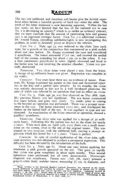
- Page 243: R a d i u m 235 3. The presence of
- Page 247 and 248: R a d i u m 239 REFERENCES 1. Wickh
- Page 249 and 250: Rajpium 241 the test of five years,
- Page 251 and 252: R a d i u m 243 comparative statist
- Page 253 and 254: R a d i u m 245 end results of 201
- Page 255 and 256: R a d i u m 247 Operation Hysterect
- Page 257 and 258: R a d i u m 'IV.) and five years, a
- Page 259 and 260: R a d i u m 251 or effect upon, the
- Page 261 and 262: R a d i u m 253 2. Early Cases.—T
- Page 263 and 264: R a d i u m 255 groups: the superfi
- Page 265 and 266: R a d i u m 257 "I am becoming more
- Page 267 and 268: R a d i u m 259 ment of 'Radiumhemm
- Page 269 and 270: R a d i u m 261 unusual symptoms an
- Page 271 and 272: R a d i u m 263 dition continued sa
- Page 273 and 274: R a d i u m 265 died from hemorrhag
- Page 275 and 276: R a d i u m 267 equals 132 millicur
- Page 277 and 278: R a d i u m 269 "The data in the li
- Page 279 and 280: R a d i u m 271 animals a diminutio
- Page 281 and 282: R a d i u m 273 rays as contrasted
- Page 283 and 284: R a d i u m 275 depression than a d
- Page 285 and 286: R a d i u m 277 repeated operations
- Page 287 and 288: R a d i u m 279 tonsil completely,
- Page 289 and 290: Radium 281 Ihe tonsil is dangerous
- Page 291 and 292: R a d i u m 283 WILLIAM H. B. AIKIN
- Page 293 and 294: DEVOTED TO THE CHEMISTRY, PHYSICS A
- Page 295 and 296:
PUBLISHED SEMI-ANNUALLY AND DEVOTED
- Page 297 and 298:
R a d i u m 3 ORAL CANCER The next
- Page 299 and 300:
lymph nodes in even case, but when
- Page 301 and 302:
adiation up to 2400 milligram hours
- Page 303 and 304:
R a d i u m o radium to relieve pat
- Page 305 and 306:
R a d i u m 11 Uterine Fibroids The
- Page 307 and 308:
R a d i u m i3 by some operators. X
- Page 309 and 310:
R a d i u m 15 from the operation h
- Page 311 and 312:
R a d i u m n tions, the distance s
- Page 313 and 314:
R a d i u m 19 APPENDIX RADIUM AND
- Page 315 and 316:
Radium 21 BASIC AND CONTBIBUTING FA
- Page 317 and 318:
RA.PIUM 23 X-RAY AND RADIUM THERAPY
- Page 319 and 320:
R a d i u m 25 Since, by the impact
- Page 321 and 322:
R a d i u m 27 duccd which were cov
- Page 323 and 324:
R a d i u m 29 In other cases the g
- Page 325 and 326:
R a d i u m 31 The recital of this
- Page 327 and 328:
R a d i u m 33 index of articles re
- Page 329 and 330:
R a d i u m 35 Block, F. B., and Cl
- Page 331 and 332:
R a d i u m ?,- J. R. Ranson) Color
- Page 333 and 334:
R a d i u m :;:» —Epithelioma of
- Page 335 and 336:
R a d i u m 4I Forsdike. S.-Cancer
- Page 337 and 338:
R a d i u m 43 Hot Springs, Arkansa
- Page 339 and 340:
R a d i u m 45 Lee. B. J., and Here
- Page 341 and 342:
R a d i u m 47 Mineral Waters, exam
- Page 343 and 344:
R a d i u m 49 —Treatment of inop
- Page 345 and 346:
R a d i u m 51 - Metabolic changes
- Page 347 and 348:
R a d i u m 58 Radium, emanation—
- Page 349 and 350:
R a d i u m .>.) —Physiological a
- Page 351 and 352:
R a d i u m 57 —Experimental rese
- Page 353 and 354:
R a d i u m 59 Spleen, physiology
- Page 355 and 356:
R a d i u m 6l —Animal tumor cell
- Page 357 and 358:
R a d i u m es —Treatment of uter
- Page 359 and 360:
PUBLISHED SEMI-ANNUALLY AND DEVOTED
- Page 361 and 362:
R a d i u m e? CONTRAINDICATIONS TO
- Page 363 and 364:
R a d i u m 69 in addition to the i
- Page 365 and 366:
Radium 71 the most common, in the o
- Page 367 and 368:
R a d i u m ?:; nexae is a positive
- Page 369 and 370:
R a d i u m 76 essential to success
- Page 371 and 372:
R a d i u m 77 Our technique is to
- Page 373 and 374:
carcinoma of the CERVIX R a d i u m
- Page 375 and 376:
R a d i u m 81 a bleeding, cauliflo
- Page 377 and 378:
R a d i u m HS Fibrinous patch on e
- Page 379 and 380:
R a d i u m sr, mal 7 days later. A
- Page 381 and 382:
R a d i u m ST idly develops and me
- Page 383 and 384:
R a d i u m 89 2. Cervical cancer o
- Page 385 and 386:
R a d i u m 91 irradiation treatmen
- Page 387 and 388:
R a d i u m 93 by a study of curett
- Page 389 and 390:
R a d i u m 96 parenchyma eventuall
- Page 391 and 392:
R a d i u m 97 ihe lowest (2) was t
- Page 393 and 394:
R a d i u m 99 3. The treatment int
- Page 395 and 396:
R a d i u m 101 harder and shows on
- Page 397 and 398:
a d i u m 103 Sloughy Not sloughy .
- Page 399 and 400:
R a d i u m 105 But 2 of 15 cases h
- Page 401 and 402:
R a d i u m 107 Sice (not of tumor,
- Page 403 and 404:
IUM 109 Time well Number of cases i
- Page 405 and 406:
R a d i u m 111 died of a secondary
- Page 407 and 408:
R a d i u m 113 epithelioma of the
- Page 409 and 410:
R a d i u m 115 This writer also fo
- Page 411 and 412:
R a d i u m 117 as held by many wri
- Page 413 and 414:
R a d i u m 119 of foods, particula
- Page 415 and 416:
R a d i u m 121 which, in the last
- Page 417 and 418:
R a d i u m 123 "It has long been r
- Page 419 and 420:
R a d i u m 125 OBITUARY JAMES C. G
- Page 422:
• 3 1812 04298 6001


