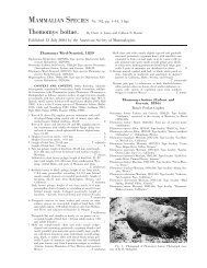
Brugia Malayi - Clark Science Center - Smith College
Brugia Malayi - Clark Science Center - Smith College
Brugia Malayi - Clark Science Center - Smith College

Create successful ePaper yourself
Turn your PDF publications into a flip-book with our unique Google optimized e-Paper software.
Effects of Surface Chemistry on Biofilm Colonization<br />
Minhee Kim, Jinglin Huang and Jing Zhang<br />
The effect of surface chemistry on biofilm colonization has become a popular research subject in medical and environmental<br />
fields, with a growing public concern on biofilm coating on medical devices and water pipelines. 1, 2 Our summer research<br />
continued our previous study on the responses of Pseudomonas aeruginosa, a model organism in biofilm studies, to nanoscale surfaces<br />
with different physical characters.<br />
Two types of nanoscale surface topography, flat hydrophobic and flat hydrophilic, were constructed and coated by P.<br />
aeruginosa, followed by a visual observation of biofilm growth under fluorescence microscope. The qualitative analysis on the<br />
arabinose-induced fluorescence, emitted by the attached bacteria, suggested that layers of bacteria start to accumulate and<br />
are discernible for measurement after six hours of incubation. Three ways to quantify the biofilm growth were performed to<br />
distinguish bacteria’s response to two studied surface types. The first one is through TECAN Infinite M1000, a microplate reader<br />
capable of detecting and quantifying the fluorescence intensity emitted from areas of interest. TECAN data collected from three<br />
sets of experiments demonstrated a distinguishable difference between biofilm growth on flat hydrophobic and hydrophilic<br />
surfaces after six hours of incubation. The three-dimensional modeling of the spatial distribution of bacteria on the surface was<br />
enabled by Magellan, a software developed by TECAN Group. To prove the validity of results obtained through TECAN, the<br />
second quantification means was proposed, through which microscopic images were sampled from areas of interest and analyzed<br />
by an image-processing program named Image J. 4 Sonication was carried out as a third way to assess the growing performance of<br />
P. aeruginosa. An ultrasound water bath 5 was applied to remove attached bacterial cells from the surfaces and after centrifugation,<br />
cells were reintroduced in aliquots to growth media. Cell colonies were counted and the difference in colony-forming units<br />
appeared indicated that more cells were detached from flat hydrophobic samples compared to flat hydrophilic ones after six hours<br />
of incubation.<br />
In continuation of our studies, the protocols developed from our summer research should be repeated for multiple times until<br />
substantial amount of data are gathered to draw the conclusion. To investigate the effects of surface roughness, protocols should<br />
be tested on rough hydrophobic and hydrophilic surfaces, in comparison to flat topography. (Supported by the Howard Hughes<br />
Medical Institute)<br />
Advisors: Kate Queeney and Rob Dorit<br />
References:<br />
1<br />
Mack D, Rohde H, Harris LG, Davies AP, Horstkotte MA, Knobloch JK. (2006). Biofilm formation in medical device-related infection. International Journal of<br />
Artificial Organs. 29 (4): 343-59.<br />
2<br />
Pavanello G, Faimali M, Pittore M, Mollica A, Mollica A, Mollica A. (2011). Exploiting a new electrochemical sensor for biofilm monitoring and water treatment<br />
optimization. Water Research. 45 (4): 1651-1658. 3 The information of TECAN Group Ltd is available at www.tecan.com.<br />
4<br />
The information of Image J is available at http://rsbweb.nih.gov/ij/.<br />
5<br />
The information of Branson model 2510 sonicating water bath is available at http://www.emersonindustrial.com/en-US/branson/Products/precision-cleaning/<br />
bench-top-cleaners/bransonics/Pages/default.aspx.<br />
2012<br />
4