Brugia Malayi - Clark Science Center - Smith College
Brugia Malayi - Clark Science Center - Smith College
Brugia Malayi - Clark Science Center - Smith College

You also want an ePaper? Increase the reach of your titles
YUMPU automatically turns print PDFs into web optimized ePapers that Google loves.
Creatine Kinase during Muscle Development<br />
Hailun Li<br />
Creatine kinase (CK) plays a major role in catalyzing the conversion of phosphocreatine and ADP into creatine and ATP, which<br />
provides nearly instantaneous ATP regeneration to many cell types with a high energy demand. Thus, it is not surprising that CK<br />
is found in mature muscle cells, but when does it appear in the development of muscle cells? This summer, I quantified CK in the<br />
three developmental stages of the C2C12 cell line, myoblast (M), early myotube (EM), and late myotube (LM).<br />
Several techniques were used: cell culture; cell lysis; sample protein estimation (Lowry assay); protein separation through SDSgel<br />
electrophoresis, along with a CK standard curve; immunoblotting to a PVDF membrane, which was probed using an anti-CK<br />
primary antibody at 4°C overnight; followed by quantitative gel densitometric scanning.<br />
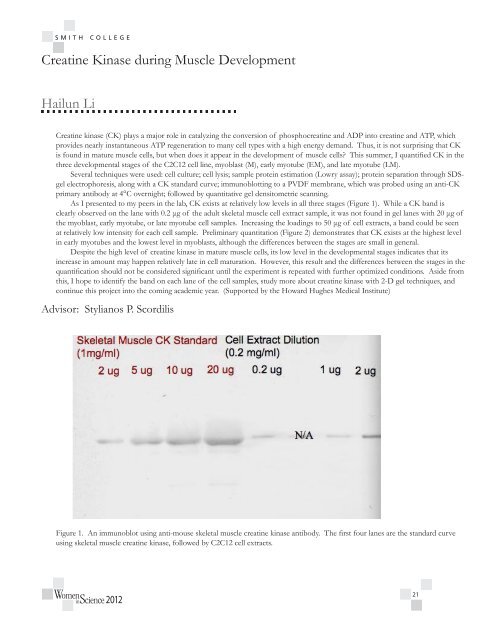
As I presented to my peers in the lab, CK exists at relatively low levels in all three stages (Figure 1). While a CK band is<br />
clearly observed on the lane with 0.2 μg of the adult skeletal muscle cell extract sample, it was not found in gel lanes with 20 μg of<br />
the myoblast, early myotube, or late myotube cell samples. Increasing the loadings to 50 μg of cell extracts, a band could be seen<br />
at relatively low intensity for each cell sample. Preliminary quantitation (Figure 2) demonstrates that CK exists at the highest level<br />
in early myotubes and the lowest level in myoblasts, although the differences between the stages are small in general.<br />
Despite the high level of creatine kinase in mature muscle cells, its low level in the developmental stages indicates that its<br />
increase in amount may happen relatively late in cell maturation. However, this result and the differences between the stages in the<br />
quantification should not be considered significant until the experiment is repeated with further optimized conditions. Aside from<br />
this, I hope to identify the band on each lane of the cell samples, study more about creatine kinase with 2-D gel techniques, and<br />
continue this project into the coming academic year. (Supported by the Howard Hughes Medical Institute)<br />
Advisor: Stylianos P. Scordilis<br />
Figure 1. An immunoblot using anti-mouse skeletal muscle creatine kinase antibody. The first four lanes are the standard curve<br />
using skeletal muscle creatine kinase, followed by C2C12 cell extracts.<br />
2012<br />
21