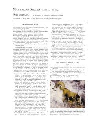
Brugia Malayi - Clark Science Center - Smith College
Brugia Malayi - Clark Science Center - Smith College
Brugia Malayi - Clark Science Center - Smith College

You also want an ePaper? Increase the reach of your titles
YUMPU automatically turns print PDFs into web optimized ePapers that Google loves.
Function of Slit and Robo in Axon Guidance<br />
Abigail Antoine and Jin Sook Park<br />
Axon-glia interactions in the developing brain are caused by Roundabout receptors detecting the presence of Slit proteins. This<br />
signal-receptor pathway guides them to form proper connections within the central nervous system. What happens when one or<br />
more of these signals are missing? Using zebrafish as our model system, we were able to study embryonic forebrains to determine<br />
if specific mutations resulted in particular phenotypes, specifically in the formation of the post-optic commissure. Out of the<br />
many different mutant fish lines we had available to us, we focused on the Slit 2 and Slit 3 mutants, as well as Astray/Twitch-twice<br />
double mutants.<br />
As the embryo brain develops, glia act as a substrate on which axons projecting from the retina move across in order to form<br />
the post-optic and anterior commissures. The Slit 1a protein acts as a chemoattractant, positively guiding the axons along the right<br />
path, while Slit 2 and Slit 3 act in the opposite manner, as a chemorepellant, preventing axons from straying too far from where<br />
they are supposed to be. 1 In a mutant fish, one or more of these proteins or receptors are missing or nonfunctional, and this<br />
would affect formation of the commissures.<br />
To pinpoint a specific phenotype within each mutant group, we examined developing embryos and tried to observe if there<br />
were any abnormal behaviors or physical characteristics. At about 32 hours post fertilization, specific proteins were labelled within<br />
the embryo using immunohistochemistry, which would fluorescently label for axons and glia. This labeling allows us to examine<br />
the commissure formations and compare the forebrain images to a control group in order to determine the existence of mutant<br />
phenotype.<br />
Since Slit 2 and Slit 3 were believed to have similar roles in axon guidance, it was unclear whether or not a loss of function in<br />
only the Slit 3 protein would show any distinct abnormalities or whether Slit 2 proteins would be able to compensate for the loss<br />
of the other chemorepellant. This examination process was made more difficult by the fact that the genotyping protocols for the<br />
Slit 2 and Slit 3 mutants were not working properly at this time, which inhibited us from processing a group of confirmed mutants<br />
for either fish line. In groups of potential Slit 3 mutants, there were instances of defasciculation of the commissure. Potential<br />
Slit 2 mutant embryos were separated by observed behaviors, such as a circular swimming pattern or deafness, and analysis of the<br />
commissures will be continued during the semester. Genotyping would be the next step in order to identify confirmed mutants.<br />
However, further examination of both mutant lines must be completed in the year to follow in order to confirm phenotypes.<br />
(Supported by the Blakeslee Fund in the Biological <strong>Science</strong>s)<br />
Advisor: Michael Barresi<br />
References:<br />
1<br />
Barresi, M.J. 2005. Hedgehog regulated Slit expression determines commissure and glial cell position in the zebrafish forebrain. Development 132, 3643-3656.<br />
2012<br />
10