Untitled - Red Temática de investigación cooperativa en cáncer
Untitled - Red Temática de investigación cooperativa en cáncer
Untitled - Red Temática de investigación cooperativa en cáncer

You also want an ePaper? Increase the reach of your titles
YUMPU automatically turns print PDFs into web optimized ePapers that Google loves.
Programa Ci<strong>en</strong>tífico 7<br />
Resum<strong>en</strong>es <strong>de</strong> Comunicaciones Orales 15<br />
Listado <strong>de</strong> Posters 37<br />
Resum<strong>en</strong>es <strong>de</strong> Posters 43<br />
Listado <strong>de</strong> Pon<strong>en</strong>tes 93<br />
© 2012 <strong>Red</strong> Temática <strong>de</strong> Investigación Cooperativa <strong>en</strong> Cáncer<br />
Imprime: Jet Print SL<br />
Diseño y maquetación: Detrazos Diseño e Internet<br />
www.<strong>de</strong>trazos.es<br />
5
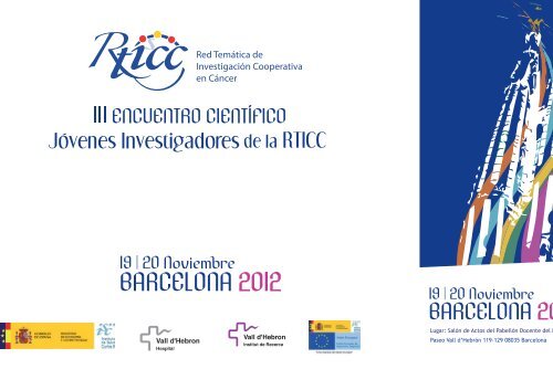
Lugar: Salón <strong>de</strong> Actos <strong>de</strong>l Pabellón Doc<strong>en</strong>te <strong>de</strong>l Hospital Vall d’Hebron<br />
Paseo Vall d’ Hebron 119 - 129 08035 Barcelona - España<br />
7
10:00 h Recepción <strong>de</strong> los Participantes<br />
10:00 h Montaje <strong>de</strong> Pósters<br />
10:30 h Inauguración<br />
Dr. Antonio L. Andreu Périz / Subdirector G<strong>en</strong>eral <strong>de</strong> Evaluación y Fom<strong>en</strong>to <strong>de</strong> la<br />
Investigación <strong>de</strong>l Instituto <strong>de</strong> Salud Carlos III (ISCIII)<br />
Dr. José Jerónimo Navas / Ger<strong>en</strong>te <strong>de</strong>l Hospital Vall d’Hebron<br />
Dr. Joan Comella / Director <strong>de</strong>l Vall d’Hebron Institut <strong>de</strong> Recerca (VHIR)<br />
Dr. Eug<strong>en</strong>io Santos / C<strong>en</strong>tro <strong>de</strong> Investigación <strong>de</strong>l Cáncer <strong>de</strong> Salamanca y Coordinador<br />
Nacional <strong>de</strong> la <strong>Red</strong> Tematica <strong>de</strong> Investigación Cooperativa <strong>en</strong> Cáncer (RTICC)<br />
Dr. Luis Montu<strong>en</strong>ga / C<strong>en</strong>tro <strong>de</strong> Investigación Médica Aplicada (CIMA), Universidad <strong>de</strong><br />
Navarra y Coordinador <strong>de</strong>l Programa <strong>de</strong> Formación y Movilidad <strong>de</strong> la RTICC<br />
Dr. Jaume Rev<strong>en</strong>tos / Unidad <strong>de</strong> Investigación Biomédica y Oncología Traslacional i<br />
Pediátrica <strong>de</strong>l Vall d’Hebron Institut <strong>de</strong> Recerca (VHIR)<br />
11.00 h. Confer<strong>en</strong>cia Inaugural<br />
“IDENTIFICATION OF NOVEL DRIVER MUTATIONS IN CHRONIC LYMPHOCYTIC LEUKEMIA THROUGH WHOLE<br />
GENOME SEQUENCING”<br />
Dr. Xose S. Pu<strong>en</strong>te<br />
Departm<strong>en</strong>t of Biochemistry, University of Oviedo<br />
12:00 h Descanso / Coffee Break<br />
Lunes 19 <strong>de</strong> Noviembre<br />
12:50 h O-02. “p21 LOSS INDUCES AN INFLAMMATORY SKIN DISEASE AND SPONTANEOUS<br />
TUMOR DEVELOPMENT IN MICE BEARING EPIDERMAL SPECIFIC pRb ABLATION”<br />
Cristina Saiz La<strong>de</strong>ra / C<strong>en</strong>tro <strong>de</strong> Investigaciones Energéticas, Medioambi<strong>en</strong>tales y<br />
Tecnológicas (CIEMAT), Madrid<br />
Grupo RTICC: RD06/0020/0029<br />
13:10 h O-03. “DOWN-REGULATION OF FOXP1 IS REQUIRED DURING GERMINAL CENTER B CELL<br />
FUNCTION”<br />
Aitziber Ainara Sagardoy Rodrigo / C<strong>en</strong>tro <strong>de</strong> Investigación Médica Aplicada (CIMA),<br />
Pamplona<br />
Grupo RTICC: RD06/0020/0088<br />
13:30 h O-04. “EPIGENETIC PROFILE IN CHRONIC LYMPHOCYTIC LEUKEMIA USING<br />
METHYLATION-SPECIFIC MULTIPLEX LIGATION-DEPENDENT PROBE AMPLIFICATION”<br />
Ana Mª Cosialls Castel / Instituto <strong>de</strong> Investigación Biomédica <strong>de</strong> Bellvitge (IDIBELL)-<br />
Universitat <strong>de</strong> Barcelona, Barcelona<br />
Grupo RTICC: RD06/0020/0097<br />
13:50 h O-05. “CRYPTIC DUPLICATIONS AND DELETIONS TO IMPROVE RISK GROUPS IN<br />
CHILDHOOD ACUTE LYMPHOBLASTIC LEUKEMIA”<br />
Elixabet López López / Universidad <strong>de</strong>l País Vasco/Euskal Herriko Unibertsitatea (UPV/EHU),<br />
Lejona (Vizcaya<br />
Grupo RTICC: RD06/0020/0048<br />
14:10 h O-06. “CCND2 REARRANGEMENTS ARE THE MOST FREQUENT GENETIC EVENTS IN<br />
CYCLIN D1-NEGATIVE MANTLE CELL LYMPHOMA”<br />
Cristina Royo Mor<strong>en</strong>o / Hospital Clínic -IDIBAPS, Barcelona<br />
Grupo RTICC: RD06/0020/0039<br />
Sesión mañana<br />
14:30 h Comida<br />
Mo<strong>de</strong>radores: Dra. Dolors Colomer / Hospital Clínic-IDIBAPS<br />
Dr. Andreas Doll / Institut <strong>de</strong> Recerca-Hospital Universitari Vall d’Hebron<br />
15:30 h Sesión <strong>de</strong> Pósters<br />
12:30 h O-01.“THE MULTI-KINASE INHIBITOR SORAFENIB BLOCKS MIGRATION, BCR SURVI-<br />
VAL SIGNALS, PROTEIN TRANSLATION AND STROMA-MEDIATED BORTEZOMIB RESISTANCE IN<br />
MANTLE CELL LYMPHOMA”<br />
Sílvia Xargay Torr<strong>en</strong>t / Hospital Clínic-IDIBAPS, Barcelona<br />
Grupo RTICC: RD06/0020/0014<br />
8 9
Sesión <strong>de</strong> Tar<strong>de</strong><br />
Mo<strong>de</strong>radores: Dr. Joan Gil / Instituto <strong>de</strong> Investigación Biomédica <strong>de</strong> Bellvitge (IDIBELL)<br />
Dra. Mireia Olivan / Institut <strong>de</strong> Recerca-Hospital Universitari Vall d’Hebron<br />
16:50 h O-07. “A DOMINANT-NEGATIVE N-TERMINAL FRAGMENT OF HER2 FREQUENTLY<br />
EXPRESSED IN BREAST CANCERS”<br />
Beatriz Morancho Armis<strong>en</strong> / Vall D’Hebrón Institut D’Oncologia (VHIO), Barcelona<br />
Grupo RTICC: RD06/0020/0022<br />
17:10 h O-08. “MITOTIC ARREST DOWN-REGULATES FLIP EXPRESSION AND SENSITIZES<br />
TUMOR CELLS TO THE EXTRINSIC PATHWAY OF APOPTOSIS”<br />
Tania Sánchez Pérez / C<strong>en</strong>tro Andaluz <strong>de</strong> Biología Molecular y Medicina Reg<strong>en</strong>erativa<br />
(CABIMER), Sevilla<br />
Grupo RTICC: RD06/0020/0068<br />
17:30 h O-09. “microRNA-BASED THERAPY FOR HIGH-RISK NEUROBLASTOMA”<br />
Aroa Soriano Fernán<strong>de</strong>z / Institut <strong>de</strong> Recerca Hospital Universitari Vall d’Hebron,<br />
Barcelona<br />
Grupo RTICC: RD06/0020/1021<br />
17:50 h O-10. “INFLUENCE OF LKB1/STK11 IN GENE EXPRESSION PATTERNS INDUCED BY<br />
ENERGY STRESS IN LUNG CANCER CELL LINES”<br />
Rossana Gonçalves Restani / Instituto <strong>de</strong> Investigación Biomédica <strong>de</strong> Bellvitge (IDIBELL),<br />
Barcelona<br />
Grupo RTICC: RD06/0020/0062<br />
18:10 h O-11. “IDENTIFICATION OF DIFFERENTIAL GENOMIC AND PROTEOMIC PROFILES<br />
ASSOCIATED WITH BONE METASTASES IN PROSTATE CANCER”<br />
Marta Garcia López / Institut <strong>de</strong> Recerca-Hospital Universitari Vall d’Hebron, Barcelona<br />
Grupo RTICC: RD06/0020/0058<br />
18:30 h O-12. “DIOXIN RECEPTOR CONTROLS CSK-BINDING PROTEIN SIGNALING TO CAVEOLIN<br />
1 AND Β1 INTEGRIN TO MODULATE CELL ADHESION AND MIGRATION”<br />
Martes 20 <strong>de</strong> Noviembre<br />
Sesión <strong>de</strong> Mañana<br />
Mo<strong>de</strong>radores:<br />
Dr. Francesc Sole Ristol / Institut <strong>de</strong> Recerca Contra la Leucèmia Josep Carreras (IJC)<br />
Dra. Marina Rigau / Institut <strong>de</strong> Recerca-Hospital Universitari Vall d’Hebron<br />
09:10 h O-13. “CD44 POSITIVITY CONFERS RESISTANCE TO SORAFENIB-INDUCED CELL DEATH<br />
IN HEPATOCELLULAR CARCINOMA (HCC) CELLS”<br />
Joan Fernando Peidró / Instituto <strong>de</strong> Investigación Biomédica <strong>de</strong> Bellvitge (IDIBELL),<br />
Barcelona<br />
Grupo RTICC: RD06/0020/0097<br />
09:30 h O-14. “ANALYSIS OF NEW GENES DIRECTLY REGULATED BY FOXE1 IN THYROID CELLS”<br />
Lara Paula Fernán<strong>de</strong>z Álvarez / Instituto <strong>de</strong> Investigaciones Biomédicas “Alberto Sols” IIB,<br />
Madrid<br />
Grupo RTICC: RD06/0020/0060<br />
09:50 h O-15. “MOLECULAR SUBSETS OF MANTLE CELL LYMPHOMA DEFINED BY THE IGHV MUTA-<br />
TIONAL STATUS AND SOX11 EXPRESSION HAVE DISTINCT BIOLOGICAL AND CLINICAL FEATURES”<br />
Alba Navarro Lopez / Instituto <strong>de</strong> Investigación Biomédica <strong>de</strong> Bellvitge (IDIBELL), Barcelona<br />
Grupo RTICC: RD06/0020/0039<br />
10:10 h O-16. “ETV5 AND FOXM1 TRANSCRIPTION FACTORS ARE OVEREXPRESSED IN OVARIAN<br />
CANCER AND REGULATE CELLULAR ADHESION AND PROLIFERATION CONTRIBUTING TO TUMOR<br />
DISSEMINATION”<br />
Marta Llauradó Fernán<strong>de</strong>z / Institut <strong>de</strong> Recerca-Hospital Universitari Vall d’Hebron,<br />
Barcelona<br />
Grupo RTICC: RD06/0020/0058<br />
10:30 h O-17. “COUNTERACTING AUTOPHAGY OVERCOMES RESISTANCE TO EVEROLIMUS IN<br />
MANTLE CELL LYMPHOMA”<br />
Laia Rosich Moya / Hospital Clínic-IDIBAPS, Barcelona<br />
Grupo RTICC: RD06/0020/0014<br />
Javier Rey Barroso / Universidad <strong>de</strong> Extremadura, Badajoz<br />
Grupo RTICC: RD06/0020/1016<br />
18:50 h Fin <strong>de</strong> las Sesiones<br />
10:50 h O-18. “PIK3CA ALTERATIONS IN BLADDER CANCER RECURRENCE”<br />
Mónica Martínez Fernán<strong>de</strong>z / C<strong>en</strong>tro <strong>de</strong> Investigaciones Energéticas, Medioambi<strong>en</strong>tales y<br />
Tecnológicas (CIEMAT), Madrid<br />
Grupo RTICC: RD06/0020/0029<br />
10 11
11:10 h O-19. “PAPEL DEL CORREPRESOR NcoR EN LA INHIBICIÓN DE LA INVASIVIDAD Y FOR-<br />
MACIÓN DE METÁSTASIS POR TR”<br />
Olaia Antia Martinez Iglesias / Instituto <strong>de</strong> Investigaciones Biomédicas “Alberto Sols” IIB,<br />
Madrid<br />
Grupo RTICC: RD06/0020/0036<br />
11:30 h O-20. “DIFFERENTIAL FUNCTIONS OF EVI1 SPLICE ISOFORMS IN CELL VIABILITY AND<br />
TRANSCRIPTIONAL REGULATION. THE EVI1 HUMAN PROTEIN REGULATES ITS OWN TRANSCRIP-<br />
TION”<br />
Mir<strong>en</strong> Maicas Irigaray / C<strong>en</strong>tro <strong>de</strong> Investigación Médica Aplicada (CIMA), Pamplona<br />
Grupo RTICC: RD06/0020/0078<br />
12:00 h Descanso / Coffee Break<br />
12:00 h Retirada <strong>de</strong> Pósters<br />
12.30 h. Confer<strong>en</strong>cia Coordinador Nacional <strong>de</strong> la RTICC<br />
Dr. Eug<strong>en</strong>io Santos / Coordinador Nacional <strong>de</strong> la RTICC, C<strong>en</strong>tro <strong>de</strong> Investigación <strong>de</strong>l Cáncer<br />
<strong>de</strong> Salamanca<br />
12:40 h / Entrega PREMIO DE INVESTIGACIÓN COOPERATIVA EN ONCOLOGÍA RTICC<br />
2012<br />
12:50 h / Exposición <strong>de</strong>l galardonado con el PREMIO DE INVESTIGACIÓN COOPERATI-<br />
VA EN ONCOLOGÍA RTICC 2012<br />
13:35 h / Clausura<br />
Dr. Eug<strong>en</strong>io Santos / C<strong>en</strong>tro <strong>de</strong> Investigación <strong>de</strong>l Cáncer <strong>de</strong> Salamanca<br />
Dr. Luis Montu<strong>en</strong>ga / C<strong>en</strong>tro <strong>de</strong> Investigación Médica Aplicada (CIMA), Universidad <strong>de</strong><br />
Navarra<br />
Dr. Jaume Rev<strong>en</strong>tos / Unidad <strong>de</strong> Investigación Biomédica y Oncología Traslacional i Pediátrica<br />
<strong>de</strong>l Vall d’Hebron Institut <strong>de</strong> Recerca (VHIR)<br />
CONFERENCIA MAGISTRAL<br />
IDENTIFICATION OF NOVEL DRIVER MUTATIONS IN CHRONIC LYMPHOCYTIC<br />
LEUKEMIA THROUGH WHOLE GENOME SEQUENCING<br />
XOSE S. PUENTE<br />
DEPARTMENT OF BIOCHEMISTRY, UNIVERSITY OF OVIEDO<br />
Chronic lymphocytic leukemia (CLL), the most frequ<strong>en</strong>t leukemia in adults in Western countries,<br />
is a B-cell neoplasm clinically and biologically very heterog<strong>en</strong>eous. The differ<strong>en</strong>ces<br />
in the behavior of the disease have be<strong>en</strong> associated with two major molecular subtypes<br />
characterized by high and no or low number of somatic mutations in the variable region of<br />
immunoglobulin g<strong>en</strong>es (IGHV-mutated and IGHV-unmutated, respectively). However, the<br />
g<strong>en</strong>etic and molecular alterations leading to the <strong>de</strong>velopm<strong>en</strong>t and progression of the disease<br />
are still poorly un<strong>de</strong>rstood. The aim of the CLL Cancer G<strong>en</strong>ome Consortium is to g<strong>en</strong>erate<br />
a compreh<strong>en</strong>sive catalogue of g<strong>en</strong>etic alterations relevant to the pathog<strong>en</strong>esis and clinical<br />
evolution of the disease using next g<strong>en</strong>eration sequ<strong>en</strong>cing combined with other g<strong>en</strong>omic<br />
technologies in a large series of well characterized tumors.<br />
We have completed the sequ<strong>en</strong>cing and analysis of 109 cases of CLL using either whole<br />
g<strong>en</strong>ome sequ<strong>en</strong>cing (4 cases) or whole exome sequ<strong>en</strong>cing (105 cases). This has led us to<br />
the i<strong>de</strong>ntification of about 1,000 somatic mutations per tumor g<strong>en</strong>ome in non-repetitive<br />
regions, and a median of 45 somatic mutations per case wh<strong>en</strong> using exome sequ<strong>en</strong>cing.<br />
Interestingly, IGHV-mutated tumors had more protein-altering mutations than CLL tumors<br />
without mutations in the IGHV region, as well as an increased number of A>C/T>G transversions.<br />
In addition, global analysis of these data resulted in the i<strong>de</strong>ntification of 78 g<strong>en</strong>es<br />
which are recurr<strong>en</strong>tly mutated in CLL. They inclu<strong>de</strong> NOTCH1 and SF3B1, which are mutated<br />
in more than 10% of cases, or POT1, CHD2 and LRP1B, mutated in about 5% of tumors.<br />
There is a clear differ<strong>en</strong>ce in the frequ<strong>en</strong>cy of these mutations betwe<strong>en</strong> IGHV-mutated<br />
and IGHV–unmutated CLL tumors, ext<strong>en</strong>ding at the g<strong>en</strong>omic level the clinical differ<strong>en</strong>ces<br />
observed in these two CLL subtypes. Together with SF3B1, which <strong>en</strong>co<strong>de</strong>s a subunit of the<br />
spliceosomal U2 small nuclear ribonucleoprotein, at least 30 g<strong>en</strong>es involved in RNA maturation<br />
have mutations in CLL, suggesting that RNA splicing and maturation might constitute<br />
an important mechanism driving this disease. Finally, further analysis of more than 250 CLL<br />
cases showed that pati<strong>en</strong>ts with mutations in either NOTCH1 or SF3B1 had faster disease<br />
progression and shorter overall survival. Together, these data illustrate the heterog<strong>en</strong>eity<br />
of the disease and the utility of g<strong>en</strong>ome-wi<strong>de</strong> studies to i<strong>de</strong>ntify the molecular mechanisms<br />
driving the <strong>de</strong>velopm<strong>en</strong>t of CLL.<br />
This project is fun<strong>de</strong>d by the Spanish Ministry of Sci<strong>en</strong>ce and Innovation through the Institute<br />
of Health Carlos III.<br />
12 13
O-01<br />
THE MULTI-KINASE INHIBITOR SORAFENIB BLOCKS MIGRATION, BCR SURVIVAL<br />
SIGNALS, PROTEIN TRANSLATION AND STROMA-MEDIATED BORTEZOMIB RESIS-<br />
TANCE IN MANTLE CELL LYMPHOMA<br />
XARGAY-TORRENT S 1 , LÓPEZ-GUERRA M 1 , SABORIT-VILLARROYA I 2 , ROSICH L 1 , NAVARRO A 2 ,<br />
VILLAMOR N 1 , AYMERICH M 1 , PÉREZ-GALÁN P 1 , ROUÉ G 1 , CAMPO E 2 , COLMER D 1<br />
1<br />
HOSPITAL CLÍNIC-IDIBAPS (RD06/0020/0014)<br />
2<br />
HOSPITAL CLÍNIC-IDIBAPS (RD06/0020/0039)<br />
Mantle cell lymphoma (MCL) is a B-lymphoma harboring the t(11;14)(q13;q32) translocation<br />
which leads to the overexpression of cyclin D1, and is characterized by bad prognosis<br />
and an aggressive course. Unfortunately, curr<strong>en</strong>t therapies have shown limited efficacy and<br />
relapses occur early, thus our purpose was to evaluate the antitumoral properties of the<br />
multikinase inhibitor soraf<strong>en</strong>ib in MCL.<br />
We analyzed the effects of soraf<strong>en</strong>ib in 9 MCL cell lines and 17 primary MCL samples, where<br />
soraf<strong>en</strong>ib induced a time- and dose-<strong>de</strong>p<strong>en</strong><strong>de</strong>nt apoptosis. Both in cell lines and primary MCL<br />
cells, soraf<strong>en</strong>ib induces rapid <strong>de</strong>phosphorylation of the BCR (B-Cell Receptor)-associated<br />
tyrosine kinases, SYK and LYN, as well as of FAK, a downstream SRC target involved in focal<br />
adhesion. In line with this, we <strong>de</strong>monstrate a strong synergy wh<strong>en</strong> combining soraf<strong>en</strong>ib<br />
with the SYK inhibitor, R406. In parallel, we show that soraf<strong>en</strong>ib also blocks Mcl-1 and cyclin<br />
D1 translation, which promotes an unbalance betwe<strong>en</strong> pro- and anti-apoptotic proteins<br />
and facilitates the release of Bax from cyclin D1. This process leads to the induction of the<br />
mitochondrial apoptotic pathway and caspase-<strong>de</strong>p<strong>en</strong><strong>de</strong>nt and in<strong>de</strong>p<strong>en</strong><strong>de</strong>nt mechanisms.<br />
Moreover, soraf<strong>en</strong>ib inhibits MCL cell migration as well as actin polymerization in response<br />
to CXCL12. FAK siRNA-mediated knockdown partially prev<strong>en</strong>ts this inhibitory effect, indicating<br />
that FAK is a relevant target for the action of soraf<strong>en</strong>ib in MCL cells. Importantly,<br />
this compound res<strong>en</strong>sitizes MCL cells cocultured with bone marrow-<strong>de</strong>rived stromal and<br />
follicular <strong>de</strong>ndritic-like cells to bortezomib-induced apoptosis, thus soraf<strong>en</strong>ib antagonizes<br />
stroma-mediated resistance in MCL.<br />
In conclusion, we provi<strong>de</strong> first evi<strong>de</strong>nce on the molecular mechanism of action of soraf<strong>en</strong>ib<br />
in MCL. We propose that this compound inhibits cell migration and stroma-mediated bortezomib<br />
resistance by interfering BCR signaling and protein translation. These results suggest<br />
that soraf<strong>en</strong>ib, alone or in combination with bortezomib-based therapies, may repres<strong>en</strong>t a<br />
promising approach for the treatm<strong>en</strong>t of MCL pati<strong>en</strong>ts.<br />
Observaciones:<br />
Research funding<br />
This work was supported in part by grants RD06/0020/0014 from <strong>Red</strong> Temática <strong>de</strong> Investigación<br />
Cooperativa <strong>en</strong> Cáncer (RTICC), Instituto <strong>de</strong> Salud Carlos III (ISCIII), Spanish<br />
Ministry of Economy and Competitiv<strong>en</strong>ess and SAF 09/9503 to D.C. S.X-T. is a recipi<strong>en</strong>t of<br />
predoctoral fellowship from Spanish Ministry of Sci<strong>en</strong>ce and Education (FPU) and M.L-G.<br />
holds a contract from Fundación Ci<strong>en</strong>tífica <strong>de</strong> la Asociación Española contra el Cáncer.<br />
17
O-02<br />
p21 LOSS INDUCES AN INFLAMMATORY SKIN DISEASE AND SPONTANEOUS TU-<br />
MOR DEVELOPMENT IN MICE BEARING EPIDERMAL SPECIFIC pRb ABLATION<br />
SAIZ-LADERA C 1 , LARA MF, GARÍN M 1 , RUIZ S, SANTOS M 1 , LORZ C 1 , GARCIA ESCUDERO R 1 ,<br />
BRAVO A 2 , FERNANDEZ-CAPETILLO O 3 , SEGRELLES C 1 PARAMIO J 1<br />
1<br />
CENTRO DE INVESTIGACIONES ENERGÉTICAS, MEDIOAMBIENTALES Y TECNOLÓGICAS (CIEMAT),<br />
MADRID (RD06/0020/0029)<br />
2<br />
FACULTAD DE VETERINARIA, USC<br />
3<br />
CENTRO NACIONAL DE INVESTIGACIONES ONCOLÓGICAS (CNIO), MADRID<br />
The ablation of the retinoblastoma g<strong>en</strong>e product in epi<strong>de</strong>rmis results in hyperplasia and<br />
hyperkeratosis, due to increased proliferation and altered differ<strong>en</strong>tiation, but not to sporadic<br />
tumors.<br />
Giv<strong>en</strong> the wi<strong>de</strong> functions of p21 in epi<strong>de</strong>rmis and the fact that p21 can control proliferation<br />
and differ<strong>en</strong>tiation, ev<strong>en</strong> in the abs<strong>en</strong>ce of pRb, we have now g<strong>en</strong>erated mice doubly<br />
<strong>de</strong>fici<strong>en</strong>t in pRb and p21 (pRb Epi p21-/-). In these mice, we found that p21 comp<strong>en</strong>sates<br />
for some of the effects observed in the abs<strong>en</strong>ce of pRb, as the ph<strong>en</strong>otypic abnormalities<br />
of Rb-<strong>de</strong>fici<strong>en</strong>t epi<strong>de</strong>rmis become more severe with the consecutive loss of Cdkn1a alleles.<br />
In addition, we also observed the <strong>de</strong>velopm<strong>en</strong>t of an inflammatory ph<strong>en</strong>otype and the<br />
spontaneous <strong>de</strong>velopm<strong>en</strong>t of tumors of epithelial origin, in particular affecting oral tissues.<br />
G<strong>en</strong>e profiling studies in conjunction with biochemical analyses revealed changes affecting<br />
p53 and DNA damage response in pRb Epi p21-/- mouse skin, and i<strong>de</strong>ntified a significant<br />
overlap betwe<strong>en</strong> <strong>de</strong>regulated g<strong>en</strong>es in mouse and human squamous cancers.<br />
Our findings support a mo<strong>de</strong>l in which pRb, in conjunction with p21, plays a c<strong>en</strong>tral role<br />
in regulating multiple epithelial processes leading to specific tumor suppressor functions.<br />
O-03<br />
DOWN-REGULATION OF FOXP1 IS REQUIRED DURING GERMINAL CENTER B CELL<br />
FUNCTION<br />
SAGARDOY A 1 , MARTÍNEZ-FERRANDIS J 1,2 , ROA S 1 , BUNTING K 2 , AZNAR A 1 , ELEMENTO O 2 ,<br />
SHAKNOVICH R 2 , FONTÁN L 1 , FRESQUET V 1 , ROBLES EF 1 , SAGAERT X 3 , MELNICK A 2 , MARTÍNEZ-<br />
CLIMENT JA 1<br />
1<br />
CENTRO DE INVESTIGACIÓN MÉDICA APLICADA (CIMA),PAMPLONA (RD06/0020/0088)<br />
2<br />
WEILL CORNELL MEDICAL COLLEGE NY (USA)<br />
3<br />
UNIVERSITY OF LEUVEN, BEGIUM<br />
B cell maturation, germinal c<strong>en</strong>ter (GC) formation and terminal differ<strong>en</strong>tiation are <strong>de</strong>p<strong>en</strong><strong>de</strong>nt<br />
on the interplay betwe<strong>en</strong> BCL6 and other key transcriptional regulators. Forkhead box<br />
protein 1 (FOXP1) is a transcription factor that coordinates transitions betwe<strong>en</strong> differ<strong>en</strong>tiation<br />
stages at early B cell <strong>de</strong>velopm<strong>en</strong>t. FOXP1 expression has be<strong>en</strong> associated with poor<br />
prognosis in pati<strong>en</strong>ts with diffuse large B cell lymphoma (DLBCL), but whether it plays a<br />
role in mature B cells is unknown.<br />
Analyses of human tonsilar B cell subpopulations helped us to reveal that FOXP1 shows<br />
opposite expression pattern to BCL6, suggesting that FOXP1 may play a role during late<br />
B cell <strong>de</strong>velopm<strong>en</strong>t in the course of B cell activation and GC formation. Chromatin immunoprecipitation<br />
and g<strong>en</strong>e expression assays on human tonsilar samples and DLBCL cells<br />
indicated that FOXP1 may act as both a transcriptional activator and repressor of a network<br />
of g<strong>en</strong>es typically involved in the GC reaction, half of which are also BCL6 targets.<br />
To study FOXP1 function in vivo, we <strong>de</strong>veloped transg<strong>en</strong>ic mice expressing human<br />
FOXP1 in the lymphoid lineage. These mice exhibited irregular expansion of GCs in the<br />
sple<strong>en</strong>, showing a mo<strong>de</strong>rate increase in naïve (B220 + IgM hi IgM hi/lo ) and marginal-zone B<br />
cells (B220 + CD21 + CD23 - ) that was accompanied by a significant <strong>de</strong>crease in GC B cells<br />
(B220 + Fas + PNA hi /GL7 hi ).<br />
Furthermore, aberrant expression of FOXP1 impaired the transcription of non-coding 1<br />
germline transcripts (GLTs) and inhibited effici<strong>en</strong>t class-switching to the IgG1 isotype.<br />
Tak<strong>en</strong> together, these studies reveal that FOXP1 is physiologically down-regulated in GC B<br />
cells and that aberrant expression of FOXP1 may impair the mechanisms triggered by B<br />
cell activation, pot<strong>en</strong>tially antagonizing some of the actions of BCL6 during the regulation<br />
of the GC reaction.<br />
18 19
O-04<br />
EPIGENETIC PROFILE IN CHRONIC LYMPHOCYTIC LEUKEMIA USING METHYLA-<br />
TION-SPECIFIC MULTIPLEX LIGATION-DEPENDENT PROBE AMPLIFICATION<br />
COSIALLS AM 1 , SANTIDRIÁN AF 1 , COLL-MULET LL 1 , IGLESIAS-SERRET D 1 , GONZÁLEZ-GIRONÈS<br />
DM 1 , PÉREZ-PERARNAU A 1 , RUBIO-PATIÑO C 1 , GONZÁLEZ-BARCA E 2 , ALONSO E 3 , PONS G 1 , GIL J 1<br />
1<br />
DEPARTAMENT DE CIÈNCIES FISIOLÒGIQUES II, INSTITUT D’INVESTIGACIÓ BIOMÈDICA DE BELL-<br />
VITGE (IDIBELL)-UNIVERSITAT DE BARCELONA, L’HOSPITALET DE LLOBREGAT, BARCELONA, SPAIN<br />
(RD06/0020/0097)<br />
2<br />
DEPARTAMENT D’HEMATOLOGIA CLÍNICA, IDIBELL-INSTITUT CATALÀ D’ONCOLOGIA, L’HOSPITALET<br />
DE LLOBREGAT, BARCELONA, SPAIN (RD06/0020/1040)<br />
3<br />
SERVEI D’HEMATOLOGIA, IDIBELL-HOSPITAL DE BELLVITGE, L’HOSPITALET DE LLOBREGAT, BAR-<br />
CELONA, SPAIN<br />
Aims: The contribution of epig<strong>en</strong>etic sil<strong>en</strong>cing mechanisms in the pathog<strong>en</strong>esis of chronic<br />
lymphocytic leukemia (CLL) has gained importance in rec<strong>en</strong>t years. However, the methods<br />
used so far are labor int<strong>en</strong>sive which makes it difficult to implem<strong>en</strong>t the methylation status<br />
assessm<strong>en</strong>t in clinical practice. Here we analyze the methylation status of 35 tumor suppressor<br />
g<strong>en</strong>es using methylation-specific multiplex ligation-<strong>de</strong>p<strong>en</strong><strong>de</strong>nt probe amplification<br />
(MS-MLPA) in CLL.<br />
Materials & Methods: DNA of 37 samples from pati<strong>en</strong>ts with CLL, 6 healthy donors and<br />
Jurkat and Ramos cells lines was analyzed by MS-MLPA.<br />
Results: Our results confirm that hypermethylation is a common and not randomly distributed<br />
ev<strong>en</strong>t in CLL, and some g<strong>en</strong>es like WT1, CDH13, IGSF4/TSLC1, GATA5, DAPK1 or<br />
RARB are hypermethylated in more than 25% of the analyzed samples. Importantly, MS-<br />
MLPA also <strong>de</strong>tected hypermethylation of some g<strong>en</strong>es not reported previously in CLL, and<br />
their methylation status was confirmed by bisulfite sequ<strong>en</strong>cing.<br />
Conclusions: These results indicate that MS-MLPA is a useful technique for the <strong>de</strong>tection<br />
of methylation in CLL samples with automated data processing at a low cost per sample.<br />
Selecting CLL-specific methylation targets in or<strong>de</strong>r to g<strong>en</strong>erate a CLL-specific MS-MLPA<br />
probe set could <strong>en</strong>hance its usefulness as a tool in studies of risk stratification and guiding<br />
the best therapeutic <strong>de</strong>cision.<br />
Observaciones: Este trabajo se ha realizado <strong>en</strong> colaboración <strong>en</strong>tre los grupos <strong>de</strong> la<br />
RTICC <strong>de</strong>l Dr. Joan Gil Santano y la Dra. Eva María González Barca (códigos <strong>de</strong> grupo<br />
RD06/0020/0097 y RD06/0020/1040).<br />
O-05<br />
CRYPTIC DUPLICATIONS AND DELETIONS TO IMPROVE RISK GROUPS IN CHILD-<br />
HOOD ACUTE LYMPHOBLASTIC LEUKEMIA<br />
LÓPEZ-LÓPEZ E 1 , PUIGGROS A 2 , PIÑAN MA 3 , NAVAJAS A 4 , SOLÉ F 2 , GARCÍA-ORAD A 1<br />
1<br />
DEPARTMENT OF GENETICS, PHYSIC ANTHROPOLOGY AND ANIMAL PHYSIOLOGY, UNIVERSITY OF THE<br />
BASQUE COUNTRY, BILBAO (RD06/0020/0048)<br />
2<br />
GROUP OF TRANSLATIONAL RESEARCH IN HEMATOLOGIC NEOPLASIAS, IMIM HOSPITAL DEL MAR,<br />
BARCELONA (RD07/0020/2004)<br />
3<br />
DEPARTMENT OF HEMATOLOGY AND HEMOTHERAPY, UNIVERSITY HOSPITAL CRUCES, BILBAO<br />
(RD06/0020/0048)<br />
4<br />
DEPARTMENT OF ONCOHEMATOLOGY, UNIVERSITY HOSPITAL CRUCES, BILBAO (RD06/0020/0048)<br />
Acute lymphoblastic leukemia (B-ALL) is the most common pediatric malignancy and a<br />
major cause of <strong>de</strong>ath by disease in childr<strong>en</strong>. Survival has increased adapting therapy to risk<br />
groups, characterized by prognostic markers that inclu<strong>de</strong> cytog<strong>en</strong>etic alterations. Therapy<br />
is int<strong>en</strong>sified in higher risk pati<strong>en</strong>ts. However, some pati<strong>en</strong>ts do not respond properly to<br />
treatm<strong>en</strong>t and have to be moved to higher risk groups. This could mean that the risk groups<br />
are not well <strong>de</strong>fined, due in part to un<strong>de</strong>tected cryptic alterations. Therefore, in this study<br />
we wanted to i<strong>de</strong>ntify novel cryptic <strong>de</strong>letions and duplications that could allow the recognition<br />
of the pati<strong>en</strong>ts that may be receiving less treatm<strong>en</strong>t than they need.<br />
Methods<br />
We analyzed DNA samples from 23 pati<strong>en</strong>ts diagnosed with B-ALL from the differ<strong>en</strong>t risk<br />
groups (7 standard risk, 10 high risk, 1 very high risk, and 5 that changed risk). We used the<br />
Cytog<strong>en</strong>etics Whole-G<strong>en</strong>ome 2.7M platform (Affymetrix) and Chromosome Analysis Suite<br />
program.<br />
Results<br />
Some recurr<strong>en</strong>t aberrations were pres<strong>en</strong>t in pati<strong>en</strong>ts from differ<strong>en</strong>t risk groups and may be<br />
associated with the leukemic process (<strong>de</strong>letions at 1q42.3, 3q13.2, 3q26.3, 3p14.2, 7q34,<br />
8q12.1, 9p21.3, 12p13.2, 14q11.2, 17q21.3, 22q11.2). Interestingly, we <strong>de</strong>tected new recurr<strong>en</strong>t<br />
aberrations that distinguish the standard risk (7p14.1 and 14q24.2 <strong>de</strong>letions) and<br />
high risk pati<strong>en</strong>ts (12q23.1 <strong>de</strong>letion). We also <strong>de</strong>tected alterations (1q21.3 and 1q25.1 duplication,<br />
5q33.3 alteration, 10q25.1-q25.2 and 12q12 <strong>de</strong>letion) that distinguish standardrisk<br />
pati<strong>en</strong>ts who remain in this group from those who were changed to high-risk. These alterations<br />
could be used to improve risk groups classification and treatm<strong>en</strong>t individualization.<br />
Conclusions<br />
Risk groups classification could be improved in pati<strong>en</strong>ts with pediatric B-ALL through the<br />
analysis of new cryptic <strong>de</strong>letions and duplications.This project was supported by RETICS<br />
(RD/06/0020/0048), Basque Governm<strong>en</strong>t (GIC10/71, SAI10/03 and 2006111015) and UPV/<br />
EHU (UFI11/35). Support by SGIker (UPV/EHU) is gratefully acknowledged.<br />
20 21
O-06<br />
CCND2 REARRANGEMENTS ARE THE MOST FREQUENT GENETIC EVENTS IN CYCLIN<br />
D1-NEGATIVE MANTLE CELL LYMPHOMA<br />
SALAVERRIA I 1 , ROYO C 1 , NAVARRO A 1 , CAMPO E 1 , BEÀ S 1<br />
1<br />
HOSPITAL CLÍNICO, BARCELONA (RD06/0020/0039)<br />
Cyclin D1-negative mantle cell lymphomas (MCL) are not well characterized, in part due to<br />
the difficulties in their recognition. SOX11 has be<strong>en</strong> rec<strong>en</strong>tly i<strong>de</strong>ntified as a reliable biomarker<br />
of MCL, also expressed in the cyclin D1-negative variant. We investigated 40 lymphomas<br />
with MCL morphology and immunoph<strong>en</strong>otype, negative for cyclin D1 expression/t(11;14)<br />
(q13;q32) but SOX11-positive.<br />
These tumors pres<strong>en</strong>ted clinically with g<strong>en</strong>eralized lympha<strong>de</strong>nopathy, advanced stage, and<br />
had a poor outcome (5-year overall survival 48%). Chromosomal rearrangem<strong>en</strong>ts of the<br />
CCND2 locus were <strong>de</strong>tected in 55% of the cases, with an IG g<strong>en</strong>e as partner in 18/22 cases,<br />
in particular with light chains (10 IGK@, 5 IGL@). No mutations in the phosphorylation<br />
motifs of CCND1, CCND2 and CCND3 were <strong>de</strong>tected.<br />
The global g<strong>en</strong>omic profile and the high complexity of the 32 cyclin D1-negative SOX11-<br />
positive MCL analyzed by copy number arrays were similar to the conv<strong>en</strong>tional cyclin D1/<br />
SOX11-positive MCL. 17p <strong>de</strong>letions and high Ki67 expression conferred a significantly worse<br />
outcome to the pati<strong>en</strong>ts.<br />
This compreh<strong>en</strong>sive characterization of a large series of cyclin D1-negative MCL indicates<br />
that these tumors are clinically and biologically similar to the conv<strong>en</strong>tional cyclin D1-positive<br />
MCL and provi<strong>de</strong>s a basis for the proper i<strong>de</strong>ntification and clinical managem<strong>en</strong>t of these<br />
pati<strong>en</strong>ts.<br />
O-07<br />
A DOMINANT-NEGATIVE N-TERMINAL FRAGMENT OF HER2 FREQUENTLY EXPRES-<br />
SED IN BREAST CANCERS<br />
MORANCHO B 1 , PARRA-PALAU JL 1 , IBRAHIM YH 2 , BERNADÓ MORALES C 1 , PEG V 3 , BECH-SERRA<br />
JJ 1 , PANDIELLA A 4 , CANALS F 1 , BASELGA J 2, 5 , RUBIO I 6 , ARRIBAS J 1,7,8<br />
1<br />
PRECLINICAL RESEARCH PROGRAM, VALL D’HEBRÓN INSTITUT D’ONCOLOGIA (VHIO), BARCELONA<br />
(RD06/0020/0022)<br />
2<br />
PRECLINICAL RESEARCH PROGRAM VALL D’HEBRÓN INSTITUT D’ONCOLOGIA (VHIO), BARCELONA<br />
(RD06/0020/0075)<br />
3<br />
PATHOLOGY DEPARTMENT, VALL D’HEBRON UNIVERSITY HOSPITAL AND DEPARTMENT OF MORPHO<br />
LOGICAL SCIENCES, UNIVERSITAT AUTONOMA DE BARCELONA, BARCELONA (RD06/0020/0104)<br />
4<br />
CENTRO DE INVESTIGACIÓN DEL CÁNCER DE SALAMANCA (CIC-IBMCC; USAL-CSIC), SALAMANCA<br />
(RD06/0020/0041)<br />
5<br />
MASSACHUSETTS GENERAL HOSPITAL CANCER CENTER, MASSACHUSETTS GENERAL HOSPITAL, HAR<br />
VARD MEDICAL SCHOOL, BOSTON MA (USA)<br />
6<br />
BREAST SURGICAL ONCOLOGY, BREAST CANCER CENTER, VALL D’HEBRON UNIVERSITY HOSPITAL,<br />
BARCELONA<br />
7<br />
DEPARTMENT OF BIOCHEMISTRY AND MOLECULAR BIOLOGY, UNIVERSITAT AUTONOMA DE BARCELONA,<br />
BARCELONA<br />
8<br />
INSTITUCIÓ CATALANA DE RECERCA I ESTUDIS AVANÇATS (ICREA), BARCELONA<br />
The transmembrane tyrosine kinase HER2 (ErbB2, neu) is a prototypical biomarker for<br />
breast cancers and a therapeutic target. Although anti-HER2 therapies are remarkably<br />
effective, HER2-positive tumors are heterog<strong>en</strong>eous and some subtypes do not respond or<br />
<strong>de</strong>velop resistance to these therapies.<br />
Here we show that H2NTF, a novel Nterminalfragm<strong>en</strong>t of HER2, is expressed at variable<br />
levels in 60% of the breast cancer samples analyzed.<br />
Despite this high frequ<strong>en</strong>cy, none of the cell lines analyzed express the fragm<strong>en</strong>t at <strong>de</strong>tectable<br />
levels, while HER2-<strong>de</strong>rived pati<strong>en</strong>t-<strong>de</strong>rived x<strong>en</strong>ografts (PDX)pres<strong>en</strong>t H2NTF expression.<br />
H2NTF was isolated and the C-termini of the fragm<strong>en</strong>t was i<strong>de</strong>ntified by mass spectrometry,<br />
being located at the kinase domain.<br />
Characterization of H2NTF shows that it is <strong>de</strong>void of the tyrosine kinase domain but it readily<br />
interacts with full-l<strong>en</strong>gth HER2 and other HER receptors. As a consequ<strong>en</strong>ce, H2NTF acts<br />
as a dominant negative, att<strong>en</strong>uating the signaling triggered by full-l<strong>en</strong>gth HER receptors.<br />
Expression of H2NTF results in resistance to the treatm<strong>en</strong>t with low conc<strong>en</strong>trations of trastuzumab<br />
in vitro. However, cells expressing H2NTF and non-expressing cells have similar<br />
s<strong>en</strong>sitivity to trastuzumab in vivo, likely because H2NTF/trastuzumab complexes trigger<br />
antibody-<strong>de</strong>p<strong>en</strong><strong>de</strong>nt cell-mediated cytotoxicity.<br />
22 23
O-08<br />
MITOTIC ARREST DOWN-REGULATES FLIP EXPRESSION AND SENSITIZES TUMOR<br />
CELLS TO THE EXTRINSIC PATHWAY OF APOPTOSIS<br />
SÁNCHEZ-PÉREZ T 1 , MEDEMA RH 2 , LÓPEZ RIVAS A 1<br />
1<br />
CENTRO ANDALUZ DE BIOLOGÍA MOLECULAR Y MEDICINA REGENERATIVA (CABIMER), SEVILLA<br />
(RD06-0020-0068)<br />
2<br />
NETHERLANDS CANCER INSTITUTE, NETHERLANDS<br />
Cell cycle <strong>de</strong>regulation is a feature of tumor cells. Most of the curr<strong>en</strong>t therapeutic strategies<br />
are based on the perturbation of the cell cycle, especially during mitosis. “Anti-mitotic<br />
drugs” target differ<strong>en</strong>t mitotic proteins impeding the proper assembly of the mitotic spindle<br />
which triggers mitotic checkpoint activation, mitotic arrest and ev<strong>en</strong>tually cell <strong>de</strong>ath.<br />
However, after several hours of mitotic arrest, cells may exit mitosis by “slippage”. This<br />
failure to die during mitotic arrest seems to be the main mechanism of resistance to these<br />
treatm<strong>en</strong>ts.<br />
We have observed that mitotic arrest-induced by these treatm<strong>en</strong>ts causes the down-regulation<br />
of the anti-apoptotic protein FLIP and consequ<strong>en</strong>tly, a s<strong>en</strong>sitization to TRAIL-induced<br />
apoptosis in tumor cell resistant to this ligand. Consequ<strong>en</strong>tly, the combination of TRAIL with<br />
either of these anti-mitotic drugs is much more effective in killing tumor cells and prev<strong>en</strong>ting<br />
the slippage process than either of these treatm<strong>en</strong>ts alone. Rec<strong>en</strong>tly, in an attempt to<br />
avoid the process of slippage, targeting mitotic exit has be<strong>en</strong> proposed as a better strategy<br />
to kill tumor cells.<br />
Our results <strong>de</strong>monstrate that FLIP down-regulation induced by mitotic arrest is in<strong>de</strong>p<strong>en</strong><strong>de</strong>nt<br />
of checkpoint activation. In this respect, treatm<strong>en</strong>ts based on combination of mitotic arrestinducing<br />
regimes with TRAIL are much more effective against tumor cells. The mechanism<br />
un<strong>de</strong>rlying FLIP down-regulation seems to involve the CDK1/Cyclin B complex but not other<br />
mitotic kinases. Thus, the use of the CDK1 specific inhibitor RO-3306 <strong>de</strong>monstrates that<br />
FLIP levels inversely correlate with CDK1 activity in mitosis. In contrast, the use of a specific<br />
inhibitor of Plk-1 does not block FLIP down-regulation during mitotic arrest.<br />
Collectively, our results indicate that mitotic arrest s<strong>en</strong>sitizes tumor cells to the extrinsic<br />
pathway of apoptosis by down-regulating FLIP expression and highlight the pot<strong>en</strong>tial therapeutic<br />
b<strong>en</strong>efit of the combination of mitotic exit-inhibiting treatm<strong>en</strong>ts and TRAIL against<br />
tumor cells.<br />
O-09<br />
MICRORNA-BASED THERAPY FOR HIGH-RISK NEUROBLASTOMA<br />
SORIANO A 1 , JUBIERRE L 1 , ROMA J 1 , REVENTÓS J 2 , GALLEGO S 1 , SEGURA MF 1 , SÁNCHEZ DE<br />
TOLEDO J 1<br />
1<br />
VALL D’HEBRON INSTITUT DE RECERCA (VHIR), BARCELONA (RD06/0020/1021)<br />
2<br />
VALL D’HEBRON INSTITUT DE RECERCA (VHIR), BARCELONA (RD06/0020/0058)<br />
Neuroblastoma (NBL) is a neoplasm of the sympathetic nervous system, and is the most<br />
common solid tumor in infants. Furthermore, it accounts for 8% to 10% of all childhood<br />
cancers and approximately 15% of cancer <strong>de</strong>aths in childr<strong>en</strong>.<br />
Neuroblastomas are very heterog<strong>en</strong>eous, with prognosis ranging from spontaneous regression<br />
to aggressive clinical course, and oft<strong>en</strong> become refractory to all curr<strong>en</strong>t forms of<br />
treatm<strong>en</strong>t. Pati<strong>en</strong>ts are assigned to three differ<strong>en</strong>t risk groups. Overall survival for low-risk<br />
pati<strong>en</strong>ts is excell<strong>en</strong>t. Minimal interv<strong>en</strong>tion is required and sometimes only surgery alone<br />
suffices. Intermediate-risk pati<strong>en</strong>ts g<strong>en</strong>erally have good prognosis and are treated with<br />
surgery and standard chemotherapy.<br />
However, high-risk pati<strong>en</strong>ts have poor prognosis and need int<strong>en</strong>se chemotherapy. Despite<br />
the aggressive treatm<strong>en</strong>t, only 30-40% of pati<strong>en</strong>ts will be cured. Acquired drug resistance is<br />
oft<strong>en</strong> characterized by multiple drug resistance (MDR) <strong>de</strong>fined as coinstantaneous existing<br />
resistance to several structurally and functionally differ<strong>en</strong>t drugs and composed of diverse<br />
cellular factors and signal transduction pathways such as the DNA Damage Response<br />
process. Owing to the multiple differ<strong>en</strong>t mechanisms that lead to NBL chemoresistance,<br />
targeting single compon<strong>en</strong>ts of a pathway may not suffice to restore chemos<strong>en</strong>sitivity. In<br />
this respect, it is <strong>de</strong>sirable to find molecules that can regulate multiple cellular processes,<br />
thereby overcoming MDR and improving the therapeutic response.<br />
In this project, we int<strong>en</strong>d to use a new type of epig<strong>en</strong>etic regulators (microRNAs) to target<br />
the oncog<strong>en</strong>ic properties of MDR neuroblastomas and r<strong>en</strong><strong>de</strong>r them vulnerable to the<br />
curr<strong>en</strong>tly used chemotherapeutic drugs. We have found a substantial number of g<strong>en</strong>es<br />
implicated in the DNA Damage Response <strong>de</strong>regulated in high-risk NBL.<br />
Bioinformatic analysis revealed that most of them can be pot<strong>en</strong>tially regulated by microR-<br />
NAs. Functional analysis in chemoresistant NBL cell lines i<strong>de</strong>ntified several microRNAs that<br />
regulate proliferation and s<strong>en</strong>sitivity to the chemotherapeutic drug melphalan. These microRNAs<br />
are promising candidates for NBL therapy.<br />
24 25
O-10<br />
INFLUENCE OF LKB1/STK11 IN GENE EXPRESSION PATTERNS INDUCED BY ENER-<br />
GY STRESS IN LUNG CANCER CELL LINES<br />
GONÇALVES RESTANI R 1 , RODRÍGUEZ NIETO S 1 , SÁNCHEZ CESPEDES M 1<br />
1<br />
PROGRAMA EPIGENETICA I BIOLOGIA DEL CANCER-PEBC/BELLVITGE BIOMEDICAL RESEARCH INSTI<br />
TUTE-IDIBELL BARCELONA (RD06/0020/0062)<br />
The LKB1 g<strong>en</strong>e, also known as STK11 is frequ<strong>en</strong>tly mutated or <strong>de</strong>leted in several types of<br />
cancer. Rec<strong>en</strong>tly, it has be<strong>en</strong> shown that one third of a<strong>de</strong>nocarcinomas carry LKB1 somatic<br />
mutations.<br />
One of the key downstream kinases of LKB1 is the AMP-activated protein kinase (AMPK),<br />
a serine/threonine kinase that serves as a master regulator of <strong>en</strong>ergy metabolism. In this<br />
work, we show that upon induced <strong>en</strong>ergy stress either by glucose <strong>de</strong>pletion or using AICAR<br />
(5-aminoimidazole-4-carboxami<strong>de</strong>-1-β-d-riboruranosi<strong>de</strong>), the pres<strong>en</strong>ce of functional LKB1<br />
is required to regulate the expression of differ<strong>en</strong>t g<strong>en</strong>es, including the zinc finger transcriptional<br />
activator KLF15 which is involved in regulating g<strong>en</strong>e expression during growth,<br />
differ<strong>en</strong>tiation, adipog<strong>en</strong>esis and gluconeog<strong>en</strong>esis.<br />
We observed that only those lung cancer cell lines expressing wild type LKB1 are able regulate<br />
the expression of KLF15 un<strong>de</strong>r <strong>en</strong>ergetic stress, while mutant cell lines do not have<br />
this capability.<br />
Further, we performed either down-regulation or ectopic expression of LKB1 in wild type or<br />
mutant cell lines, respectively, and confirmed these observations.<br />
In addition, upon <strong>en</strong>ergy stress conditions, only in LKB1 wild-type cell lines were able to<br />
restore the LKB1/AMPK pathway as evi<strong>de</strong>nced by the phosphorylation of acetyl-CoA carboxylase<br />
(ACC), a target of AMPK. These observations were confirmed after ectopically<br />
restituting LKB1 in mutant cell lines.<br />
Finally, ectopic expression of a dominant negative form of AMPK’s catalytic subunit (alpha-<br />
K95R) prev<strong>en</strong>ted the up-regulation KLF15, <strong>de</strong>monstrating that the pathway requires a<br />
functional AMPK to control the levels of expression of KLF15. Altogether, these results show<br />
the relationship betwe<strong>en</strong> LKB1, <strong>en</strong>ergy metabolism and KLF15.<br />
O-11<br />
IDENTIFICATION OF DIFFERENTIAL GENOMIC AND PROTEOMIC PROFILES ASSO-<br />
CIATED WITH BONE METASTASES IN PROSTATE CANCER<br />
GARCÍA M 1 , RIGAU M 1 , GOMIS R 2 , MOROTE J 3 , REVENTÓS J 1 , DOLL A 1<br />
1<br />
RESEARCH INSTITUTE VALL HEBRON UNIVERSITY BARCELONA (RD06/0020/0058)<br />
2<br />
INSTITUTE FOR RESEARCH IN BIOMEDICINE, BARCELONA<br />
3<br />
VALL HEBRON HOSPITAL, BARCELONA (RD06/0020/0058)<br />
Bone metastases are a frequ<strong>en</strong>t and <strong>de</strong>vastating complication in cancer pati<strong>en</strong>ts, occurring<br />
predominantly in pati<strong>en</strong>ts with prostate or breast cancer, but also in lung, thyroid, kidney,<br />
and stomach cancer pati<strong>en</strong>ts. Prostate cancer (PCa) is the most common diagnosed cancer<br />
in m<strong>en</strong> in western world, and skeletal metastases occur in as many as 90% of pati<strong>en</strong>ts with<br />
advanced PCa. Bone metastatic disease, usually incurable, also has serious clinical manifestations<br />
like severe pain, pathologic fractures, hypercalcemia and spinal cord compression,<br />
making this <strong>de</strong>vastating cancer complication an important and costly health issue.<br />
Objective: To i<strong>de</strong>ntify novel molecules responsible for the bone metastatic PCa process at<br />
the miRNA, g<strong>en</strong>e and protein level.<br />
Methodology: We serially performed intracardiacal injections (i.c.) of human PCa (PC-3)<br />
stably luciferase expressing cells in male nu<strong>de</strong> mice in or<strong>de</strong>r to select a highly bone metastatic<br />
PCa cell population. Cells from bone metastasis were isolated, expan<strong>de</strong>d in culture<br />
and re-injected again into another group of mice. We repeated this step three times. We<br />
used the IVIS System to monitor tumor growth and <strong>de</strong>tect metastases in the living animal.<br />
G<strong>en</strong>omic and proteomic approaches were done comparing the highly bone metastatic PCa<br />
cell population to the par<strong>en</strong>tal one.<br />
Results: The highly bone metastatic PCa cell population obtained by in vivo selection<br />
showed an increased bone metastasis pattern wh<strong>en</strong> they were injected in vivo.<br />
Differ<strong>en</strong>tial g<strong>en</strong>omic profiles of specific miRNAs, g<strong>en</strong>es and proteins with a FC>2 and p-<br />
value
O-12<br />
DIOXIN RECEPTOR CONTROLS CSK-BINDING PROTEIN SIGNALING TO CAVEOLIN 1<br />
AND Β1 INTEGRIN TO MODULATE CELL ADHESION AND MIGRATION<br />
REY-BARROSO J 1 , COLÓ GP 2 , ÁLVAREZ-BARRIENTOS A 1 , REDONDO-MUÑOZ J 2 , CARVAJAL-GONZÁ-<br />
LEZ JM 3 , MULERO-NAVARRO S 3 , GARCÍA-PARDO A 2 , TEIXIDÓ J 2 ,FERNÁNDEZ-SALGUERO PM 1<br />
1<br />
UNIVERSIDAD DE EXTREMADURA, BADAJOZ (RD06/0020/1016)<br />
2<br />
CENTRO DE INVESTIGACIONES BIOLÓGICAS (CIB), MADRID (RD06/0020/0011)<br />
3<br />
MOUNT SINAI SCHOOL OF MEDICINE<br />
O-13<br />
CD44 POSITIVITY CONFERS RESISTANCE TO SORAFENIB-INDUCED CELL DEATH IN<br />
HEPATOCELLULAR CARCINOMA (HCC) CELLS<br />
FERNANDO J 1 , ÁLVAREZ-BARRIENTOS A 2 , FERNÁNDEZ-SALGUERO PM 2 , CEPEDA EB 1 , FERNÁNDEZ-<br />
RODRIGUEZ CM 3 , SANCHO P 1 , FABREGAT I 1<br />
1<br />
IDIBELL, BARCELONA (RD06/0020/0097)<br />
2<br />
UNIVERSIDAD DE EXTREMADURA BADAJOZ (RD06/0020/1016)<br />
3<br />
HOSPITAL UNIVERSITARIO FUNDACIÓN ALCORCÓN, MADRID<br />
Rec<strong>en</strong>t studies strongly support a regulatory role for the dioxin receptor (AhR) in cell adhesion<br />
and migration.<br />
Following our previous work, we report here that the C-terminal Src kinase-binding protein<br />
(Cbp)-Csk-Src pathway sustains migration of transformed fibroblasts (T-FGM) and regulates<br />
β1 integrin activation andcaveolin-1 (Cav1) tyrosine phosphorylation, and that such<br />
mechanism is AhR <strong>de</strong>p<strong>en</strong><strong>de</strong>nt. T-FGM AhR-/-fibroblasts had higher integrin β1 activation<br />
that coinci<strong>de</strong>d with higher Cbp expression, increased fibronectin secretion and impaired<br />
directional migration.<br />
Notably, interfering Cbp/Pag1 expression in AhR-/- fibroblasts rescued β1 integrin activation,<br />
migration and cell morphology. c-Src activation (Tyr 416) was inhibited, probably<br />
because the increase in Cbp/Pag1 levels produced an accumulation of inhibitory Csk- Cbp/<br />
Pag1 complexes. Consist<strong>en</strong>tly, focal adhesion kinase (FAK) phosphorylation was reduced at<br />
Tyr 576 –Tyr 577.<br />
The c-Src target Cav1was hypophosphorylated at Tyr 14, and its association to c-Src diminished<br />
in AhR-/- cells. Interestingly, AhR was pres<strong>en</strong>t at the plasma membrane in <strong>de</strong>terg<strong>en</strong>t<br />
resistant microdomains (DRM; rafts) and co-immunoprecipitated with Cav1 un<strong>de</strong>r basal cell<br />
conditions, revealing a functional link by which AhR could alter Cav1 distribution betwe<strong>en</strong><br />
DRM and non-DRM domains.<br />
Furthermore, AhR interfer<strong>en</strong>ce impaired directional migration and Cav1distribution. Thus,<br />
fibroblasts migration requires AhR for proper regulation of the Cbp/Pag1-<strong>de</strong>p<strong>en</strong><strong>de</strong>nt signaling<br />
to β1 integrin and Cav1.<br />
As Hepatocellular carcinoma (HCC) is the fifth cause of cancer-associated mortality in the<br />
West and one of the leading worldwi<strong>de</strong> causes of <strong>de</strong>ath, exists an urge to <strong>de</strong>velop efficacious<br />
tumor-targeted ag<strong>en</strong>ts [1-4]. Exist<strong>en</strong>ce of Cancer Stem Cells (CSCs) or Tumor Initiating<br />
Cells (TICs) was proposed forty years ago, but they have only be<strong>en</strong> i<strong>de</strong>ntified rec<strong>en</strong>tly<br />
in liver cancers. These cells show a highly-effici<strong>en</strong>t self r<strong>en</strong>ewal, ability to differ<strong>en</strong>tiate into<br />
differ<strong>en</strong>t lineages, drug-resistance and tumor reg<strong>en</strong>erating capacity [5]. Their pres<strong>en</strong>ce<br />
might change therapeutic targeting, since effective approaches could overlook TICs elimination.<br />
In this work we have examined the expression of TIC markers, such as EpCAM,<br />
CD44 or CD90, in differ<strong>en</strong>t HCC cell lines to establish correlations, if they exist, with the<br />
responsiv<strong>en</strong>ess to the first line targeted therapeutic actual option for HCC, i.e., the multikinase<br />
inhibitor soraf<strong>en</strong>ib [6].<br />
Analysis of TICs-related markers allowed us to i<strong>de</strong>ntify a group of differ<strong>en</strong>t HCC cell lines<br />
that were EpCAM+/CD44- and another one that were EpCAM-/CD44+. Expression of CD44<br />
correlated with a more <strong>de</strong>-differ<strong>en</strong>tiated and invasive ph<strong>en</strong>otype . Interestingly, CD44+<br />
cells were much less responsive to soraf<strong>en</strong>ib in terms of cell <strong>de</strong>ath. Targeting knock-down<br />
of CD44 with specific siRNA s<strong>en</strong>sitized cells to this drug, indicating the direct role played by<br />
CD44 in conferring soraf<strong>en</strong>ib resistance. These results op<strong>en</strong> the possibility for a better fitness<br />
prediction to soraf<strong>en</strong>ib treatm<strong>en</strong>t and suggest a pot<strong>en</strong>tial targeting of CD44 combined<br />
with the use of soraf<strong>en</strong>ib as a strategy on CD44-positive pati<strong>en</strong>ts. Furthermore, we found<br />
that soraf<strong>en</strong>ib could be interfering in TIC markers pres<strong>en</strong>tation, as the inhibitor <strong>de</strong>creased<br />
the perc<strong>en</strong>tage of CD90+ cells, but simultaneously increased EpCAM expression in those<br />
cells that previously expressed it. Future experim<strong>en</strong>ts will reveal the relevance of these<br />
effects.<br />
Refer<strong>en</strong>ces:<br />
1. Andrisani, O.M., L. Studach, and P. Merle. Semin Cancer Biol. 21(1): p. 4-9.<br />
2. Worns, M.A. and P.R. Galle. Expert Opin Investig Drugs. 19(5): p. 615-29.<br />
3. Luo, J.H., et al. Hepatology. 44(4): p. 1012-24.<br />
4. Villanueva, A., et al. Annu Rev Med. 61: p. 317-28.<br />
5. Rountree, C.B., L. Mishra, and H. Will<strong>en</strong>bring. Hepatology. 55(1): p. 298-306.<br />
6. Llovet, J.M., et al. N Engl J Med. 359(4): p. 378-90.<br />
We acknowledge Ministerio <strong>de</strong> Economía y Competitividad, Spain, AGAUR-G<strong>en</strong>eralitat <strong>de</strong><br />
Catalunya and IDIBELL for providing research funds and also the <strong>Red</strong> Temática <strong>de</strong> Investigación<br />
Cooperativa <strong>en</strong> Cáncer, RTICC-ISCIII, for funding a mobility program that ma<strong>de</strong> this<br />
work possible. We are also <strong>de</strong>eply grateful to Bayer Schering Pharma AG (Berlin,Germany)<br />
for providing soraf<strong>en</strong>ib.<br />
28 29
O-14<br />
ANALYSIS OF NEW GENES DIRECTLY REGULATED BY FOXE1 IN THYROID CELLS<br />
FERNÁNDEZ LP 1 , SANTISTEBAN P 1<br />
1<br />
INSTITUTO DE INVESTIGACIONES BIOMÉDICAS “ALBERTO SOLS”, MADRID (RD06/0020/0060)<br />
Variations on FOXE1 g<strong>en</strong>e have be<strong>en</strong> ext<strong>en</strong>sively associated with several types of cancer<br />
susceptibility, including papillary thyroid cancer. FOXE1 was i<strong>de</strong>ntified as a thyroid-specific<br />
transcription factor that has a pioneer role during thyroid <strong>de</strong>velopm<strong>en</strong>t and differ<strong>en</strong>tiation,<br />
due to its intrinsic capacity to initiate chromatin-op<strong>en</strong>ing ev<strong>en</strong>ts. Its function, is ess<strong>en</strong>tial<br />
for thyroid gland <strong>de</strong>velopm<strong>en</strong>t, as well as for the maint<strong>en</strong>ance of the thyroid differ<strong>en</strong>tiated<br />
state in adults. It is known that FOXE1 recognizes and binds to a DNA sequ<strong>en</strong>ce pres<strong>en</strong>t in<br />
thyroglobulin (Tg) and thyroperoxidase (Tpo) promoters, two thyroid differ<strong>en</strong>tiation g<strong>en</strong>es.<br />
However, FOXE1 binding to other DNA sequ<strong>en</strong>ces differ<strong>en</strong>t than Tg and Tpo promoters<br />
remains almost unexplored.<br />
In or<strong>de</strong>r to investigate FOXE1 downstream targets in thyroid cells, we performed a g<strong>en</strong>ome-wi<strong>de</strong><br />
scre<strong>en</strong>ing that provi<strong>de</strong>d us insights into FOXE1 transcriptional networks in differ<strong>en</strong>tiated<br />
thyroid cells. For this purpose, we used PCCl3 cells, a continuous line of rat<br />
differ<strong>en</strong>tiated thyroid follicular cells and we performed whole g<strong>en</strong>ome microarray analysis<br />
after knocking-down FOXE1. Microarray data analysis, allowed us to obtain 55 differ<strong>en</strong>tially<br />
regulated g<strong>en</strong>es (17 of them were up-regulated and 38 were down-regulated).<br />
O-15<br />
MOLECULAR SUBSETS OF MANTLE CELL LYMPHOMA DEFINED BY THE IGHV MUTATIONAL STA-<br />
TUS AND SOX11 EXPRESSION HAVE DISTINCT BIOLOGICAL AND CLINICAL FEATURES<br />
NAVARRO A 1 , CLOT G 1 , ROYO C 1 , JARES P 1 , HADZIDIMITRIOU A 2 , AGATHANGELIDIS A 2 , BIKOS V 2 ,<br />
DARZENTAS N 2 , PAPADAKI T 2 , SALAVERRIA I 3 , PINYOL M 1 , PUIG X 4 , PALOMERO J 1 , VEGLIANTE MC 1 ,<br />
AMADOR V 1 , MARTÍNEZ A 1 , STEFANCIKOVA L 5 , WIESTNER A 6 , WILSON W 6 , POTT C 7 , CALASANZ<br />
MJ 8 , TRIM N 9 , ERBER W 10 , SANDER B 11 , OTT G 12 , ROSENWALD A 13 , COLOMER D 14 , GINÉ E 15 ,<br />
SIEBERT R 16 , LÓPEZ-GUILLERMO A 15 , STAMATOPOULOS K 2 , BEÀ S 1 , CAMPO E 1<br />
1<br />
PATHOLOGY AND HEMATOLOGY DEPARTMENTS, HOSPITAL CLÍNIC, UNIVERSITY OF BARCELONA, INSTITUTE OF BIOMEDICAL<br />
RESEARCH AUGUST PI I SUNYER (IDIBAPS), BARCELONA (RD06/0020/0039)<br />
2<br />
INSTITUTE OF AGROBIOTECHNOLOGY, CENTER FOR RESEARCH AND TECHNOLOGY HELLAS, THESSALONIKI, GREECE<br />
3<br />
INSTITUTE OF BIOMEDICAL RESEARCH AUGUST PI I SUNYER (IDIBAPS), BARCELONA (RD06/0020/0039)<br />
4<br />
DEPARTMENT OF STATISTICS, TECHNICAL UNIVERSITY OF CATALONIA, BARCELONA<br />
5<br />
DEPARTMENT OF EXPERIMENTAL BIOLOGY, FACULTY OF SCIENCES, MASARYK UNIVERSITY, BRNO, CZECH REPUBLIC<br />
6<br />
HEMATHOLOGY BRANCH, NATIONAL HEART, LUNG, AND BLOOD INSTITUTE, BETHESDA, MD, USA<br />
7<br />
DEPARTMENT OF MEDICINE, UNIVERSITY MEDICAL CENTER SCHLESWIG-HOLSTEIN, KIEL, GERMANY<br />
8<br />
DEPARTMENT OF GENETICS, UNIVERSITY OF NAVARRA, PAMPLONA, (RD06/0020/0078)<br />
9<br />
ADDENBROOKE’S HOSPITAL, CAMBRIDGE, UK<br />
10<br />
SCHOOL OF PATHOLOGY AND LABORATORY MEDICINE, THE UNIVERSITY OF WESTERN AUSTRALIA, NEDLANDS, AUSTRALIA<br />
11<br />
DEPARTMENT OF LABORATORY MEDICINE, KAROLINSKA INSTITUTET, STOCKHOLM, SWEDEN<br />
12<br />
DEPARTMENT OF CLINICAL PATHOLOGY, ROBERT-BOSCH-KRANKENHAUS AND DR.MARGARETE FISCHER-BOSCH<br />
INSTITUTE OF CLINICAL PHARMACOLOGY, STUTTGART, GERMANY<br />
13<br />
UNIVERSITY OF WÜRZBURG, WÜRZBURG, GERMANY<br />
With the aim of investigating new FOXE1 direct targets, we searched for a specific FOXE1<br />
binding sequ<strong>en</strong>ce in promoter regions of FOXE1 statistically significant regulated g<strong>en</strong>es<br />
(+/-1000pb). FOXE1 binding sequ<strong>en</strong>ce was pres<strong>en</strong>ted in promoter regions of 26 out of<br />
55 differ<strong>en</strong>tially FOXE1 <strong>de</strong>regulated g<strong>en</strong>es. In five of these promoter sequ<strong>en</strong>ces, we have<br />
performed chromatin inmunoprecipitation analysis (ChIP).<br />
From this study we conclu<strong>de</strong> that in vivo, FOXE1 binds to three regulatory regions, located<br />
close to two g<strong>en</strong>es involved in thyroid differ<strong>en</strong>tiation and consequ<strong>en</strong>tly, in transformation.<br />
They are new direct downstream targets of FOXE1. Furthermore, this study point out the<br />
important role of FOXE1 in regulation of a large number of g<strong>en</strong>es in thyroid cells, which<br />
need to be evaluated by further studies.<br />
14<br />
PATHOLOGY AND HEMATOLOGY DEPARTMENTS, HOSPITAL CLÍNIC, UNIVERSITY OF BARCELONA, INSTITUTE OF<br />
BIOMEDICAL RESEARCH AUGUST PI I SUNYER (IDIBAPS), BARCELONA (RD06/0020/0014)<br />
15<br />
PATHOLOGY AND HEMATOLOGY DEPARTMENTS, HOSPITAL CLÍNIC, UNIVERSITY OF BARCELONA, INSTITUTE OF<br />
BIOMEDICAL RESEARCH AUGUST PI I SUNYER (IDIBAPS), BARCELONA (RD06/0020/0051)<br />
16<br />
INSTITUTE OF HUMAN GENETICS, UNIVERSITY KIEL, KIEL, GERMANY<br />
Mantle cell lymphoma (MCL) is a heterog<strong>en</strong>eous disease with most pati<strong>en</strong>ts following an aggressive<br />
clinical course while others have an indol<strong>en</strong>t behavior. We performed an integrative and<br />
multidisciplinary analysis of 177 MCL to <strong>de</strong>termine whether the immunog<strong>en</strong>etic features of the<br />
clonotypic B cell receptors may i<strong>de</strong>ntify differ<strong>en</strong>t subsets of tumors. ‘Truly unmutated’ (100%<br />
i<strong>de</strong>ntity) IGHV g<strong>en</strong>es were found in 24% cases, 40% were ‘minimally/bor<strong>de</strong>rline mutated’ (99.9-<br />
97%), 19% ‘significantly mutated’ (96.9-95%) and 17% ‘hypermutated’ (
O-16<br />
ETV5 AND FOXM1 TRANSCRIPTION FACTORS ARE OVEREXPRESSED IN<br />
OVARIAN CANCER AND REGULATE CELLULAR ADHESION AND PROLIFE-<br />
RATION CONTRIBUTING TO TUMOR DISSEMINATION<br />
LLAURADÓ M 1 , MAJEM B 2 , CASTELLVÍ J 2 , CABRERA S 3 , PÉREZ-BENAVENTE A 3 , COLÁS E 1 , RIGAU<br />
M 1 , OLIVÁN M 1 , DOLL A 1 , ABAL M 1 , DOLCET X 4 , MATÍAS-GUIU X 4 , GIL-MORENO A 3 , XERCAVINS<br />
J 3 , RUIZ A 1 , REVENTÓS J 1<br />
O-17<br />
COUNTERACTING AUTOPHAGY OVERCOMES RESISTANCE TO EVEROLI-<br />
MUS IN MANTLE CELL LYMPHOMA<br />
ROSICH L 1 , XARGAY-TORRENT S 1 , G. LÓPEZ-GUERRA M 1 , CAMPO E 2 , COLOMER D 1 , ROUE G 1<br />
1<br />
HOSPITAL CLÍNIC–IDIBAPS, BARCELONA (RD06/0020/0014)<br />
2<br />
HOSPITAL CLÍNIC–IDIBAPS, BARCELONA (RD06/0020/0039)<br />
1<br />
INSTITUT RECERCA VALL HEBRÓN, BARCELONA (RD06/0020/0058)<br />
2<br />
PATOLOGÍA HOSPITAL VALL HEBRÓN, BARCELONA (RD06/0020/0058)<br />
3<br />
GINECOLOGÍA HOSPITAL VALL HEBRÓN BARCELONA (RD06/0020/0058)<br />
4<br />
IRB LLEIDA (RD06/0020/1034)<br />
Background and aims: Epithelial ovarian cancer is the most lethal gynecological malignancy<br />
and the fifth leading cause of cancer <strong>de</strong>aths in wom<strong>en</strong> in the Western world. ETS transcription<br />
factors are known to act as positive or negative regulators of the expression of g<strong>en</strong>es that are<br />
involved in various biological processes. We have characterized the upregulation of the ETV5<br />
g<strong>en</strong>e (an ETS transcription factor) in <strong>en</strong>dometrial cancer (Monge M 2005, 2007, 2009; Planagumà<br />
J 2011; Colás E 2012). In the pres<strong>en</strong>t study we have investigated the role of ETV5 in epithelial<br />
ovarian cancer.<br />
Methods: We analyzed ETV5 expression by quantitative RT-PCR and immunohistochemistry in<br />
ovarian tumor samples and controls. We examined the biological effects of modulating ETV5<br />
expression in two differ<strong>en</strong>t human ovarian cancer cell lines. We analyzed cell adhesion proteins<br />
by using immunofluoresc<strong>en</strong>ce and Western blot, and we performed proliferation, migration, adhesion<br />
and apoptosis assays. Moreover, we analyzed by g<strong>en</strong>e expression microarray technology<br />
those g<strong>en</strong>es whose expression was altered in an ovarian cancer cell line with a stable downregulation<br />
of ETV5.<br />
Results: We found ETV5 upregulated in ovarian tumor samples compared to ovarian tissue<br />
controls. The in vitro inhibition of ETV5 <strong>de</strong>creased cell proliferation in serum-<strong>de</strong>prived conditions,<br />
induced EMT and <strong>de</strong>creased cell adhesion to extracellular matrix compon<strong>en</strong>ts. ETV5 inhibition<br />
also <strong>de</strong>creased cell-cell adhesion and induced apoptosis in anchorage in<strong>de</strong>p<strong>en</strong><strong>de</strong>nt conditions.<br />
Accordingly, ETV5 upregulation induced the expression of cell adhesion molecules and <strong>en</strong>hanced<br />
cell survival in a spheroid mo<strong>de</strong>l. The analysis of the g<strong>en</strong>es and signaling pathways un<strong>de</strong>r the control<br />
of ETV5 in OV90 cells has unraveled new signaling pathways that interact with ETV5, among<br />
them the cell cycle progression and the TGFβ signaling pathway. In addition, we found that the<br />
down-regulation of ETV5 reduced the expression of the oncog<strong>en</strong>ic transcription factor FOXM1.<br />
Consist<strong>en</strong>tly, FOXM1 was over-expressed in ovarian tumor samples, and its transcriptional levels<br />
increased with ETV5 transcription in ovarian tumor samples. Moreover, FOXM1 expression levels<br />
increased with tumor gra<strong>de</strong>, suggesting a role in the progression of ovarian cancer.<br />
Conclusions: Our findings suggest that the overexpression of ETV5 <strong>de</strong>tected in ovarian cancer<br />
cells may contribute to ovarian tumor progression through the ability of ETV5 to <strong>en</strong>hance ovarian<br />
cancer cell proliferation and ovarian cancer cell dissemination and metastasis into the peritoneal<br />
cavity by regulating the expression of cell adhesion molecules as E-cadherin as well as other<br />
transcription factors as FOXM1.<br />
Refer<strong>en</strong>ces: Llauradó et al., Int J Cancer 2011; Llauradó et al., Mol Cancer Res 2012.<br />
Mantle cell lymphoma (MCL) is an aggressive B-lymphoid neoplasm with poor response to conv<strong>en</strong>tional<br />
chemotherapy and short survival. The phosphatidylinositol 3-kinase/Akt/mTOR survival<br />
pathway is constitutively activated in MCL cells, thereby making the mTOR inhibition an attractive<br />
therapeutic strategy. The first clinical studies of everolimus (RAD001), an mTOR inhibitor, in relapsed<br />
MCL pati<strong>en</strong>ts have reported a significant response. Our aim was to analyze the mechanism<br />
related to everolimus resistance/s<strong>en</strong>sitivity in MCL cells.<br />
S<strong>en</strong>sitivity to everolimus was analyzed in MCL cell lines and primary MCL cells. Everolimus mechanism<br />
of action was <strong>de</strong>termined by flow cytometry, and western blot. Particularly, autophagy was<br />
studied by LC3BI/II expression, autophagolysosomes <strong>de</strong>tection by flow cytometry and fluoresc<strong>en</strong>ce<br />
microscopy, and siRNA-mediated g<strong>en</strong>e sil<strong>en</strong>cing.<br />
Everolimus exerted antitumoral effect on MCL cells while sparing normal cells. In MCL cell lines<br />
this ph<strong>en</strong>om<strong>en</strong>on was associated to G1 cell-cycle arrest, <strong>de</strong>phosphorylation of the mTOR downstream<br />
targets, 4E-BP1 and S6RP, and rephosphorylation of Akt. A synergistic cytotoxic effect was<br />
observed betwe<strong>en</strong> everolimus and an Akt inhibitor, which overcame the comp<strong>en</strong>satory reactivation<br />
within the mTOR signaling pathway. Interestingly, MCL cells with low response to this combination<br />
showed high levels of autophagy. Accordingly, selective triple knockdown of the autophagy<br />
g<strong>en</strong>es ATG7, ATG5 and ATG3, and pre-treatm<strong>en</strong>t with the autophagy inhibitor hydroxychloroquine<br />
effici<strong>en</strong>tly overcame the resistance to Akt/mTOR inhibitors, leading to the activation of the mitochondrial<br />
apoptotic pathway.<br />
These results suggest that autophagy induction protects MCL cells from Akt/mTOR targeting<br />
and counteracting autophagy may repres<strong>en</strong>t an attractive strategy for s<strong>en</strong>sitizing MCL cells to<br />
everolimus-based therapy.<br />
This work was supported (in part) by grants from Fondo <strong>de</strong> Investigación Sanitaria (PI09/0060)<br />
(to GR), Ministerio <strong>de</strong> Ci<strong>en</strong>cia e Innovación (SAF 09/9503) (to DC), <strong>Red</strong> Temática <strong>de</strong> Investigación<br />
Cooperativa <strong>en</strong> Cáncer (RTICC), Instituto <strong>de</strong> Salud Carlos III (ISCIII), Spanish Ministry of<br />
Economy and Competitiv<strong>en</strong>ess (RED 2006-20-014 to DC and 2006-20-039 to EC) and G<strong>en</strong>eralitat<br />
<strong>de</strong> Catalunya (2009SGR967 to DC). LR and SX-T are recipi<strong>en</strong>ts of predoctoral fellowships from<br />
IDIBAPS and Ministerio Ci<strong>en</strong>cia e Innovación (FPU), respectively. ML-G has a contract from RED<br />
2006-20-014.<br />
32 33
O-18<br />
PIK3CA ALTERATIONS IN BLADDER CANCER RECURRENCE<br />
MARTÍNEZ-FERNÁNDEZ M 1 , DUEÑAS M 1 , GARCÍA ESCUDERO R 1 , VILLACAMPA F 2 , DUARTE J 2 ,<br />
MARTÍNEZ V 2 , GÓMEZ MJ 2 , MARTÍN ML 2 , FERNÁNDEZ M 2 , CASTELLANO D 2 , RODRÍGUEZ-PERALTO<br />
JL 2 , DE LA ROSA F 2 , PARAMIO J 1<br />
1<br />
CENTRO DE INVESTIGACIONES ENERGÉTICAS, MEDIOAMBIENTALES Y TECNOLÓGICAS (CIEMAT),<br />
MADRID (RD06/0020/0029)<br />
2<br />
HOSPITAL 12 DE OCTUBRE, MADRID<br />
The phosphatidylinositol 3 kinase (PI3K) pathway is one of the major pathways modulating<br />
cell growth, proliferation, metabolism, survival, and angiog<strong>en</strong>esis. Interestingly, hyperactivation<br />
of this pathway is one of the most frequ<strong>en</strong>t occurr<strong>en</strong>ces in human cancer and is thus<br />
an obvious target for treatm<strong>en</strong>t of this disease.<br />
Here, we study its role in blad<strong>de</strong>r cancer, the fifth most common cancer in the world. It is<br />
a complex disease caused by both g<strong>en</strong>etic and <strong>en</strong>vironm<strong>en</strong>tal factors, and constitutes an<br />
important problem for health care systems due to its frequ<strong>en</strong>t recurr<strong>en</strong>ces, being one of<br />
the most costly malignancies.<br />
We have analyzed the mutation, copy gain and expression of PIK3CA g<strong>en</strong>e, which <strong>en</strong>co<strong>de</strong>s<br />
the p110α catalytic subunit of PI3K, in a series of human blad<strong>de</strong>r cancer samples and their<br />
corresponding healthy mucosas. Our data indicate a high frequ<strong>en</strong>cy of alterations in this<br />
g<strong>en</strong>e that seems to <strong>de</strong>termine a reduced likelihood of recidive. G<strong>en</strong>e expression studies<br />
indicated that increased recurr<strong>en</strong>ce probability is associated reduced expression of g<strong>en</strong>es,<br />
mediated by polycomb repressive complex, which showed increased expression in tumors<br />
bearing PIK3CA g<strong>en</strong>e alterations. Therefore, our data support that PIK3CA status may constitute<br />
a good prognostic factor in recurr<strong>en</strong>ce of blad<strong>de</strong>r cancer and revealed a previously<br />
non reported inverse correlation betwe<strong>en</strong> PIK3CA g<strong>en</strong>e alteration and polycomb repressive<br />
complex function that might help to i<strong>de</strong>ntify new therapeutic av<strong>en</strong>ues for blad<strong>de</strong>r cancer<br />
pati<strong>en</strong>ts.<br />
O-19<br />
PAPEL DEL CORREPRESOR NcoR EN LA INHIBICIÓN DE LA INVASIVIDAD<br />
Y FORMACIÓN DE METÁSTASIS POR TRβ<br />
MARTÍNEZ IGLESIAS O 1 , MARTÍN OROZCO R 1 , ARANDA IRIARTE A 1<br />
1<br />
INSTITUTO DE INVESTIGACIONES BIOMÉDICAS “ALBERTO SOLS”, MADRID (RD06/0020/0036)<br />
Los receptores <strong>de</strong> hormonas tiroi<strong>de</strong>as (TRs) <strong>de</strong>sempeñan un papel fundam<strong>en</strong>tal <strong>en</strong> procesos<br />
<strong>de</strong> proliferación y homeostasis y se expresan ubicuam<strong>en</strong>te <strong>en</strong> los tejidos normales. Sin<br />
embargo, <strong>en</strong> células tumorales esta expresión se <strong>en</strong>cu<strong>en</strong>tra reducida y se han <strong>de</strong>tectado<br />
alteraciones y mutaciones.<br />
Resultados previos <strong>de</strong> nuestro laboratorio han <strong>de</strong>scrito que la expresión <strong>de</strong> TRβ inhibe<br />
la tumorigénesis, invasividad y formación <strong>de</strong> metastasis, al m<strong>en</strong>os <strong>en</strong> parte, mediante la<br />
inhibición <strong>de</strong> la expresión <strong>de</strong> una amplia variedad <strong>de</strong> g<strong>en</strong>es prometastásicos como COX2,<br />
MMP9 e ID1.<br />
Varios mecanismos podrían explicar este efecto supresor tumoral y metastásico <strong>de</strong>l TRβ.<br />
En este trabajo analizamos la posibilidad <strong>de</strong> que la inhibición <strong>de</strong> g<strong>en</strong>es prometastasicos,<br />
invasividad y metastasis sea <strong>de</strong>bida al reclutami<strong>en</strong>to <strong>de</strong> complejos correpresores.<br />
Hemos observado que la expresión <strong>de</strong>l correpresor NcoR se <strong>en</strong>cu<strong>en</strong>tra aum<strong>en</strong>tada tanto <strong>en</strong><br />
la línea celular <strong>de</strong> hepatocarcinoma sk-hep1 que expresa TRβ (SK-TRβ) como <strong>en</strong> los tumores<br />
formados por estas células <strong>en</strong> ratones inmunosuprimidos. A<strong>de</strong>más, el sil<strong>en</strong>ciami<strong>en</strong>to <strong>de</strong><br />
NcoR mediante técnicas <strong>de</strong> RNA <strong>de</strong> interfer<strong>en</strong>cia produce un claro aum<strong>en</strong>to <strong>en</strong> los niveles<br />
<strong>de</strong> expresión <strong>de</strong> los g<strong>en</strong>es prometastásicos COX2, MMP9 e ID1. Este sil<strong>en</strong>ciami<strong>en</strong>to también<br />
produce un aum<strong>en</strong>to <strong>en</strong> la invasividad <strong>en</strong> <strong>en</strong>sayos in vitro a través <strong>de</strong> matrigel y aum<strong>en</strong>ta<br />
la extravasación <strong>de</strong> las células tumorales <strong>en</strong> pulmón cuando son inoculadas <strong>en</strong> la v<strong>en</strong>a <strong>de</strong><br />
la cola <strong>de</strong> ratones inmunosuprimidos.<br />
Por otro lado, la represión <strong>de</strong> la expresión génica por los TRs pue<strong>de</strong> <strong>de</strong>berse a una union<br />
directa a HRE negativos o a mecanismos <strong>de</strong> cross-talk con otras vías <strong>de</strong> señalización. Mediante<br />
transfecciones transitorias hemos localizado sitios CRE, AP1 y SP1 <strong>en</strong> los promotores<br />
<strong>de</strong> los g<strong>en</strong>es prometastásicos que parec<strong>en</strong> responsables <strong>de</strong>l efecto inhibitorio <strong>de</strong>l TRβ.<br />
Todos estos datos corroboran el papel <strong>de</strong>l correpresor NcoR <strong>en</strong> la inhibición <strong>de</strong> g<strong>en</strong>es prometastasicos,<br />
invasividad y extravasación por parte <strong>de</strong> TRβ.<br />
34 35
O-20<br />
DIFFERENTIAL FUNCTIONS OF EVI1 SPLICE ISOFORMS IN CELL VIABI-<br />
LITY AND TRANSCRIPTIONAL REGULATION. THE EVI1 HUMAN PROTEIN<br />
REGULATES ITS OWN TRANSCRIPTION<br />
MAICAS M 1 , VÁZQUEZ I 1 , VICENTE C 1 , MARCOTEGUI N 1 , URQUIZA L 1 , CALASANZ MJ 1 , ODERO<br />
MD 1<br />
1<br />
CENTRO DE INVESTIGACIÓN MÉDICA APLICADA (CIMA), PAMPLONA (RD06/0020/0078)<br />
Overexpression of EVI1 (3q26) leads to aggressive forms of human acute myeloid leukemia<br />
(AML). Numerous studies have addressed the biological functions of this protein; however,<br />
the mechanism by which EVI1 operates in the transformation of hematopoietic cells remains<br />
unclear. The MDS1 and EVI1 complex locus (MECOM) co<strong>de</strong>s for at least 4 differ<strong>en</strong>t<br />
proteins: EVI1-145kDa, EVI1-RP-9, Δ324, and MDS1EVI1.<br />
Our aim was to study these still uncharacterized isoforms, in or<strong>de</strong>r to better un<strong>de</strong>rstand the<br />
biological activities of EVI1 in AML.<br />
Analysis at mRNA and protein level of cell lines and AML pati<strong>en</strong>t samples showed diverse<br />
expression patterns of these isoforms, and functional experim<strong>en</strong>ts showed that EVI1-<br />
145kDa and EVI1-RP-9 increased cell viability by reducing the number of apoptotic cells,<br />
and MDS1EVI1 induced cell apoptosis. Moreover, bioinformatic analysis, ChIP assays and<br />
luciferase experim<strong>en</strong>ts showed that the EVI1 transcription factor acts as a regulator of its<br />
own expression: the EVI1-RP-9, Δ324 and MDS1EVI1 isoforms repress its transcription,<br />
while EVI1-145kDa acts as an activator. Finally, we found that EVI1-145kDa, EVI1-RP-9,<br />
and Δ324 activate the transcription of GATA2, a well-known target of EVI1, while MDS1EVI1<br />
represses it.<br />
Our results support the complex functional role of this protein and op<strong>en</strong> new directions to<br />
further un<strong>de</strong>rstand the mechanisms of EVI1 overexpressing leukemias<br />
36 37
P-01 “HOMEOBOX NKX2-3 PROTEIN INDUCES ONCOGENIC TRANSFORMATION IN<br />
B-CELL LYMPHOMA”<br />
MARIA MENA VARAS. CENTRO DE INVESTIGACIÓN MÉDICA APLICADA (CIMA), PAMPLONA (RD06/0020/0088)<br />
P-02 “THE DIOXIN RECEPTOR SUPPRESSES MELANOMA GROWTH AND METAS-<br />
TASIS BY DIFFERENTIALLY ACTING ON THE TUMOR CELL AND THE MICROENVIRON-<br />
MENT”<br />
MARIA CONTADOR TROCA. UNIVERSIDAD DE EXTREMADURA, BADAJOZ (RD06/0020/1016)<br />
P-03 “TETRACHLORODIBENZO-P-DIOXIN DISRUPTS INTRACELLULAR CALCIUM<br />
HOMEOSTASIS IN HUMAN NEURONAL CELL LINE SHSY5Y”<br />
ANTONIO MORALES HERNÁNDEZ. UNIVERSIDAD DE EXTREMADURA, BADAJOZ (RD06/0020/1016)<br />
P-04 “CHILDHOOD SOLID TUMOURS SURVIVAL IN SPAIN 1980-2006:TIME TRENDS<br />
AND VARIATIONSBY AGE AND SEX”<br />
SARAY FELIPE GARCÍA. REGISTRO NACIONAL DE TUMORES INFANTILES. UNIVERSITAT DE VALENCIA (RD06/0020/0033)<br />
P-05 “SOX11 REGULATES PAX5 EXPRESSION AND BLOCKS TERMINAL B-CELL DI-<br />
FFERENTIATION IN AGGRESSIVE MANTLE CELL LYMPHOMA”<br />
MARIA CARMELA VEGLIANTE. INSTITUT D’INVESTIGACIONS AUGUST PI I SUNYER – IDIBAPS (RD06/0020/0039)<br />
P-06 “VEGF-R2: ¿NUEVO PREDICTOR DE METÁSTASIS EN CARCINOMA DE MER-<br />
KEL?”<br />
SILVIA CARNICERO CÁCERES. HOSPITAL UNIVERSITARIO MARQUÉS DE VALDECILLA (RD06/0020/0107)<br />
P-07 “DIOXIN RECEPTOR KNOCK-DOWN PROMOTES PRIMARY AND TGF-INDUCED<br />
EPITHELIAL-MESENCHYMAL TRANSITION”<br />
EVA MARÍA RICO LEO. UNIVERSIDAD DE EXTREMADURA, BADAJOZ (R006/0020/1016)<br />
P-08 “ANÁLISIS DE LAS ALTERACIONES CITOGENÉTICAS EN 2131 PACIENTES CON<br />
LLC Y LBM. ESTUDIO MULTICÉNTRICO DEL GCECGH Y GELLC”<br />
ANNA PUIGGROS METJE. HOSPITAL DEL MAR, PARC DE SALUT MAR BARCELONA (RD07/0020/2004)<br />
P-09 “GENETIC ABERRATIONS IN PEDIATRIC FOLLICULAR LYMPHOMA”<br />
IDOA MARTÍN GUERRERO. UNIVERSIDAD DEL PAÍS VASCO, BILBAO (RD06/0020/0048)<br />
P-10 “MOLECULAR PATHWAYS REGULATED BY ETV5 TRANSCRIPTION FACTOR IN<br />
THE INVASION OF ENDOMETRIAL CARCINOMA”<br />
NURIA PEDROLA MONTERO. INSTITUT RECERCA VALL HEBRÓN, BARCELONA (RD06/0020/0058)<br />
P-11 “ANALISIS DE LA EXPRESION DE MKP1 Y MKP3 EN MUESTRAS DE PACIENTES<br />
DE CANCER DE MAMA”<br />
EDUARDO TORMO MARTÍN. FUNDACIÓN INVESTIGACIÓN CLÍNICO VALENCIA / INCLIVA (RD06/0020/0080)<br />
P-13 “ACADESINE EXERTS ANTITUMORAL ACTIVITY AND COOPERATES WITH RI-<br />
TUXIMAB IN IN VITRO AND IN VIVO MODELS OF MANTLE CELL LYMPHOMA”<br />
ARNAU MONTRAVETA SIMON. HOSPITAL CLÍNIC-IDIBAPS, BARCELONA (RD06/0020/0014)<br />
P-14 “AICAR INDUCES BAK/BAK-DEPENDENT APOPTOSIS THROUGH UP-REGULA-<br />
TION OF THE BH3-ONLY PROTEINS BIM AND NOXA IN MOUSE EMBRYO FIBROBLASTS”<br />
CRISTINA MONCUNILL MASSAGUER. INSTITUT D’INVESTIGACIÓ BIOMÈDICA DE BELLVITGE (IDIBELL)–UNIVERSITAT DE<br />
BARCELONA, BARCELONA (RD06/0020/0097)<br />
P-15 “DETERMINATION OF SNAIL1 PARACRINE FUNCTIONS: IMPLICATION IN<br />
PRO-TUMOROGENIC ABILITIES ON COLORECTAL EPITHELIAL CELLS LINES”<br />
ALBERTO HERRERA TEJERO. HOSPITAL UNIVERSITARIO PUERTA DE HIERRO-MAJADAHONDA (RD06/0020/0020)<br />
P-16 “MEAT CONSUMPTION AND OESOPHAGEAL ADENOCARCINOMA RISK IN THE<br />
EPIC COHORT”<br />
LEILA LUJÁN BARROSO. INSTITUT CATALÀ D’ONCOLOGIA/IDIBELL, BARCELONA (RD06/0020/0091)<br />
P-17 “ΔNp73 UPREGULATION CORRELATES WITH POOR PROGNOSIS OF COLON<br />
CANCER PATIENTS”<br />
CORAL SAN MILLÁN BLANCO. HOSPITAL UNIVERSITARIO PUERTA DE HIERRO-UAM, MADRID (RD06/0020/0020)<br />
P-18 “DIFFERENT EXOSOME CARGO FROM PLASMA/BRONCHOALVEOLAR LAVAGE<br />
IN NON-SMALL-CELL LUNG CANCER”<br />
MARTA RODRÍGUEZ MORENO. FUNDACIÓN PARA LA INVESTIGACIÓN BIOMÉDICA DEL HOSPITAL UNIVERSITARIO PUERTA DE<br />
HIERRO-MAJADAHONDA MADRID (RD06/0020/0020)<br />
P-19 “GENE EXPRESSION PROFILE ASSOCIATED WITH OVARIAN CANCER DISSE-<br />
MINATION. A COMPARATIVE STUDY OF OVARIAN PRIMARY TUMORS, ASCITES AND<br />
METASTASES”<br />
BLANCA MAJEM CAVALLER. INSTITUT RECERCA VALL HEBRÓN, BARCELONA (RD06/0020/0058)<br />
P-20 “THE POLARITY GENE PARD3 IS INACTIVATED IN SQUAMOUS CELL CARCINO-<br />
MAS OF THE LUNG”<br />
ESTER BONASTRE LLORT. INSTITUT D’INVESTIGACIÓ BIOMÈDICA DE BELLVITGE (IDIBELL), BARCELONA<br />
(RD06/0020/0062)<br />
P-21 “FUNCTIONAL ANALYSIS OF THE GATA2 PROMOTER SHOWS THAT MUTA-<br />
TIONS OF GATA2 IMPAIR ITS OWN TRANSCRIPTIONAL REGULATION”<br />
XABIER CORTÉS LAVAUD. CENTRO DE INVESTIGACIÓN MÉDICA APLICADA (CIMA), PAMPLONA (RD06/0020/0078)<br />
P-22 “ÍNDICE DE RIESGO BASADO EN GENES ANGIOGÉNICOS. VALOR PRONÓSTI-<br />
CO EN PACIENTES CON CÁNCER DE PULMÓN NO MICROCÍTICO (CPNM) RESECABLE”<br />
MARTA USÓ MARCO. HOSPITAL GENERAL UNIVERSITARIO DE VALENCIA, VALENCIA (RD06/0020/1024)<br />
P-12 “HETEROGENEITY OF CANCER-ASSOCIATED FIBROBLASTS FROM HUMAN<br />
PRIMARY COLON TUMORS IN THEIR PRO-TUMOROGENIC ABILITIES ON CANCER<br />
CELLS”<br />
P-23 “ETV5 REGULATES IGCAMS SUPERFAMILY DURING MYOMETRIAL INVASION”<br />
LAURA DEVIS JAUREGUI. INSTITUT RECERCA VALL HEBRÓN, BARCELONA (RD06/0020/0058)<br />
MERCEDES HERRERA TORRES. HOSPITAL UNIVERSITARIO PUERTA DE HIERRO-MAJADAHONDA (RD06/0020/0020)<br />
38 39
P-24 “NOX4 PLAYS A TUMOR SUPRESSOR ROLE IN HEPATOCELLULAR CARCINOMA<br />
CELLS”<br />
EVA CROSAS MOLIST. INSTITUT D’INVESTIGACIÓ BIOMÈDICA DE BELLVITGE (IDIBELL), BARCELONA<br />
(RD06/0020/0097)<br />
P-25 “AhR BINDING TO Alu ELEMENTS X14S, X36S AND X45S MODULATES THE<br />
EXPRESSION OF STEMNESS-RELEVANT GENES Oct4, Nanog, Shh AND Sox2”<br />
FRANCISCO JAVIER GONZÁLEZ RICO. UNIVERSIDAD DE EXTREMADURA, BADAJOZ (R006/0020/1016)<br />
P-26 “SECUENCIACIÓN DE ALTO RENDIMIENTO PARA LA CARACTERIZACIÓN<br />
DEL MIRNAOMA EN CPNM EN ESTADIOS INICIALES. ANÁLISIS DE EXPRESIÓN<br />
DIFERENCIAL”<br />
SANDRA GALLACH GARCIA. HOSPITAL GENERAL UNIVERSITARIO DE VALENCIA, VALENCIA (RD06/0020/1024)<br />
P-27 “ISOLATION AND CHARACTERISATION OF RHABDOMYOSARCOMA-INITIA-<br />
TING CELLS”<br />
ANA ALMAZÁN MOGA. INSTITUT DE RECERCA HOSPITAL UNIVERSITARI VALL D’HEBRON, BARCELONA (RD06/0020/1021)<br />
P-28 “EFFECTS OF GAMMA-SECRETASE INHIBITORS IN RHABDOMYOSARCOMA<br />
MOUSE MODELS”<br />
CARLA MOLIST MUÑOZ. INSTITUT DE RECERCA HOSPITAL UNIVERSITARI VALL D’HEBRON, BARCELONA (RD06/0020/1021)<br />
P-29 “MOLECULAR EVENTS IN ENDOMETRIAL CARCINOSARCOMAS AND THE ROLE<br />
OF HMGA2 IN ENDOMETRIAL CARCINOGENESIS”<br />
LAURA ROMERO PÉREZ. INSTITUTO DE BIOMEDICINA DE SEVILLA (IBIS), SEVILLA (RD06/0020/0013)<br />
P-30 “GENETIC VARIANTS IN MIRNAS: NEW PREDICTIVE MARKERS IN THE<br />
ORIGIN OF PEDIATRIC ACUTE LYMPHOBLASTIC LEUKEMIA”<br />
ANGELA GUTIÉRREZ CAMINO. UNIVERSIDAD DEL PAÍS VASCO, BILBAO (RD06/0020/0048)<br />
P-31 “ACTIVITY OF LENALIDOMIDE IN IN VITRO AND IN VIVO MODELS OF<br />
BORTEZOMIB-RESISTANT MANTLE CELL LYMPHOMA”<br />
ALEXANDRA MOROS SANZ. HOSPITAL CLÍNIC-IDIBAPS (RD06/0020/0014)<br />
P-32 “VALOR PRONÓSTICO DE LA EXPRESIÓN DEL RECEPTOR CANNABINOIDE<br />
TIPO 2, CB2, EN CÁNCER COLORRECTAL HUMANO”<br />
ESTHER MARTÍNEZ MARTÍNEZ. HOSPITAL UNIVERSITARIO PUERTA DE HIERRO-MAJADAHONDA, MADRID<br />
(RD06/0020/0020)<br />
P-33 “MNT, EL PRIMO GRANDE DE LA RED MYC”<br />
Mª CARMEN LAFITA NAVARRO. INSTITUTO DE BIOMEDICINA Y BIOTECNOLOGÍA DE CANTABRIA (IBBTEC), SANTANDER<br />
(RD06/0020/0017)<br />
P-34 “CARACTERIZACIÓN CLÍNICA Y BIOLÓGICA DEL LINFOMA DE LA ZONA<br />
MARGINAL ESPLÉNICO CON Y SIN LINFOCITOS VELLOSOS CIRCULANTES”<br />
ANNA CALULL BAGÓ. HOSPITAL DEL MAR, PARC DE SALUT MAR BARCELONA (RD07/0020/2004)<br />
P-35 “ROLE OF AhR IN MURINE SPERMATOGENESIS THROUGH REGULATION OF<br />
THE SINE B1-X35S RETROTRANSPOSON”<br />
40 41<br />
NURIA MORENO MARÍN. UNIVERSIDAD DE EXTREMADURA, BADAJOZ (RD06/0020/1016)<br />
P-36 “DIETARY IRON, HEME IRON AND BODY IRON STATUS AND CANCER RISK – A<br />
SYSTEMATIC REVIEW”<br />
ANA CELESTE FONSECA NUNES. INSTITUTO CATALÁN DE ONCOLOGÍA (ICO)-IDIBELL, BARCELONA (RD06/0020/0091)<br />
P-37 “IDENTIFICATION OF PROTEIN BIOMARKERS TO IMPROVE EARLY DIAGNO-<br />
SIS IN ENDOMETRIAL CANCER”<br />
ELENA MARTÍNEZ GARCÍA. HOSPITAL VALL D´HEBRÓN, BARCELONA (RD06/0020/0058)<br />
P-38 “URINE EXOSOME-DERIVED BIOMARKERS FOR THE EARLY DETECTION OF<br />
PROSTATE CANCER”<br />
TAMARA SEQUEIROS FONTÁN. INSTITUT RECERCA VALL HEBRÓN (VHIR), BARCELONA (RD06/0020/0058)<br />
P-39 “NOVEL IDENTIFICATION OF EXOSOMES IN UTERINE ASPIRATES AND ITS<br />
UTILITY AS A SOURCE FOR NEW BIOMARKERS”<br />
IRENE CAMPOY MONCAYO. HOSPITAL VALL D´HEBRÓN, BARCELONA (RD06/0020/0058)<br />
P-40 “INHIBITORY MECHANISMS OF THE RESPONSE TO TGF-B BY THE THYROID<br />
HORMONE RECEPTORS”<br />
ELVIRA ALONSO MERINO. INSTITUTO DE INVESTIGACIONES BIOMÉDICAS “ALBERTO SOLS”, MADRID (RD06/0020/0036)<br />
P-41 “LA FOSFORILACIÓN DE eIF4E AUMENTA LA SUPERVIVENCIA Y EL CRECI-<br />
MIENTO CELULAR BAJO SITUACIONES DE ESTRÉS”<br />
ALBA MARTÍNEZ DÍAZ. INSTITUT RECERCA VALL HEBRÓN (VHIR), BARCELONA (RD06/0020/0104)<br />
P-42 “EXPRESIÓN DE GENES INMUNOREGULADORES EN CÁNCER DE PULMÓN NO<br />
MICROCÍTICO”<br />
ADRIÁN ALMAGRO LARROSA. HOSPITAL GENERAL UNIVERSITARIO DE VALENCIA, VALENCIA (RD06/0020/1024)<br />
P-43 “ESTUDIO DE CAMBIOS FOCALES EN EL NÚMERO DE COPIAS DE DNA EN<br />
NEUROBLASTOMA MEDIANTE ARRAYS DE SNP”<br />
ANA PILAR BERBEGALL BELTRÁN. FACULTAD DE MEDICINA –UNIVERSIDAD DE VALENCIA, VALENCIA (RD06/0020/0102)<br />
P-44 “MicroRNAs IN URINE AS BIOMARKERS FOR PROSTATE CANCER”<br />
MELÀNIA MONTES PÉREZ. INSTITUT RECERCA VALL HEBRÓN (VHIR), BARCELONA (RD06/0020/0058)<br />
P-45 “METHODS FOR RNA AND miRNA PURIFICATION IN EXOSOMES FROM URINE<br />
OF PROSTATE CANCER PATIENTS”<br />
MIREIA OLIVAN RIERA. INSTITUT RECERCA VALL HEBRÓN (VHIR), BARCELONA (RD06/0020/0058)<br />
P-46 “P95HER2/90-100kDa INDUCED SIGNALING LEADS TO APOPTOSIS ELUCIDA-<br />
TING A POSSIBLE MOLECULAR SWITCH BETWEEN OIS AND APOPTOSIS”<br />
CRISTINA BERNADÓ MORALES. VALL D’HEBRÓN INSTITUT D’ONCOLOGIA (VHIO), BARCELONA (RD06/0020/0022)<br />
P-47 “ACTIVATION OF THE CLASSICAL COMPLEMENT PATHWAY IN LUNG CANCER:<br />
A NOVEL BIOMARKER FOR DIAGNOSIS AND PROGNOSIS”<br />
DANIEL AJONA MARTÍNEZ-POLO. CENTRO DE INVESTIGACIÓN MÉDICA APLICADA (CIMA), PAMPLONA<br />
(RD06/0020/0066)
,<br />
43
P-01<br />
HOMEOBOX NKX2-3 PROTEIN INDUCES ONCOGENIC TRANSFORMATION<br />
IN B-CELL LYMPHOMA<br />
MENA M 1 , ROBLES EF 1 , ALDAZ B 1 , AKASAKA T 2, MACRI-PELLIZZERI L 3<br />
1<br />
CENTRO DE INVESTIGACIÓN MÉDICA APLICADA (CIMA), PAMPLONA (RD06/0020/0088)<br />
2<br />
UNIVERSITY OF LEICESTER<br />
3<br />
CENTRO DE INVESTIGACIÓN MÉDICA APLICADA (CIMA), PAMPLONA (RD06/0020/0111)<br />
Molecular cloning of chromosomal translocation t(10;14)(q24;q32) in B-cell lymphoma cells<br />
i<strong>de</strong>ntified NKX2.3, a g<strong>en</strong>e <strong>en</strong>coding a homeobox transcription factor, fused to IGH, which<br />
resulted in an increase of g<strong>en</strong>e expression with respect normal lymphocytes. In addition,<br />
NKX2-3 over-expression was <strong>de</strong>tected in 49 of 170 (29%) pati<strong>en</strong>ts with spl<strong>en</strong>ic and extranodal<br />
marginal-zone (MZ) lymphomas and diffuse large B-cell lymphoma (DLBCL) but not<br />
in other mature B-cell malignancies. To <strong>de</strong>termine the pot<strong>en</strong>tial oncog<strong>en</strong>ic role of NKX2-3,<br />
transg<strong>en</strong>ic mice with restricted expression of human NKX2-3 to lymphocytes were g<strong>en</strong>erated.<br />
Young mice <strong>de</strong>veloped partial bone marrow (BM) blocka<strong>de</strong> in the pre-B1-to-pre-B2<br />
transition accompanied by a <strong>de</strong>crease in the number of circulating B lymphocytes. From<br />
~12 months of age, mice started to <strong>de</strong>velop clinical signs of disease and were euthanized,<br />
showing massive spl<strong>en</strong>omegaly (5-10 times larger than normal controls) in all cases<br />
(n=46). Immunohistochemical studies showed that the infiltrating cells were mature B<br />
lymphocytes, with reactive CD3+ T lymphocytes. In 55% of the mice, additional extranodal<br />
tumors involving the lungs, liver and kidneys were <strong>de</strong>tected, showing infiltrates of small<br />
mature B lymphocytes. Using g<strong>en</strong>e expression microarray analysis, a significant overlap<br />
was found betwe<strong>en</strong> transcriptional signatures of the mouse NKX2-3 spl<strong>en</strong>ic lymphomas<br />
and human SMZLs, including g<strong>en</strong>es known to be involved in human SMZL pathog<strong>en</strong>esis.<br />
Moreover, these tumors showed activation of JNK and JAK-STAT3 pathways, and non-canonical<br />
NF-kB signaling. Tak<strong>en</strong> together, these data indicate that the murine tumors closely<br />
resembled human spl<strong>en</strong>ic and extranodal marginal-zone (MALT) lymphomas. Furthermore,<br />
analysis of spl<strong>en</strong>ic and extranodal lymphomas from mice ol<strong>de</strong>r than 18 months revealed<br />
areas of high-gra<strong>de</strong> transformation to DLBCL, further highlighting the parallelism betwe<strong>en</strong><br />
spl<strong>en</strong>ic and human lymphomas. In conclusion, NKX2-3 protein is over-expressed in a subset<br />
of pati<strong>en</strong>ts with SMZL, MALT lymphoma and DLBCL, and that the ectopic expression of<br />
NKX2-3 in mouse B lymphocytes recapitulates the main features of the human lymphoma<br />
counterparts.<br />
P-02<br />
THE DIOXIN RECEPTOR SUPPRESSES MELANOMA GROWTH AND METAS-<br />
TASIS BY DIFFERENTIALLY ACTING ON THE TUMOR CELL AND THE MI-<br />
CROENVIRONMENT<br />
CONTADOR M 1 , ÁLVAREZ A 1 , BARRASA E 1 , RICO EM 1 , CATALINA FERNÁNDEZ I 2 , MENACHO M 3 ,<br />
BUSTELO XR 3 , GÓMEZ DURÁN A 2 , SAEZ-SANTAMARÍA J 2 ,FERNÁNDEZ-SALGUERO P 1<br />
1<br />
UNIVERSIDAD DE EXTREMADURA, BADAJOZ (RD06/0020/1016)<br />
2<br />
HOSPITAL INFANTA CRISTINA<br />
3<br />
CENTRO DE INVESTIGACIÓN DEL CÁNCER DE SALAMANCA (CIC-IBMCC; USAL-CSIC),<br />
SALAMANCA (RD06/0020/0001)<br />
Metastatic melanoma is steadily rising in the population with poor rate of pati<strong>en</strong>t survival.<br />
Finding prognostic and therapeutic targets for melanoma is therefore a major issue in<br />
cancer. The dioxin receptor (AhR) has a role in cell proliferation and migration. Yet, our<br />
knowledge of how AhR affects tumor growth and dissemination remains largely limited.<br />
Here we report that AhR suppresses melanoma growth and metastasis in vivo. As a consequ<strong>en</strong>ce,<br />
B16F10 cells lacking AhR expression (sh-AhR) displayed exacerbated melanoma<br />
primary tumorig<strong>en</strong>esis and lung metastasis wh<strong>en</strong> introduced in the stroma of wild type<br />
recipi<strong>en</strong>t mice. By contrast, sh-AhR B16F10 cells failed to <strong>en</strong>hance melanoma progression<br />
and metastasis wh<strong>en</strong> introduced in AhR-/- mice. Thus, AhR knock-down in the melanoma<br />
cell requires stromal AhR expression to reach maximum tumor progression and metastasis.<br />
Consist<strong>en</strong>tly, constitutive activation of AhR in B16F10 cells suppressed melanoma progression<br />
regardless of stromal AhR.<br />
The intrinsic tumor suppressor-like functions of AhR in melanoma cells are connected to<br />
its role in the blockage of stem cell populations and of molecular intermediates of the promotility<br />
(1 integrins) and anti-motility (caveolin1) machinery. Consist<strong>en</strong>t with this tumor<br />
inhibitory role of AhR, we observed that AhR expression is reduced in human melanoma<br />
tumors versus nevi lesions. Moreover, the pattern of AhR down-modulation in malignant<br />
melanoma concurs with that of the putative tumor suppressor caveolin1.<br />
Therefore, AhR could serve as a molecular marker in melanoma and its activation in the<br />
tumor and stromal compartm<strong>en</strong>ts should be tak<strong>en</strong> into consi<strong>de</strong>ration.<br />
44 45
P-03<br />
TETRACHLORODIBENZO-P-DIOXIN DISRUPTS INTRACELLULAR<br />
CALCIUM HOMEOSTASIS IN HUMAN NEURONAL CELL LINE SHSY5Y<br />
MORALES-HERNÁNDEZ A 1 , SÁNCHEZ-MARTÍN F J 1 , M P HORTIGÓN VINAGRE M P 1 , HENAO F 1 ,<br />
MERINO JM 1<br />
1<br />
UNIVERSIDAD DE EXTREMADURA, BADAJOZ (RD06/0020/1016)<br />
The tumorig<strong>en</strong>ic ag<strong>en</strong>t 2,3,7,8-tetrachlorodib<strong>en</strong>zo-p-dioxin (TCDD) induces neurotoxic<br />
effects that alters neuro<strong>de</strong>velopm<strong>en</strong>t and behavior both during <strong>de</strong>velopm<strong>en</strong>t and adulthood.<br />
There are many ongoing efforts to <strong>de</strong>termine the molecular mechanisms of TCDDmediated<br />
neurotoxicity, the signaling pathways involved and its molecular targets in neurons.<br />
In this work, we have used SHSY5Y human neuroblastoma cells to characterize the<br />
TCDD-induced toxicity. TCDD produces a loss of viability (EC50 = 292 ± 28 nM) linked to an<br />
increased caspase-3 activity, PARP-1 fragm<strong>en</strong>tation, DNA lad<strong>de</strong>ring and nuclear fragm<strong>en</strong>tation,<br />
in a similar way than staurosporine, a prototypical molecule of apoptosis induction.<br />
In addition, TCDD produces a <strong>de</strong>crease of mitochondrial membrane pot<strong>en</strong>tial and an increase<br />
of intracellular calcium conc<strong>en</strong>tration. Finally, based on the high lipophilic properties of<br />
the dioxin, we test the TCDD effect on the membrane integrity using sarcoplasmic reticulum<br />
vesicles as a mo<strong>de</strong>l. TCDD produces calcium efflux through the membrane and an anisotropy<br />
<strong>de</strong>crease that reflects an increase in membrane fluidity. Altogether these results support<br />
the hypothesis that TCDD toxicity in SHSY5Y neuroblastoma cells provokes the disruption<br />
of calcium homeostasis, probably affecting membrane structural integrity, leading to an<br />
apoptotic process.<br />
P-04<br />
CHILDHOOD SOLID TUMOURS SURVIVAL IN SPAIN 1980-2006:TIME<br />
TRENDS AND VARIATIONSBY AGE AND SEX<br />
FELIPE-GARCÍA S 1 , SÁNCHEZ DE TOLEDO-CODINA J 2 , ACHA GARCÍA T 3 , SASTRE–URGELLES A 4 ,<br />
NAVAJAS GUTÍERREZ A 5 , PERIS-BONET R 1<br />
1<br />
REGISTRO NACIONAL DE TUMORES INFANTILES. UNIVERSITAT DE VALENCIA (RD06/0020/0033)<br />
2<br />
SERVICIO DE ONCOLOGÍA Y HEMATOLOGÍA PEDIÁTRICA. HOSPITAL UNIVERSITARIO VALL D’HEBRON,<br />
BARCELONA (RD06/0020/1021)<br />
3<br />
UNIDAD DE ONCOLOGÍA PEDIÁTRICA. HOSPITAL MATERNO INFANTIL CARLOS HAYA, MÁLAGA<br />
4<br />
SERVICIO DE ONCOLOGÍA INFANTIL. HOSPITAL LA PAZ, MADRID<br />
5<br />
UNIDAD DE ONCOLOGÍA INFANTIL. HOSPITAL DE CRUCES. BARACALDO, BIZCAIA<br />
Objectives: To docum<strong>en</strong>t time tr<strong>en</strong>ds and variations by age and sex of 5-year survival in<br />
childhood solid tumours in Spain.<br />
Material and methods: Retrospective hospital-based (paediatric oncology units) cohort<br />
study. Subjects were 9383 childr<strong>en</strong>, aged 0-14 years, diagnosed in 1980-2006 with an inci<strong>de</strong>nt<br />
solid tumour (Groups III to XII of the International Classification of Childhood Cancer).<br />
Periods 1980-2006 and 1990-2006 were analyzed for tr<strong>en</strong>ds; and 1995-2004 for comparisons<br />
by sex and age. Observed survival was estimated by Kaplan-Meier method for cohorts<br />
<strong>de</strong>fined by year of inci<strong>de</strong>nce. 5-year follow-up after diagnosis was active. Differ<strong>en</strong>ces by sex<br />
and age, and tr<strong>en</strong>ds were tested with the log-rank and the log-rank for tr<strong>en</strong>d respectively.<br />
Results: Overall survival of solid tumours increased significantly (p
P-05<br />
SOX11 REGULATES PAX5 EXPRESSION AND BLOCKS TERMINAL B-CELL<br />
DIFFERENTIATION IN AGGRESSIVE MANTLE CELL LYMPHOMA<br />
VEGLIANTE MC 1 , PALOMERO J 1 , NAVARRO A 1 , CAMPO E 1 , AMADOR V 1<br />
1<br />
INSTITUT D’INVESTIGACIONS AUGUST PI I SUNYER – IDIBAPS (RD06/0020/0039)<br />
Mantle cell lymphoma (MCL) is one of the most aggressive lymphoid neoplasms whose<br />
pathog<strong>en</strong>esis is not fully un<strong>de</strong>rstood. The neural transcription factor SOX11 is constantly<br />
overexpressed in virtually all aggressive MCLs, and at lower levels in a subgroup of Burkitt<br />
lymphomas and acute lymphoblastic lymphomas but not in other lymphoid neoplasms or<br />
normal lymphoid cells. In contrast to aggressive MCL, SOX11 is totally abs<strong>en</strong>t in pati<strong>en</strong>ts<br />
with an indol<strong>en</strong>t clinical outcome of the disease.<br />
Its highly specific expression in aggressive MCL suggests that it may be an important elem<strong>en</strong>t<br />
in the <strong>de</strong>velopm<strong>en</strong>t and progression of this tumour.<br />
In the pres<strong>en</strong>t study, we show that SOX11 promotes tumor growth in a MCL-x<strong>en</strong>otransplant<br />
mouse mo<strong>de</strong>l.<br />
G<strong>en</strong>ome-wi<strong>de</strong> ChIP-chip analysis and g<strong>en</strong>e expression profiling (GEP),after SOX11 sil<strong>en</strong>cing<br />
in MCL cell lines, revealed target g<strong>en</strong>es and transcriptional programs regulated by SOX11<br />
including the block of mature B-cell differ<strong>en</strong>tiation, modulation of cell cycle, apoptosis and<br />
stem cell <strong>de</strong>velopm<strong>en</strong>t. PAX5 emerges as one of the major SOX11 direct targets. SOX11<br />
sil<strong>en</strong>cing downregulates PAX5, induces BLIMP1 expression and promotes the shift from a<br />
mature B-cell into the initial plasmacytic differ<strong>en</strong>tiation ph<strong>en</strong>otype, in both primary tumor<br />
cells and in an in vitro mo<strong>de</strong>l.<br />
Our results suggest that SOX11 contributes to tumor <strong>de</strong>velopm<strong>en</strong>t and aggressive behavior<br />
of MCL by altering the terminal B-cell differ<strong>en</strong>tiation program. These findings provi<strong>de</strong> an<br />
improved un<strong>de</strong>rstanding of the molecular mechanisms contributing to the pathog<strong>en</strong>esis of<br />
MCL and may have clinical implications in the diagnosis of pati<strong>en</strong>ts and selection of therapeutic<br />
strategies more adapted to the molecular diversity of this tumor.<br />
P-06<br />
VEGF-R2: ¿NUEVO PREDICTOR DE METÁSTASIS EN CARCINOMA DE MER-<br />
KEL?<br />
CARNICERO CÁCERES S 1 , GONZÁLEZ VELA MC 1 , VAQUÉ DÍEZ JP 2 , PIRIS PINILLA MA 1<br />
1<br />
HOSPITAL UNIVERSITARIO MARQUÉS DE VALDECILLA (RD06/0020/0107)<br />
2<br />
IFIMAV-FUNDACIÓN MARQUÉS DE VALDECILLA ( RD06/0020/0107)<br />
El carcinoma <strong>de</strong> Merkel es un agresivo carcinoma neuro<strong>en</strong>docrino <strong>de</strong> la piel. Suele aparecer<br />
<strong>en</strong> las zonas expuestas al sol y afecta a personas mayores e inmuno<strong>de</strong>primidos. Aunque<br />
su frecu<strong>en</strong>cia es rara, su inci<strong>de</strong>ncia está aum<strong>en</strong>tando <strong>en</strong> los últimos años. Frecu<strong>en</strong>tem<strong>en</strong>te<br />
pres<strong>en</strong>ta metástasis a nivel ganglionar <strong>en</strong> el mom<strong>en</strong>to <strong>de</strong>l diagnóstico y ti<strong>en</strong>e un alto índice<br />
<strong>de</strong> recurr<strong>en</strong>cia local, por lo que se han propuesto varios indicadores pronósticos, si bi<strong>en</strong> los<br />
datos pres<strong>en</strong>tes <strong>en</strong> la literatura son conflictivos.<br />
Material y Métodos: Se recogieron 33 casos <strong>de</strong> los archivos <strong>de</strong>l Departam<strong>en</strong>to <strong>de</strong> Patología<br />
<strong>de</strong>l Hospital Universitario Marqués <strong>de</strong> Val<strong>de</strong>cilla, <strong>de</strong>s<strong>de</strong> el año 1992 hasta 2011, y se<br />
analizan tanto las características clínicopatológicas (sexo, edad, localización, tamaño, estadio,<br />
tratami<strong>en</strong>to) como histológicas (patrón, exist<strong>en</strong>cia o no <strong>de</strong> ulceración o necrosis, número<br />
<strong>de</strong> mitosis, infiltrado linfocitario, invasión vascular, distancia a márg<strong>en</strong>es quirúrgicos)<br />
e inmunohistoquímicas <strong>de</strong> los tumores (CK20, cromogranina A, sinaptofisina, TTF-1, NM23,<br />
VEGF-R2, p53, p63 y Ki67). El estudio <strong>de</strong> IHQ se realizó <strong>en</strong> tejido parafinado, mediante el<br />
sistema EnVision sistema (Dako, Glostrup, Dinamarca). El estudio estadístico se realizó<br />
usando using SPSS (V15.0) soft-ware (SPSS Inc., Chicago, IL, USA).<br />
Resultados: Se realizó seguimi<strong>en</strong>to <strong>de</strong> 31 <strong>de</strong> los 33 paci<strong>en</strong>tes, con una media <strong>de</strong> 29,3<br />
meses (rango 1-144). Diecinueve casos eran mujeres (57,6%) y 14 varones (42,4%); la<br />
edad media fue <strong>de</strong> 77,5 años (rango:42-93).<br />
La afectación <strong>de</strong> márg<strong>en</strong>es quirúrgicos, angioinvasión e inmunoreactividad para VEGF-R2<br />
se correlacionaron con mayor recurr<strong>en</strong>cia local y / o metástasis a distancia, si bi<strong>en</strong> mediante<br />
análisis multivariante únicam<strong>en</strong>te el VEGF-R2 pres<strong>en</strong>tó una asociación estadística significativa,<br />
convirtiéndole <strong>en</strong> un factor predictor in<strong>de</strong>p<strong>en</strong>di<strong>en</strong>te.<br />
Los resultados <strong>de</strong> este estudio sugier<strong>en</strong> un posible papel <strong>de</strong> la vía <strong>de</strong> señalización <strong>de</strong> VE-<br />
GF-R2 <strong>en</strong> la progresión <strong>de</strong>l MCC y el posible b<strong>en</strong>eficio <strong>de</strong>l uso <strong>de</strong> los tratami<strong>en</strong>tos dirigidos<br />
contra VEGF-R2.<br />
48 49
P-07<br />
DIOXIN RECEPTOR KNOCK-DOWN PROMOTES PRIMARY AND TGF-INDU-<br />
CED EPITHELIAL-MESENCHYMAL TRANSITION<br />
RICO-LEO EM 1 , ALVÁREZ-BARRIENTOS A 1 , FERNÁNDEZ-SALGUERO PM 1<br />
1<br />
UNIVERSIDAD DE EXTREMADURA, BADAJOZ (R006/0020/1016)<br />
Rec<strong>en</strong>t studies have highlighted the role of the dioxin receptor (AhR) in maintaining cell<br />
morphology, adhesion and migration. Overall, those AhR functions <strong>de</strong>p<strong>en</strong>d on the cell ph<strong>en</strong>otype<br />
and while AhR expression maintains mes<strong>en</strong>chymal fibroblasts migration it also inhibits<br />
keratinocytes motility.<br />
These observations prompted us to investigate whether AhR modulates the epithelial-tomes<strong>en</strong>chymal<br />
transition (EMT) in primary AhR + / + and AhR-/- keratinocytes and in NMuMG<br />
cells <strong>en</strong>gineered to knock-down AhR (sh-AhR) or to express a constitutively active receptor<br />
(CA-AhR). Both AhR-/- keratinocytes and sh-AhR NMuMG cells had increased migration and<br />
reduced levels of epithelial markers E-cadherin, -cat<strong>en</strong>in together with increased amounts<br />
of mes<strong>en</strong>chymal markers Slug/Snai2, Snail, vim<strong>en</strong>tin, N-cadherin and -smooth muscle actin.<br />
Consist<strong>en</strong>tly, CA-AhR NMuMG cells had reduced migration and more pronounced expression<br />
of epithelial markers. TGF exacerbated the pro-migratory mes<strong>en</strong>chymal ph<strong>en</strong>otype in<br />
both AhR-expressing and AhR-<strong>de</strong>pleted cells, although the effects on the latter were more<br />
significant.<br />
Rescuing AhR expression in sh-AhR cells reduced migration and Slug/Snai2 and Snail expression<br />
and restored E-cadherin levels. Human HaCaT cells, interfered for AhR (si-AhR),<br />
further supported the AhR-<strong>de</strong>fici<strong>en</strong>t EMT ph<strong>en</strong>otype.<br />
Interestingly, co-immunoprecipitation and immunofluoresc<strong>en</strong>ce assays showed that AhR<br />
associates with E-cadherin and -cat<strong>en</strong>in in common protein complexes, arguing for the<br />
exist<strong>en</strong>ce of non-nuclear roles of AhR in cell migration. Thus, reducing AhR expression in<br />
epithelial cells favors EMT, a ph<strong>en</strong>otypical change pot<strong>en</strong>tially relevant in normal <strong>de</strong>velopm<strong>en</strong>t<br />
and in such diseases as metastatic cancer.<br />
P-08<br />
ANÁLISIS DE LAS ALTERACIONES CITOGENÉTICAS EN 2131 PACIENTES<br />
CON LLC Y LBM. ESTUDIO MULTICÉNTRICO DEL GCECGH Y GELLC<br />
PUIGGROS A 1 , COLLADO R 2 , DELGADO J 3 , HERNÁNDEZ JA 4 , HERNÁNDEZ JM 5 , SOLÉ F 1 , CARBO-<br />
NELL F 2 , ESPINET B 1<br />
1<br />
HOSPITAL DEL MAR, PARC DE SALUT MAR BARCELONA (RD07/0020/2004)<br />
2<br />
HOSPITAL GENERAL UNIVERSITARIO DE VALENCIA, VALENCIA<br />
3<br />
HOSPITAL CLÍNIC, BARCELONA<br />
4<br />
HOSPITAL UNIVERSITARIO INFANTA LEONOR<br />
5<br />
HOSPITAL UNIVERSITARIO DE SALAMANCA, SALAMANCA (RD06/0020/0006)<br />
Introducción: Las alteraciones citog<strong>en</strong>éticas ti<strong>en</strong><strong>en</strong> valor pronóstico <strong>en</strong> la leucemia linfática<br />
crónica (LLC), si<strong>en</strong>do las más frecu<strong>en</strong>tes: <strong>de</strong>l(13q), +12, <strong>de</strong>l(11q) y <strong>de</strong>l(17p). Exist<strong>en</strong><br />
pocas series amplias <strong>de</strong> citog<strong>en</strong>ética conv<strong>en</strong>cional (CC) <strong>en</strong> LLC. Respecto a la linfocitosis B<br />
monoclonal (LBM), los estudios por FISH sugier<strong>en</strong> alteraciones similares a la LLC.<br />
Objetivos: Valorar la inci<strong>de</strong>ncia <strong>de</strong> alteraciones recurr<strong>en</strong>tes por CC y/o FISH, e integrarla<br />
con datos clínico-biológicos. Desarrollar estudios <strong>de</strong>rivados que analic<strong>en</strong> específicam<strong>en</strong>te<br />
alteraciones concretas.<br />
Paci<strong>en</strong>tes y Métodos: En el marco <strong>de</strong>l Grupo Cooperativo Español <strong>de</strong> Citog<strong>en</strong>ética Hematológica<br />
y <strong>de</strong>l Grupo Español <strong>de</strong> LLC, se han recogido los datos clínico-biológicos <strong>de</strong> 2131<br />
paci<strong>en</strong>tes con LLC/LBM. 898 pres<strong>en</strong>taban datos <strong>de</strong> CC, 1835 datos <strong>de</strong> FISH (13q14, 12,<br />
11q22.3 y 17p13) y 698 información <strong>de</strong> ambas al diagnóstico.<br />
Resultados: Se han estudiado 1738 LLC y 393 LBM. Las principales características clínicobiológicas<br />
y <strong>de</strong> seguimi<strong>en</strong>to se recog<strong>en</strong> <strong>en</strong> la Tabla. Un 81,4% <strong>de</strong> LLC se diagnosticaron<br />
<strong>en</strong> estadío A <strong>de</strong> Binet y un 38,8% progresaron. Un 23,4% <strong>de</strong> LBM progresaron a LLC.<br />
La frecu<strong>en</strong>cia <strong>de</strong> cariotipos alterados no difería significativam<strong>en</strong>te <strong>en</strong>tre LLC y LBM (36%<br />
vs 34%). Las anomalías más frecu<strong>en</strong>tes <strong>de</strong>tectadas <strong>en</strong> LLC por CC eran: +12, alt. 13q,<br />
<strong>de</strong>l(11q), t(18q21), t(14q32), <strong>de</strong>l(6q) y +18, +19. Por FISH, ambas <strong>en</strong>tida<strong>de</strong>s pres<strong>en</strong>taron<br />
porc<strong>en</strong>tajes similares <strong>de</strong> +12 y <strong>de</strong>l(13q). La <strong>de</strong>tección <strong>de</strong> <strong>de</strong>l(11q) y <strong>de</strong>l(17p) fue significativam<strong>en</strong>te<br />
inferior <strong>en</strong> la LBM.<br />
Conclusiones: 1.La frecu<strong>en</strong>cia y tipo <strong>de</strong> alteraciones por CC <strong>de</strong> la serie española son<br />
similares a los <strong>de</strong>scritos <strong>en</strong> otras series <strong>de</strong> LLC. 2.El porc<strong>en</strong>taje <strong>de</strong> <strong>de</strong>tección <strong>de</strong> anomalías<br />
por FISH es ligeram<strong>en</strong>te inferior al <strong>de</strong>scrito por Döhner y cols (2000), especialm<strong>en</strong>te<br />
<strong>de</strong> <strong>de</strong>l(11q) (9% vs 18%). 3.La inci<strong>de</strong>ncia <strong>de</strong> alteraciones <strong>en</strong> LBM es similar a la <strong>de</strong> LLC,<br />
aunque las alteraciones <strong>de</strong> 11q y 17p, <strong>de</strong> mal pronóstico, son m<strong>en</strong>os frecu<strong>en</strong>tes. 4. La ampliación<br />
<strong>de</strong> esta serie permitirá realizar estudios, algunos <strong>de</strong> los cuales ya están <strong>en</strong> marcha,<br />
que analic<strong>en</strong> el impacto <strong>de</strong> alteraciones citog<strong>en</strong>éticas específicas con un tamaño muestral<br />
a<strong>de</strong>cuado.<br />
50 51
P-09<br />
GENETIC ABERRATIONS IN PEDIATRIC FOLLICULAR LYMPHOMA<br />
MARTÍN GUERRERO I 1,2, SALAVERRIA I 2 , BURKHARDT B 3 , SZCZEPANOWSKI M 4 , GARCÍA-ORAD A 1 ,<br />
BAUDIS M 5 , BENS S 2 , DE LEVAL L 6 , HORN H 7 , LISFELD J 3 , PELLISSERY S 2 , KLAPPER W 4 , OSCHLIES<br />
I 4 , SIEBERT R 2<br />
1<br />
DEPARTMENT OF GENETICS, PHYSICAL ANTHROPOLOGY AND ANIMAL PHYSIOLOGY, UPV-EHU<br />
(RD06/0020/0048)<br />
2<br />
INSTITUTE OF HUMAN GENETICS, UNIVERSITY HOSPITAL SCHLESWIG-HOLSTEIN, KIEL, GERMANY,<br />
3<br />
NHL-BFM STUDY CENTER, DEPARTMENT OF PEDIATRIC HEMATOLOGY AND ONCOLOGY, JUSTUS-<br />
LIEBIG-UNIVERSITY, GIESSEN, GERMANY<br />
4<br />
DEPARTMENT OF PATHOLOGY, HEMATOPATHOLOGY SECTION AND LYMPH NODE REGISTRY, CHRIS<br />
TIAN-ALBRECHT UNIVERSITY, KIEL, GERMAN<br />
5<br />
INSTITUTE OF MOLECULAR LIFE SCIENCES, UNIVERSITY OF ZURICH, ZURICH, SWITZERLAND<br />
6<br />
INSTITUTE OF PATHOLOGY, CHUV, UNIVERSITY HOSPITAL OF LAUSANNE, SWITZERLAND<br />
7<br />
DEPARTMENT OF CLINICAL PATHOLOGY, ROBERT-BOSCH-HOSPITAL AND DR. MARGARETE FISCHER-<br />
BOSCH-INSTITUTE OF CLINICAL PHARMACOLOGY STUTTGART, GERMANY<br />
Pediatric follicular lymphoma (FL) is a rare disease that differs from its adult counterpart<br />
both g<strong>en</strong>etically and clinically. Excluding pediatric FL with IRF4-translocation, the g<strong>en</strong>etic<br />
ev<strong>en</strong>ts associated with pediatric FL have not yet be<strong>en</strong> <strong>de</strong>fined. The aim of this study was to<br />
perform a complete g<strong>en</strong>etic characterization of IRF4-translocation negative pediatric follicular<br />
lymphomas to elucidate the g<strong>en</strong>etic profile of these rare pediatric cases and <strong>de</strong>termine<br />
common g<strong>en</strong>etic alterations that could be associated to this ph<strong>en</strong>otype.<br />
We applied array-comparative g<strong>en</strong>omic hybridization and molecular inversion probe assay<br />
adapted to formalin-fixed paraffin-embed<strong>de</strong>d tissues from 18 pati<strong>en</strong>ts aged ≤18 years diagnosed<br />
with FL. With the exception of one case with only focal involvem<strong>en</strong>t by lymphoma,<br />
the tumor cell cont<strong>en</strong>t excee<strong>de</strong>d 50% in the evaluable samples. Elev<strong>en</strong> of 18 pati<strong>en</strong>ts were<br />
treated according to NHL-BFM group multic<strong>en</strong>ter trials whereas the remaining according to<br />
differ<strong>en</strong>t protocols. All lacked t(14;18) translocation. Mutational analysis of TNFRSF14 g<strong>en</strong>e<br />
was performed in 17 cases.<br />
Only six pediatric cases displayed chromosomal imbalances, with gain/amplification of<br />
6pter-p24.3 (including IRF4) and <strong>de</strong>letion/copy number neutral-loss of heterozygosity in<br />
1p36 (including TNFRSF14) being the most frequ<strong>en</strong>t alterations. Sequ<strong>en</strong>cing of the candidate<br />
g<strong>en</strong>e TNFRSF14 at 1p36.32 showed nine mutations in sev<strong>en</strong> cases.<br />
Combination of molecular and g<strong>en</strong>etic features differ<strong>en</strong>tiated a recurr<strong>en</strong>t pattern of g<strong>en</strong>omic<br />
imbalances as well as of TNFRSF14 mutations in pediatric FL which together with other<br />
g<strong>en</strong>etic alterations distinguishes two subsets of pediatric follicular lymphomas. The first<br />
group shows g<strong>en</strong>omic aberrations and is associated with more aggressive histopathologic<br />
and clinical features. The second group lacks g<strong>en</strong>etic alterations <strong>de</strong>tectable with the pres<strong>en</strong>t<br />
approaches and is associated with a more limited disease. Despite the abs<strong>en</strong>ce of g<strong>en</strong>omic<br />
aberrations, these cases resembled FL by their histopathological features.<br />
Molecular beacons in situ techniques will be used to address possible co-localization betwe<strong>en</strong><br />
these proteins and transcripts <strong>de</strong>rived from B1X35S and piRNAs.<br />
Agra<strong>de</strong>cimi<strong>en</strong>tos a otros autores: David Ivars, Ana Rodriguez-Vic<strong>en</strong>te, José Ángel Hernan<strong>de</strong>z, Ana<br />
Ferrer, Eva Gim<strong>en</strong>o, Maite Ardanaz, Elisa Luño, Javier Grau, Isabel Marugán, Mar Osma, Teresa González,<br />
Mª José Marco, Mª José Calasanz, Alberto Vali<strong>en</strong>te, Eva Arranz, Ana Batlle, Ana Carla Oliveira,<br />
Ismael Buño, José Cervera, Mª Teresa Vargas, Mª Angeles Piñán, Mª Teresa Giménez, María Talavera,<br />
Marcos González, Anna Av<strong>en</strong>tín, Eugènia Abella, Carm<strong>en</strong> Sanzo, Jordi Juncà, Mª José Jiménez, Miguel<br />
Sagüés, Isana B<strong>en</strong>et, Mª José Terol, Francisco Ortuño, Eug<strong>en</strong>ia Fernán<strong>de</strong>z, Intza Aoiz, Carolina Muñoz,<br />
Javier Loscertales, Carolina Martínez-Laperche, Alicia Rodríguez, <strong>en</strong> repres<strong>en</strong>tación <strong>de</strong>l Grupo Coope-<br />
52 rativo Español <strong>de</strong> Citog<strong>en</strong>ética Hematológica (GCECGH) y el Grupo Español <strong>de</strong> Leucemia Linfática<br />
53<br />
Crónica (GELLC).
P-10<br />
MOLECULAR PATHWAYS REGULATED BY ETV5 TRANSCRIPTION FACTOR<br />
IN THE INVASION OF ENDOMETRIAL CARCINOMA<br />
PEDROLA N 1 , COLAS C 1 , CASTELLVÍ J 2 , GIL-MORENO A 3 , XERCAVINS J 3 , REVENTÓS J 1 , RUIZ A 1<br />
1<br />
INSTITUT RECERCA VALL HEBRÓN, BARCELONA (RD06/0020/0058)<br />
2<br />
PATOLOGÍA HOSPITAL VALL HEBRÓN, BARCELONA (RD06/0020/0058)<br />
3<br />
GINECOLOGÍA HOSPITAL VALL HEBRÓN BARCELONA (RD06/0020/0058)<br />
Our group has <strong>de</strong>scribed the ETV5 transcription factor associated with the early steps of<br />
<strong>en</strong>dometrial cancer invasion. Here, we aim to i<strong>de</strong>ntify ETV5 downstream target g<strong>en</strong>es involved<br />
in myometrial infiltration.<br />
Material and Methods: We have performed a g<strong>en</strong>e expression analysis to compare Hec1a<br />
<strong>en</strong>dometrial cancer cells against its stable population overexpressing ETV5. Statistical and<br />
literature mining were performed to select ETV5 candidate target g<strong>en</strong>es. Selected g<strong>en</strong>es<br />
were analysed by chromatin immunoprecipitation and promoter-regulation was confirmed<br />
by luciferase reporter assays. Cell migration and invasion assays were performed to examine<br />
the role of NID1 and NUPR1g<strong>en</strong>es in Hec1A ETV5 overexpressing cells. Tumor invasion<br />
was also analysed in an orthotopic mouse mo<strong>de</strong>l. Finally, human <strong>en</strong>dometrial tumor samples<br />
were analysed by RTqPCR and IHC to <strong>de</strong>termine levels of g<strong>en</strong>e and protein expression.<br />
Results: ChIP analysis and luciferase reporter assays confirmed NID1 and NUPR1 as ETV5<br />
transcriptional targets. Inhibition of NID1 and NUPR1 in Hec1A cells overexpressing ETV5<br />
reduces cell migration and invasion both in vitro and in vivo. Expression of NID1 and NUPR1<br />
in human <strong>en</strong>dometrial tumor samples shows an increase of expression in the <strong>en</strong>dometrial<br />
invasion front.<br />
Conclusions: We have i<strong>de</strong>ntified Nidog<strong>en</strong>1 (NID1) and NUPR1 as direct transcriptional targets<br />
of ETV5. Both g<strong>en</strong>es mediate some of the migratory and invasive capabilities induced<br />
by ETV5 overexpression in <strong>en</strong>dometrial cancer cells both in vitro and in vivo. The i<strong>de</strong>ntification<br />
of new players in the myometrial infiltration step repres<strong>en</strong>ts a valuable tool for the<br />
molecular classification of pati<strong>en</strong>ts as well as for the <strong>de</strong>sign of rational target<br />
P-11<br />
ANÁLISIS DE LA EXPRESIÓN DE MKP1 Y MKP3 EN MUESTRAS DE<br />
PACIENTES DE CÁNCER DE MAMA<br />
TORMO E 1 , SÁNCHEZ L 1 , FURRIOL J 1 , FERRER J 1 , BURGUÉS O 2 , ALONSO E 1 , NAVARRO L 2 , BRAGA-<br />
DO R 3 , ROJO F 3 , ROVIRA A 4 , ALBANELL J 4 , EROLES P 1 , LLUCH A 5<br />
1<br />
FUNDACIÓN INVESTIGACIÓN CLÍNICO VALENCIA / INCLIVA (RD06/0020/0080)<br />
2<br />
SERVICIO DE PATOLOGÍA, HOSPITAL CLÍNICO UNIVERSITARIO DE VALENCIA<br />
3<br />
FUNDACIÓN JIMÉNEZ DÍAZ<br />
4<br />
CANCER RESEARCH PROGRAM, IMIM-HOSPITAL DEL MAR, BARCELONA (RD06/0020/0109)<br />
5<br />
SERVICIO DE HEMATOLOGÍA Y ONCOLOGÍA, HOSPITAL CLÍNICO UNIVERSITARIO DE VALENCIA<br />
(RD06/0020/0080)<br />
El cáncer <strong>de</strong> mama es el tumor maligno más frecu<strong>en</strong>te <strong>en</strong>tre las mujeres <strong>de</strong> todo el mundo.<br />
Entre las nuevas dianas terapéuticas reci<strong>en</strong>tem<strong>en</strong>te propuestas se <strong>en</strong>cu<strong>en</strong>tra la familia <strong>de</strong><br />
las MKPs (mitog<strong>en</strong>-activated protein kinase phospaphatases) conocidas también como fosfatasas<br />
<strong>de</strong> especificidad dual. El miembro prototipo <strong>de</strong> esta familia, MKP-1, es una fosfatasa<br />
nuclear inducible capaz <strong>de</strong> <strong>de</strong>sfosforilar MAPKs (ERK, JNK, y p-38). Las MAPKs (mitog<strong>en</strong>activated<br />
protein kinase), los sustratos <strong>de</strong> MKPs, juegan un papel importante <strong>en</strong> proliferación,<br />
respuestas a estrés, apoptosis, y respuesta inmune, y se ha visto que MKP-1 y MKP-3<br />
pue<strong>de</strong>n <strong>de</strong>sempeñar un papel importante <strong>en</strong> tumorigénesis y contrarrestar la citotoxicidad<br />
<strong>de</strong> varios ag<strong>en</strong>tes anticanceríg<strong>en</strong>os.<br />
En este trabajo se estudió la expresión <strong>de</strong> MKP-1 y MKP-3 <strong>en</strong> muestras parafinadas <strong>de</strong> paci<strong>en</strong>tes<br />
<strong>en</strong> terapia adyuvante y neoadyuvante, <strong>en</strong> tumores primarios y sus recaídas metastásicas,<br />
así como la expresión <strong>en</strong> muestras <strong>de</strong> tejido sano. Se obtuvo el cDNA y se analizó<br />
mediante PCR real-time por el método ΔCt. Los resultados se analizaron estadísticam<strong>en</strong>te<br />
mediante el test <strong>de</strong> kruskal –Wallis.<br />
Se observó un <strong>de</strong>sc<strong>en</strong>so g<strong>en</strong>eralizado <strong>de</strong> la expresión <strong>de</strong> MKP-1 y MKP-3 <strong>en</strong> las muestras<br />
<strong>de</strong> tejido tumoral respecto a las muestras <strong>de</strong> tejido sano. La expresión <strong>de</strong> MKP-1 y MKP-3<br />
aum<strong>en</strong>tó <strong>en</strong> las recaídas metastásicas respecto a sus parejas primarias. En las muestras <strong>de</strong><br />
terapia neoadyuvante, MKP-1 aum<strong>en</strong>tó significativam<strong>en</strong>te (p=0,009) <strong>en</strong> las muestras proce<strong>de</strong>ntes<br />
<strong>de</strong> cirugías respecto a sus parejas <strong>de</strong> biopsias, mi<strong>en</strong>tras que MKP-3 no pres<strong>en</strong>tó<br />
cambios significativos.<br />
Conclusiones: MKP-1 y MKP-3 se <strong>en</strong>cu<strong>en</strong>tran infraexpresadas <strong>en</strong> muestras tumorales.<br />
Las muestras proce<strong>de</strong>ntes <strong>de</strong> paci<strong>en</strong>tes tratadas, bi<strong>en</strong> sean metástasis o cirugía <strong>en</strong> el caso<br />
<strong>de</strong> tratami<strong>en</strong>to neoadyuvante, pres<strong>en</strong>tan aum<strong>en</strong>to <strong>de</strong> los niveles <strong>de</strong> expresión <strong>de</strong> MKP-1<br />
respecto a sus parejas previo tratami<strong>en</strong>to. MKP-3 increm<strong>en</strong>ta su expresión <strong>en</strong> las muestras<br />
metastásicas respecto al tumor primario.<br />
54 55
P-12<br />
HETEROGENEITY OF CANCER-ASSOCIATED FIBROBLASTS FROM HUMAN<br />
PRIMARY COLON TUMORS IN THEIR PRO-TUMOROGENIC ABILITIES ON<br />
CANCER CELLS<br />
HERRERA M 1 , HERRERA A 1 , B.M.M.K. ISLAM A 2 , BONILLA F 1 , PEÑA C 1<br />
1<br />
HOSPITAL U. PUERTA DE HIERRO-MAJADAHONDA (RD06/0020/0020)<br />
2<br />
UNIVERSIDAD DE DHAKA, DHAKA 1000, BANGLADESH<br />
Cancer associated-fibroblasts (CAFs) constitute the major compon<strong>en</strong>t of the tumor stroma.<br />
CAFs actively participate in reciprocal communication with the tumor cells and with other<br />
cell types of the micro<strong>en</strong>vironm<strong>en</strong>t, contributing to a tumor-permissive neighborhood and<br />
promoting tumor progression. The aim of this study is the characterization of CAFs from<br />
primary human colon tumors regarding their ability to promote tumorog<strong>en</strong>esis.<br />
Primary CAFs cultures were established from 15 primary human colon tumors. Purity of<br />
primary fibroblast was evaluated by the expression of differ<strong>en</strong>t epithelial and myofibroblasts<br />
specific markers by RT-PCR and inmunofluoresc<strong>en</strong>ce. CAFs were co-cultured with LIM1215<br />
and SW480-ADH tumor colon cells to evaluate their abilities to induce migration. Also,<br />
collag<strong>en</strong> gel contraction capacity and the expression of putative stem cell markers were<br />
examined in differ<strong>en</strong>t fibroblast cultures. For further classification, g<strong>en</strong>e expression profile<br />
was analyzed by microarray analysis.<br />
Heterog<strong>en</strong>eity of α-sma expression was observed in established primary colon CAFs among<br />
differ<strong>en</strong>t individuals. Co-culture assays of primary CAFs with tumor colon cells showed<br />
significant differ<strong>en</strong>ces of fibroblasts-<strong>de</strong>rive paracrine pro-migratory effects on cancer cells.<br />
Thus, primary CAFs were grouped regarding their ability to promote tumor migration. Also,<br />
an association betwe<strong>en</strong> collag<strong>en</strong> gel contraction and pro-migratory effects of primary CAFs<br />
was observed. Preliminary results from microarray data, α-sma expression and stem cells<br />
markers analyzed indicated association betwe<strong>en</strong> the groups based on the information of<br />
g<strong>en</strong>e expression and the differ<strong>en</strong>t promoting migration groups.<br />
The CAFs population from the colon tumor micro<strong>en</strong>vironm<strong>en</strong>t is heterog<strong>en</strong>eous regarding<br />
differ<strong>en</strong>t pati<strong>en</strong>ts pres<strong>en</strong>ting differ<strong>en</strong>t pro-tumorog<strong>en</strong>ic abilities, providing us the chance to<br />
search for new cross-talk mediators betwe<strong>en</strong> fibroblasts and tumor cells that finally could<br />
be blocked to inhibit tumor progression.<br />
P-13<br />
ACADESINE EXERTS ANTITUMORAL ACTIVITY AND COOPERATES WITH<br />
RITUXIMAB IN IN VITRO AND IN VIVO MODELS OF MANTLE CELL<br />
LYMPHOMA<br />
MONTRAVETA A 1 , CAMPO E 2 , ROUÉ G 1 , COLOMER D 1<br />
1<br />
HOSPITAL CLÍNIC-IDIBAPS, BARCELONA (RD06/0020/0014)<br />
2<br />
HOSPITAL CLÍNIC-IDIBAPS, BARCELONA (RD06/0020/0039)<br />
Mantle cell lymphoma (MCL) is an aggressive B-cell neoplasm characterized by the t(11;14)<br />
(q13:q32) that involves cyclin D1 overexpression. Tipically, MCL is characterized by relative<br />
short survival, and poor outcome. Despite this, some cases displays non-nodal leukemic<br />
disease with predominantly hypermutated IgVH, non-complex karyotypes and low or no<br />
expression of SOX11. This indol<strong>en</strong>t variant (iMCL) shows a less aggressive clinical course<br />
that might be managed more conservatively than the conv<strong>en</strong>tional subtype (cMCL), which<br />
treatm<strong>en</strong>t remains a chall<strong>en</strong>ge. Aca<strong>de</strong>sine is a nucleosi<strong>de</strong> analogue initially <strong>de</strong>veloped as a<br />
cardioprotective ag<strong>en</strong>t, and which has shown a wi<strong>de</strong> range of metabolic effects, including<br />
the activation of AMP-activated protein kinase (AMPK). Aca<strong>de</strong>sine was reported to induce<br />
apoptosis in primary cells from several B lymphoid neoplasms and has be<strong>en</strong> finished powerfully<br />
a phase I/II clinical trial with relapsed/refractory chronic lymphocytic leukemia (CLL)<br />
pati<strong>en</strong>ts.<br />
To evaluate the antitumoral properties of aca<strong>de</strong>sine in MCL, we exposed a set of 15 MCL<br />
primary cultures and 9 MCL cell lines for up to 48h with increasing doses of the drug. In<br />
both MCL cell lines and MCL primary cultures, we observed a heterog<strong>en</strong>eous response,<br />
with no correlation to common g<strong>en</strong>etic alterations. Among MCL primary cultures, Aca<strong>de</strong>sine<br />
showed selective cytotoxic activity against malignant B cells while sparing accompanying T<br />
cells. Of note, those cases corresponding to iMCL group showed increased s<strong>en</strong>sitivity to the<br />
drug, wh<strong>en</strong> compared to conv<strong>en</strong>tional MCL cases (p=0.014).<br />
In drug combination assays, Aca<strong>de</strong>sine showed a synergistic effect wh<strong>en</strong> combined with<br />
the anti-CD20 monoclonal antibody Rituximab. The Aca<strong>de</strong>sine-Rituximab combination also<br />
blocked tumor growth in a cell line <strong>de</strong>rived x<strong>en</strong>ograft mouse mo<strong>de</strong>l. By performing g<strong>en</strong>e<br />
profile analysis of these tumors, we found that the combination down-regulates signatures<br />
related with cell cycle, inflammatory response and cell proliferation, suggesting that<br />
Aca<strong>de</strong>sine-Rituximab combination may repres<strong>en</strong>t a new approach for this <strong>en</strong>tity.<br />
Observaciones: This work was supported by grants from Ministerio <strong>de</strong> Ci<strong>en</strong>cia y Innovación<br />
(SAF 09/9503) (to DC), <strong>Red</strong>es Temáticas <strong>de</strong> Investigacion Cooperativa <strong>de</strong> Cáncer from<br />
the Instituto <strong>de</strong> Salud Carlos III (RED 2006-20-014 and 079) (to DC and EC), and Fondo <strong>de</strong><br />
Investigación Sanitaria (CP07/00072 and PI09/00060) (to GR). AM is the recipi<strong>en</strong>t of a FPI<br />
pre-doctoral fellowship from Ministerio <strong>de</strong> Ci<strong>en</strong>cia e Innovación.<br />
The laboratory exp<strong>en</strong>ses of this study were covered in part by a research contract from<br />
Advancell Therapeutics, Advancell-Advanced In Vitro Cell Technologies S.A.<br />
56 57
P-14<br />
AICAR INDUCES BAK/BAK-DEPENDENT APOPTOSIS THROUGH UP-REGU-<br />
LATION OF THE BH3-ONLY PROTEINS BIM AND NOXA IN MOUSE EMBRYO<br />
FIBROBLASTS<br />
MONCUNILL-MASSAGUER C 1 , GONZÁLEZ-GIRONÈS D 1 ,IGLESIAS-SERRET D 1 , COSIALLS AM 1<br />
PÉREZ-PERARNAU A 1 , MARIELA-PALMERI C 1 , RUBIO-PATIÑO C 1 , VIOLLET B 2 , PONS G 1 , VILLUNGER<br />
A 3 , GIL J 1<br />
1<br />
DEPARTAMENT DE CIÈNCIES FISIOLÒGIQUES II, INSTITUT D’INVESTIGACIÓ BIOMÈDICA DE<br />
BELLVITGE (IDIBELL)–UNIVERSITAT DE BARCELONA, L’HOSPITALET DE LLOBREGAT, CATALUNYA,<br />
(RD06/0020/0097)<br />
2<br />
INSTITUT COCHIN, UNIVERSITÉ PARIS DESCARTES, CENTRE NATIONAL DE LA RECHERCHE SCIENTI-<br />
FIQUE (CNRS), PARIS, FRANCE<br />
3<br />
DIVISION OF DEVELOPMENTAL IMMUNOLOGY, BIOCENTER, INNSBRUCK MEDICAL UNIVERSITY,<br />
INNBRUCK, AUSTRIA<br />
AICAR triggers apoptosis in<strong>de</strong>p<strong>en</strong><strong>de</strong>ntly of AMPK and p53 through the upregulation of the<br />
BH3-only proteins BIM and NOXA in chronic lymphocytic leukemia (CLL) cells. However,<br />
it remains unknown how AICAR treatm<strong>en</strong>t modulates the expression of these BH3-only<br />
proteins.<br />
Here we propose mouse embryonic fibroblasts (MEFs) as a useful mo<strong>de</strong>l to analyze the<br />
mechanism of AICAR-induced apoptosis. ZMP formation is required for AICAR-induced<br />
apoptosis and it activates its main target Ampk. Nevertheless, direct Ampk activation with<br />
A-769662 failed to induce apoptosis in MEFs, and AICAR also pot<strong>en</strong>tly induced apoptosis<br />
in Ampkα1-/-/α2-/- MEFs, <strong>de</strong>monstrating an Ampk-in<strong>de</strong>p<strong>en</strong><strong>de</strong>nt mechanism of cell <strong>de</strong>ath.<br />
Notably, MEFs double knockouot for Bax and Bak were completely resistant to the apoptosis<br />
triggered by AICAR, confirming the involvem<strong>en</strong>t of the mitochondrial pathway in its mechanism<br />
of action. Apoptosis was precee<strong>de</strong>d by ZMP-<strong>de</strong>p<strong>en</strong><strong>de</strong>nt but Ampk-in<strong>de</strong>p<strong>en</strong><strong>de</strong>nt modulation<br />
of mRNA levels of differ<strong>en</strong>t Bcl-2 family members, including Noxa, Bim and Bcl-2.<br />
Bim protein levels are accumulated by AICAR treatm<strong>en</strong>t of MEF cells, suggesting its role in<br />
the apoptotic process. Strikingly, MEFs lacking both Bim and Noxa display high resistance to<br />
AICAR, in concordance with their requirem<strong>en</strong>t for AICAR-induced cell <strong>de</strong>ath B lymphocytes.<br />
Overall, we <strong>de</strong>monstrate that AICAR mechanism of apoptosis is highly similar in MEFs,<br />
mouse B lymphocytes and CLL cells and thus MEFs will be used for the complete elucidation<br />
of AICAR-induced apoptosis mechanism.<br />
P-15<br />
DETERMINATION OF SNAIL1 PARACRINE FUNCTIONS: IMPLICATION<br />
IN PRO-TUMOROGENIC ABILITIES ON COLORECTAL EPITHELIAL CELLS<br />
LINES<br />
HERRERA-TEJERO A 1 , HERRERA-TORRES M 1 ,GARCÍA DE HERREROS A 1 , BONILLA-VELASCO<br />
F 1 , PEÑA-MAROTO 1<br />
1<br />
HOSPITAL UNIV. PUERTA HIERRO-MAJADAHONDA (RD06/0020/0020)<br />
Experim<strong>en</strong>tal data support the i<strong>de</strong>a that the active recruitm<strong>en</strong>t of stromal cells by tumor<br />
cells is ess<strong>en</strong>tial for the g<strong>en</strong>eration of a micro<strong>en</strong>vironm<strong>en</strong>t that actively fosters tumor<br />
growth. Snail1 is a transcriptional factor that plays an important role in epithelial-mes<strong>en</strong>chymal<br />
transition (EMT)-mediated metastasis through the regulation of E-Cadherin expression.<br />
E-Cadherin is downregulated in human colon epithelial cells wh<strong>en</strong> co-cultured with Snail1<br />
expressing cells. Moreover, expression of Snail1 in the tumor stroma correlates with lower<br />
specific survival of cancer pati<strong>en</strong>ts. The aim of this study was to <strong>de</strong>termine the possible<br />
pro-tumorog<strong>en</strong>ic paracrine functions of Snail1 expressing cells.<br />
Cancer-Associated Fibroblasts (CAFs) and Normal Fibroblasts (NFs) were isolated from 15<br />
primary human colon tumors and 5 normal mucosa respectively. Colon cancer cell lines<br />
SW480-ADH or LIM1215 were co-cultured with a panel of Snail1 expressing colon and<br />
fibroblasts cell lines, or primary CAFs, to study migration and proliferation changes. Coculture<br />
analyses were <strong>de</strong>veloped using trans-well culture system. Proliferation assays were<br />
also analyzed with conditioned medium from fibroblasts and CAFs. The Snail1 expressing<br />
cell lines induced higher cell migration of SW480-ADH or LIM1215 compared with Mock<br />
cells. Moreover, increase of proliferation in<strong>de</strong>x in SW480-ADH cells were observed wh<strong>en</strong><br />
co-cultured with Snail1 expressing cells. In both migration and proliferation, the effect was<br />
higher wh<strong>en</strong> Snail1 was expressed by fibroblasts, regarding tumor colon cells. Analysis of<br />
primary CAFs showed higher Snail expression levels in CAFs compared with NFs. Interestingly,<br />
co-culture of CAFs with LIM1215 or SW480-ADH cells induced a significant increase in<br />
epithelial cell migration, as well as in proliferation, associated with Snail expression.<br />
Together, these experim<strong>en</strong>ts <strong>de</strong>monstrate the Snail1-<strong>de</strong>p<strong>en</strong><strong>de</strong>nt paracrine pro-tumorog<strong>en</strong>ic<br />
effects of fibroblasts on colon cancer cells. Future analysis will inclu<strong>de</strong> cytokine profile study<br />
of Snail1 expressing cells, as well as primary CAFs and NFs, to characterize the cross-talk<br />
betwe<strong>en</strong> colorectal cancer cells and fibroblasts.<br />
58 59
P-16<br />
MEAT CONSUMPTION AND OESOPHAGEAL ADENOCARCINOMA RISK IN<br />
THE EPIC COHORT<br />
LUJAN-BARROSO L 1 , JAKSZYN P 1 , AGUDO A 1 , GONZÁLEZ SVATETZ CA 1<br />
1<br />
ICO/IDIBELL, BARCELONA (RD06/0020/0091)<br />
Background: Previous analysis from our group based on only 65 oesophageal a<strong>de</strong>nocarcinomas<br />
cancer (OAC) cases suggest a positive association with processed meat and an<br />
unexpected positive association with poultry. Very little information is available regarding<br />
differ<strong>en</strong>t types of meat intake and OAC in prospective studies.<br />
Objetive: To assess the association betwe<strong>en</strong> meat intake (red, white and processed<br />
meat) and OAC. Estimate the substitution effect of differ<strong>en</strong>t types of meat.<br />
Methods: The EPIC study is a study <strong>de</strong>signed to investigate the relationships betwe<strong>en</strong> diet,<br />
lifestyle, and <strong>en</strong>vironm<strong>en</strong>tal factors and the inci<strong>de</strong>nce of cancer carried-out in 23 c<strong>en</strong>tres<br />
from 10 European countries. Meat intake was estimated based on country-specific food<br />
questionnaires. The hazard ratios (HRs) and their corresponding 95% confi<strong>de</strong>nce intervals<br />
(CIs) for the <strong>de</strong>velopm<strong>en</strong>t of OAC cancer were estimated using the Cox proportional<br />
hazards mo<strong>de</strong>l. Meat intake is pres<strong>en</strong>ted as 25g/2000kcal. All mo<strong>de</strong>ls were adjusted for<br />
BMI, sex, smoking status, educational level, total fruits and vegetables, alcohol and <strong>en</strong>ergy<br />
intake. Additive mo<strong>de</strong>l was constructed to evaluate: 1. additive effect (the risk associated<br />
with to adding the amount of red meat while white meat remains constant). 2. Substitution<br />
effect (the effect of substitution red meat for white meat while keeping total meat<br />
intake constant).<br />
Results: The association betwe<strong>en</strong> total meat intake and OAC was statically significant<br />
(HR: 1.16, 95%CI=1.07-1.27). Additive mo<strong>de</strong>ls shows a positive significant effect of white<br />
meat (HR: 1.22, 95%CI=1.03- 1.45) and processed meat (HR: 1.18, 95%CI= 1.02-1.37).<br />
None substitution effect were found for any meat-type.<br />
Conclusion: We have found a positive significant association betwe<strong>en</strong> processed meat<br />
intake and OAC and a non significant association with red meat intake. An unexpected statically<br />
significant increase of the risk due to white meat intake was observed. Further studies<br />
with large sample size are nee<strong>de</strong>d in or<strong>de</strong>r to clarify these findings<br />
P-17<br />
ΔNP73 UPREGULATION CORRELATES WITH POOR PROGNOSIS OF COLON<br />
CANCER PATIENTS<br />
SAN MILLÁN BLANCO C 1 , SOLDEVILLA NAVARRO B 1 , GIL CALDERÓN B 1 , BONILLA VELASCO F 1 ,<br />
DOMÍNGUEZ MUÑOZ G 1<br />
1<br />
HOSPITAL UNIV. PUERTA DE HIERRO-UAM, MADRID (RD06/0020/0020)<br />
Cumulative data support the role of ∆TAp73 variants in tumorig<strong>en</strong>ic processes such as drug resistance.<br />
We evaluate the impact of TP73 isoforms and their putative target g<strong>en</strong>es ABCB1, HMGB1<br />
and CASP1 on the survival of colon cancer pati<strong>en</strong>ts and the correlation betwe<strong>en</strong> their expressions.<br />
We <strong>de</strong>termined in 77 colon cancer pati<strong>en</strong>ts the expression of Ex2p73, Ex2/3p73, ΔNp73, TAp73,<br />
ABCB1, HMGB1 and CASP1 by RT-PCR. Tumor characteristics, disease-free survival (DFS) and<br />
overall survival (OS) were examined in each pati<strong>en</strong>t. Additionally, functional experim<strong>en</strong>ts were<br />
performed to check whether ectopic expression of ∆Np73 modifies the proliferation, drug resistance,<br />
migration and invasion properties of colon tumor cells and the expression of ABCB1,<br />
HMGB1 and CASP1.<br />
Positive correlations were observed betwe<strong>en</strong> the expression levels of ∆TAp73 variants and HMGB1.<br />
A tr<strong>en</strong>d was also observed for ABCB1. Over-expression of ΔEx2/3p73 and ΔNp73 isoforms significantly<br />
associated with advanced stages (p = 0.04 and p = 0.03, respectively) and predicted<br />
short<strong>en</strong>ed OS (p = 0.04 and p = 0.05, respectively). Similarly, high levels of ABCB1 and HMGB1<br />
associated with shorter OS (p = 0.04 and p = 0.05, respectively). Multivariate analysis showed<br />
that, in addition to the tumor stage, ABCB1 and HMGB1 had in<strong>de</strong>p<strong>en</strong><strong>de</strong>nt relationships with OS<br />
(p = 0.008). Ectopic expression of ∆Np73 associated with an increase in proliferation and drug<br />
resistance. The positive correlation betwe<strong>en</strong> ∆TAp73 variants and HMGB1 and ABCB1 expression<br />
supports them as TP73 targets. The fact that upregulation of ∆TAp73 isoforms associated with<br />
short<strong>en</strong>ed OS, increase in proliferation and drug resistance confirms their oncog<strong>en</strong>ic role and<br />
plausible value as prognostic markers.<br />
ABCB1 and HMGB1, putative ∆TAp73 target g<strong>en</strong>es, strongly predict OS in an in<strong>de</strong>p<strong>en</strong><strong>de</strong>nt manner,<br />
making clear the importance of studying downstream TP73 targets that could predict the outcome<br />
of colon cancer pati<strong>en</strong>ts better than ∆TAp73 themselves do.<br />
60 61
P-18<br />
DIFFERENT EXOSOME CARGO FROM PLASMA/BRONCHOALVEOLAR LAVAGE IN<br />
NON-SMALL-CELL LUNG CANCER<br />
RODRÍGUEZ MORENO M 1 , SILVA LEAL FJ 1 , SOLDEVILLA NAVARRO B 1 , HERRERA TORRES M 1 ,<br />
GARCÍA BARBERÁN V 1<br />
1<br />
FUNDACIÓN PARA LA INVESTIGACIÓN BIOMÉDICA DEL HOSPITAL UNIVERSITARIO PUERTA DE HIERRO-<br />
MAJADAHONDA MADRID (RD06/0020/0020)<br />
Exosomes were isolated and characterized in plasma and bronchoalveolar lavage from pati<strong>en</strong>ts<br />
with non-small-cell lung cancer and from pati<strong>en</strong>ts with several pulmonary diseases.<br />
Exosomes from 30 and 75 pati<strong>en</strong>ts with tumour and non-tumour pathology were quantified<br />
by acetylcholinesterase activity and characterized by Western Blot and Confocal Microscopy.<br />
Differ<strong>en</strong>ces in exosome cargo were analyzed by miRNA PCR and human protein cytokine<br />
arrays.<br />
Exosomes were <strong>de</strong>tected in greater amounts in plasma than in BAL in both pathologies<br />
(p
P-20<br />
THE POLARITY GENE PARD3 IS INACTIVATED IN SQUAMOUS CELL<br />
CARCINOMAS OF THE LUNG<br />
BONASTRE E 1 , ZONDERVAN I 2 , FACCHINETTI F 3 , CONDOM E 4 , SIDRANSKY D 5 , ROZ L 3 , SAVOLA<br />
S 2 , SÁNCHEZ-CÉSPEDES M 1<br />
1<br />
IDIBELL, BARCELONA (RD06/0020/0062)<br />
2<br />
MRC-HOLLAND, NETHERLANDS<br />
3<br />
GINECOLOGÍA HOSPITAL VALL HEBRÓN BARCELONA (RD06/0020/0058)<br />
4<br />
HOSPITAL DE BELLVITGE, BARCELONA<br />
5<br />
JOHNS HOPKINS UNIVERSITY SCHOOL<br />
In spite of the rec<strong>en</strong>t advances in cancer g<strong>en</strong>omics, the g<strong>en</strong>etics un<strong>de</strong>rlying the <strong>de</strong>velopm<strong>en</strong>t<br />
of lung squamous cell carcinomas (SCC) is still poorly un<strong>de</strong>rstood. To get insights<br />
into the g<strong>en</strong>es that contribute to lung SCCs we tested for alterations at the cell polarity<br />
related g<strong>en</strong>e, PARD3, in a panel of 58 lung cancer cell lines from various histopathologocial<br />
subtypes. The analysis confirmed an intrag<strong>en</strong>ic <strong>de</strong>letion in a SCC cell line, this and other<br />
informations circumscribed PARD3 alterations to this lung cancer type. Next, we ext<strong>en</strong><strong>de</strong>d<br />
the g<strong>en</strong>etic scre<strong>en</strong>ing, which also inclu<strong>de</strong>d the <strong>de</strong>termination of intrag<strong>en</strong>ic <strong>de</strong>letions by<br />
multiplex ligation-<strong>de</strong>p<strong>en</strong><strong>de</strong>nt probe amplification assay, to 107 primary lung SCCs. Six per<br />
c<strong>en</strong>t of the tumors <strong>en</strong>dured biallelic and tumor-specific alterations at PARD3. The abs<strong>en</strong>ce<br />
of PAR3 protein was evi<strong>de</strong>nt in those tumor carrying frameshift mutations or intrag<strong>en</strong>ic<br />
<strong>de</strong>letions, while high levels and abnormal immunostaining of PAR3 were observed in some<br />
PARD3 wild type tumors. Since some PARD3 alterations lead to aminoacid changes or predicted<br />
for shorter proteins, we <strong>de</strong>ci<strong>de</strong>d to evaluate how these affect the normal function<br />
of the PAR3 protein. Our results clearly evi<strong>de</strong>nced that, in contrast to mutants, only the<br />
wild type PAR3 proteins restituted the formation of tight junctions, clearly evi<strong>de</strong>ncing that<br />
these mutations affect the correct function of the protein. In conclusion, the pres<strong>en</strong>ce of<br />
<strong>de</strong>leterious mutations at PARD3 attest to its relevance in the <strong>de</strong>velopm<strong>en</strong>t of lung SSCs.<br />
P-21<br />
FUNCTIONAL ANALYSIS OF THE GATA2 PROMOTER SHOWS THAT MUTA-<br />
TIONS OF GATA2 IMPAIR ITS OWN TRANSCRIPTIONAL REGULATION<br />
CORTÉS LAVAUD X 1 , MAICAS M 1 , VÁZQUEZ I 1 , VICENTE C 1 , URQUIZA L 1 , GARCÍA SÁNCHEZ<br />
MA 1 , ODERO MD 1<br />
1<br />
CENTRO DE INVESTIGACIÓN MÉDICA APLICADA (CIMA), PAMPLONA (RD06/0020/0078)<br />
GATA2 <strong>en</strong>co<strong>de</strong>s a transcription factor with ess<strong>en</strong>tial functions in hematopoiesis. Rec<strong>en</strong>t<br />
studies report mutations in GATA2 in familial syndromes characterized by predisposition to<br />
myelodysplastic syndrome (MDS) and acute myeloid leukemia (AML).<br />
It has be<strong>en</strong> showed in murine mo<strong>de</strong>ls that GATA2 activates its own transcription by binding<br />
to regions located at -2.8 and -1.8 kb from the transcription start site (TSS). Thus, we hypothesized<br />
that these GATA2 mutations could alter the GATA2 autoregulatory loop, affecting<br />
the transcription of GATA2. Bioinformatic analysis showed two regions in the human GATA2<br />
promoter, with two putative GATA2 binding sites. ChIP-qPCR assays showed that GATA2<br />
binds to these sites.<br />
To assess the ability of both wild-type and GATA2 mutations to regulate its own transcription,<br />
we transfected these GATA2 g<strong>en</strong>e variants along with differ<strong>en</strong>t GATA2 promoter<br />
constructs into HEK293T cells, and performed luciferase reporter assays. Wild-type GATA2<br />
activated its transcription through the -2.4 kb site; however, it was not able to activate the<br />
full l<strong>en</strong>gth promoter construct containing both the -3.4 and -2.4 sites. CEBPA binding sites<br />
near the -3.4 site could explain these results.<br />
The T354M mutant activated GATA2 transcription in a similar manner than the GATA2 wildtype,<br />
raising the question about the complex function of T354M. On the contrary, <strong>de</strong>l355T<br />
was unable of sustain any activation of GATA2.<br />
Finally, the L359V mutation, pres<strong>en</strong>t in 10% of CML cases with blast crisis, was able to activate<br />
the GATA2 promoter, ev<strong>en</strong> the full l<strong>en</strong>gth promoter construct that contains both -3.4<br />
and -2.4 sites, supporting that L359V is a gain-of-function mutation.<br />
In summary, GATA2 mutations had differ<strong>en</strong>t effects on the GATA2 promoter that could<br />
affect the dose of GATA2 and therefore, of other GATA2 targets. Studies to further clarify<br />
these questions are in progress.<br />
64 65
P-22<br />
ÍNDICE DE RIESGO BASADO EN GENES ANGIOGÉNICOS. VALOR PRONÓS-<br />
TICO EN PACIENTES CON CÁNCER DE PULMÓN NO MICROCÍTICO (CPNM)<br />
RESECABLE<br />
SANMARTÍN E* 1 , USÓ M* 1 , MARTÍNEZ A 1 , SIRERA R 1 , MARTORELL M 1 , GUIJARRO R 1 , BLASCO A 1 ,<br />
JANTUS-LEWINTRE E 1 , CAMPS C 1<br />
1<br />
HOSPITAL GENERAL UNIVERSITARIO DE VALENCIA, VALENCIA (RD06/0020/1024)<br />
*AMBAS DEBEN CONSIDERARSE CO-AUTORAS<br />
La angiogénesis es un mecanismo clave <strong>en</strong> el crecimi<strong>en</strong>to y progresión tumoral regulada<br />
principalm<strong>en</strong>te por los miembros <strong>de</strong> la familia VEGF. En este trabajo se analizó la expresión<br />
<strong>de</strong> 11 g<strong>en</strong>es angiogénicos <strong>en</strong> una cohorte <strong>de</strong> paci<strong>en</strong>tes con CPNM y se correlacionaron los<br />
niveles <strong>de</strong> expresión con las variables clinicopatológicas y pronósticas.<br />
Material y métodos:<br />
Se analizaron 175 muestras <strong>de</strong> tumor y tejido normal adyac<strong>en</strong>te <strong>de</strong> paci<strong>en</strong>tes con CPNM, a<br />
partir <strong>de</strong> las cuales se extrajo el ARN. La cuantificación génica se realizó mediante RT-qPCR<br />
para los sigui<strong>en</strong>tes g<strong>en</strong>es: HIF1-A, PIGF, VEGF-A, VEGF-B, VEGF-C, VEGF-D, VEGFR-1, VE-<br />
GFR-2, VEGFR-3, NRP1 y NRP2. La expresión génica relativa se calculó mediante la fórmula<br />
<strong>de</strong> Pfaffl.<br />
Resultados:<br />
Las muestras tumorales pres<strong>en</strong>taron sobreexpresión <strong>de</strong> PlGF e infraexpressión <strong>de</strong> VEGF-D,<br />
VEGFR2 y VEGR3 <strong>en</strong> comparación con el tejido normal (2.76x, 0.035x, 0.417x y 0.426x,<br />
respectivam<strong>en</strong>te). Los análisis <strong>de</strong> superviv<strong>en</strong>cia revelaron que los paci<strong>en</strong>tes con niveles<br />
<strong>de</strong> expresión <strong>de</strong> VEGF-A o PlGF elevados, así como aquellos con valores <strong>de</strong> expresión <strong>de</strong><br />
VEGF-B o VEGF-D bajos pres<strong>en</strong>tan un peor pronóstico tanto <strong>en</strong> tiempo a la progresión<br />
(TLP) (p=0.024, p=0.027, p=0.020 y p=0.135 respectivam<strong>en</strong>te) como <strong>en</strong> superviv<strong>en</strong>cia<br />
global (SG) (p=0.055, p=0.048, p=0.020 y p=0.135, respectivam<strong>en</strong>te). En base a estos<br />
resultados se propone un mo<strong>de</strong>lo <strong>de</strong> Índice <strong>de</strong> Riesgo Angiogénico. Tanto el TLP como la<br />
SG son significativam<strong>en</strong>te difer<strong>en</strong>tes <strong>en</strong>tre los paci<strong>en</strong>tes con valores bajos, medios o altos<br />
<strong>de</strong> este índice (ver Tabla 1).<br />
Conclusiones:<br />
La expresión <strong>de</strong> los miembros <strong>de</strong> la familia <strong>de</strong> VEGF modula los procesos <strong>de</strong> angio y linfangiogénesis<br />
<strong>en</strong> CPNM. Se han <strong>en</strong>contrado difer<strong>en</strong>cias <strong>en</strong> la expresión <strong>de</strong> 4 g<strong>en</strong>es <strong>en</strong> tumor<br />
con respecto a estroma. A<strong>de</strong>más se ha establecido un índice <strong>de</strong> riesgo angiogénico que<br />
clasifica a los paci<strong>en</strong>tes según su pronóstico. (Financiado <strong>en</strong> parte por ISCIII: PS09-01149<br />
y RD06/0020/1024.)<br />
Tabla 1: TLP y SG según los grupos <strong>de</strong> riesgo.<br />
P-23<br />
ETV5 REGULATES IGCAMS SUPERFAMILY DURING MYOMETRIAL INVA-<br />
SION<br />
DEVIS JAUREGUI L 1 , CAMPOY I 1 , PEDROLA N 1 , ALONSO-ALCONADA L, CASTELLVÍ J 1 , ABAL M 2<br />
REVENTÒS J 1 , COLÁS E 1<br />
1<br />
INSTITUT RECERCA VALL HEBRÓN, BARCELONA (RD06/0020/0058)<br />
2<br />
CHUS, SANTIAGO DE COMPOSTELA<br />
Endometrial carcinoma (EC) is the most frequ<strong>en</strong>t among infiltrating tumors of the female<br />
g<strong>en</strong>ital tract, with myometrial invasion repres<strong>en</strong>ting an increase in the rate of recurr<strong>en</strong>ces<br />
and a <strong>de</strong>crease in survival for the most common subtype, the <strong>en</strong>dometrioid EC. We have<br />
previously i<strong>de</strong>ntified an ETS transcription factor, ETV5, associated with myometrial infiltration<br />
in human ECs through the promotion of EMT, regulation of metalloproteinases and<br />
protection against the oxidative stress.<br />
A cDNA microarray study, comparing Hec1a cells and its stable population overexpressing<br />
ETV5, analyzed g<strong>en</strong>e expression patterns on a whole-g<strong>en</strong>ome scale. Bioinformatics analysis<br />
evi<strong>de</strong>nced that ETV5 promotes EMT and pointed to cell adhesion, cell-cell contact and<br />
cellular junctions, and actin cytoskeleton reorganization. One of the main families of this<br />
molecular cells adhesions altered was the Immunoglobulin superfamily (IgCAMs). IgCAMs<br />
have be<strong>en</strong> <strong>de</strong>scribed as key mediators of cell-cell and cell-matrix adhesion. The differ<strong>en</strong>tial<br />
expression of selected g<strong>en</strong>es was validated by qRT-PCR, immunobloting and immunofluoresc<strong>en</strong>ce<br />
wh<strong>en</strong> comparing Hec1a against its stable population overexpressing ETV5.<br />
We i<strong>de</strong>ntified that ETV5 regulates the expression of the selected g<strong>en</strong>es through ChIP and<br />
luciferase-reporter assays. We performed qRT-PCR and immunohistochemistry on tissues of<br />
elev<strong>en</strong> in<strong>de</strong>p<strong>en</strong><strong>de</strong>nt non invasive and invasive ECs to characterize Ig-CAMs expression and<br />
validate ETV5 regulation through Chip-on-Chip assay. We conclu<strong>de</strong>d that ETV5 overexpression<br />
modulates the IgCAMs profile at transcriptional and protein levels binding to ICAM2,<br />
NrCAM, and ALCAM promoters both in EC cells and tissues..<br />
66 67
P-24<br />
NOX4 PLAYS A TUMOR SUPRESSOR ROLE IN HEPATOCELLULAR<br />
CARCINOMA CELLS<br />
Crosas-Molist E 1 , Bertran E 1 , Fernando J 1 , Fabregat I 1, 2<br />
1<br />
IDIBELL, BARCELONA (RD06/0020/0097)<br />
2<br />
UNIVERSIDAD DE BARCELONA, BARCELONA<br />
Introduction and AIMS: NOX4 has be<strong>en</strong> implicated in a variety of physiological processes,<br />
including cellular s<strong>en</strong>esc<strong>en</strong>ce, apoptosis, survival, migration, <strong>en</strong>doplasmic reticulum<br />
stress, and differ<strong>en</strong>tiation (Brown and Gri<strong>en</strong>dling, Free Radic Biol Med. 2009). In liver cells,<br />
NOX4 upregulation is necessary for TGF-beta-induced suppressor effects, in particular, for<br />
cell <strong>de</strong>ath (Carmona-Cu<strong>en</strong>ca et al., J Hepatol. 2008; Caja et al., Cancer Res. 2009). In or<strong>de</strong>r<br />
to analyze whether NOX4 may play a g<strong>en</strong>eral tumor suppressor role in liver tumorig<strong>en</strong>esis,<br />
we have analyzed the effect of NOX4 knock-down in human HCC (hepatocellular carcinoma)<br />
cell lines.<br />
Results: Stable sil<strong>en</strong>cing of NOX4 conferred an increased proliferative capacity to HCC<br />
cell lines, as proved through a DNA synthesis assay and by impedance measurem<strong>en</strong>t of<br />
attached cells through time (Roche xCELLig<strong>en</strong>ce System). Caspase-3 activity assay was performed<br />
in or<strong>de</strong>r to analyze basal cell <strong>de</strong>ath, showing <strong>de</strong>creased apoptosis in sil<strong>en</strong>ced cells<br />
wh<strong>en</strong> compared to control cells. Interestingly,NOX4 sil<strong>en</strong>cing increased migration capacity.<br />
This finding was in accordance to a diminished cell-ECM (Extracellular Matrix) adhesion<br />
capacity, important for individual migration. Cell-to-matrix adhesion was analyzed by impedance<br />
measurem<strong>en</strong>t in differ<strong>en</strong>t substrates such as an inert matrix, collag<strong>en</strong> I, collag<strong>en</strong><br />
IV and fibronectin. Results showed a <strong>de</strong>creased adhesion capacity of the sil<strong>en</strong>ced cells in<br />
all the substrates studied. Moreover, Vinculin and p-FAK immunofluoresc<strong>en</strong>ce showed a<br />
differ<strong>en</strong>t pattern in NOX4 knock-down cells in comparison to control cells. Finally, stable<br />
sil<strong>en</strong>cing of NOX4 increased the tumorig<strong>en</strong>ic capacity of HCC cells after subcutaneous injection<br />
of them into Balb c/Nu<strong>de</strong> mice.<br />
Conclusion: Stable sil<strong>en</strong>cing of NOX4 in HCC cells confer them a more malignant ph<strong>en</strong>otype,<br />
since they show increased cell proliferation and <strong>de</strong>creased basal apoptosis. Simultaneously,<br />
a more invasive ph<strong>en</strong>otype is observed, with <strong>de</strong>creased cell-to-cell contact and<br />
cell-to-matrix attachm<strong>en</strong>t, and increased migration. In summary, NOX4 may play a relevant<br />
tumor suppressor role in liver tumorig<strong>en</strong>esis.<br />
P-25<br />
AhR BINDING TO Alu ELEMENTS X14S, X36S AND X45S MODULATES THE<br />
EXPRESSION OF STEMNESS-RELEVANT GENES Oct4, Nanog, Shh AND Sox2<br />
GONZÁLEZ RICO FJ 1 , ÁNGEL CARLOS ROMÁN 2 , PEDRO M. FERNÁNDEZ SALGUERO 1<br />
1<br />
UNIVERSIDAD DE EXTREMADURA, BADAJOZ (RD06/0020/1016)<br />
2<br />
INSTITUTO CAJAL, CSIC<br />
Eukaryotic g<strong>en</strong>omes are organized into expression domains whose activity <strong>de</strong>p<strong>en</strong>ds on<br />
local chromatin structure. To accomplish that, functional g<strong>en</strong>e clusters are <strong>de</strong>fined by the<br />
binding of transcription factors to conserved regulatory sequ<strong>en</strong>ces. Transposable elem<strong>en</strong>ts<br />
are no longer consi<strong>de</strong>red parasite mobile sequ<strong>en</strong>ces; instead they seem to regulate g<strong>en</strong>e<br />
expression by modulating g<strong>en</strong>ome organization and stability. Such transposon functions are<br />
likely associated to their ability to bind specific transcription factors.<br />
We have previously reported that a g<strong>en</strong>ome-wi<strong>de</strong> SINE-B1 retrotransposon (B1-X35S) has<br />
a pot<strong>en</strong>t insulation activity by binding of the transcription factors dioxin receptor (AhR) and<br />
Slug (Snai2) to their cons<strong>en</strong>sus elem<strong>en</strong>ts pres<strong>en</strong>t in the transposon sequ<strong>en</strong>ce. In the human<br />
g<strong>en</strong>ome, three Alu elem<strong>en</strong>ts have be<strong>en</strong> i<strong>de</strong>ntified that are heterologous to the B1-X35S<br />
mouse transposon and that contains a conserved AhR binding site. Interestingly, these Alus<br />
(hereafter X14S, X36S and X45S) are pres<strong>en</strong>t in most stemness-relevant g<strong>en</strong>es including<br />
Oct4, Nanog, Shh, Sox2 and Notch1.<br />
In the undiffer<strong>en</strong>tiated embryonic carcinoma Ntera-2 cell line, treatm<strong>en</strong>t with retinoic acid<br />
(RA) induces differ<strong>en</strong>tiation by <strong>de</strong>creasing Oct4, Nanog and Shh expression levels. Remarkably,<br />
RA-induced differ<strong>en</strong>tiation promotes a parallel increase in AhR protein. Sil<strong>en</strong>cing<br />
of AhR by RNA interfer<strong>en</strong>ce (siRNA of shRNA) blocks the <strong>de</strong>crease in Oct4, Nanog and Shh<br />
induced by RA thus maintaining an undiffer<strong>en</strong>tiated state. Chromatin inmunoprecipitation<br />
(ChIP) experim<strong>en</strong>ts were performed to confirm AhR binding to the X14S, X36S and X45S<br />
Alu elem<strong>en</strong>ts pres<strong>en</strong>t in the promoter of such target g<strong>en</strong>es. In addition, <strong>en</strong>hancer blocking<br />
assays (EBAs) will be used to address the insulator activity of X14S, X36S and X45S Alu<br />
elem<strong>en</strong>ts. Their insulator activity will be confirmed in vivo in zebrafish.<br />
We suggest that X14S, X36S and X45S Alu elem<strong>en</strong>ts can repres<strong>en</strong>t evolutionary conserved<br />
g<strong>en</strong>ome-wi<strong>de</strong> insulators activated by the transcription factor AhR to control <strong>de</strong>velopm<strong>en</strong>tal,<br />
oncog<strong>en</strong>ic or toxicological-<strong>de</strong>p<strong>en</strong><strong>de</strong>nt processes.<br />
This work was supported by grants from Ministerio <strong>de</strong> Economía y Competitividad<br />
(BFU2009-07219 and ISCIII-RTICC: RD06/0020/0097) and G<strong>en</strong>eralitat <strong>de</strong> Catalunya<br />
(2009SGR312). EC is recipi<strong>en</strong>t of a FPU fellowship from Ministerio <strong>de</strong> Educación, Cultura<br />
y Deporte<br />
68 69
P-26<br />
SECUENCIACIÓN DE ALTO RENDIMIENTO PARA LA CARACTERIZACIÓN<br />
DEL MIRNAOMA EN CPNM EN ESTADIOS INICIALES. ANÁLISIS DE EXPRE-<br />
SIÓN DIFERENCIAL<br />
GALLACH S 1 , JANTUS-LEWINTRE E 1 , USÓ M 1 , FIGUEROA S 1 , MONTANER D 2 , MARTINEZ CHAN N 1 ,<br />
HERNANDO C 1 , CAMPS C 1<br />
1<br />
HOSPITAL GENERAL UNIVERSITARIO DE VALENCIA, VALENCIA (RD06/0020/1024)<br />
2<br />
CENTRO INVESTIGACIÓN PRÍNCIPE FELIPE, VALENCIA<br />
Introducción: Los miRNAs están si<strong>en</strong>do estudiados como posibles biomarcadores <strong>en</strong> cáncer.<br />
La secu<strong>en</strong>ciación masiva es una herrami<strong>en</strong>ta útil para estudiar el miRNAoma <strong>en</strong> tumores<br />
sólidos, por lo que utilizamos secu<strong>en</strong>ciación <strong>de</strong> alto r<strong>en</strong>dimi<strong>en</strong>to (NGS), para estudiar la<br />
expresión <strong>de</strong> miRNAs <strong>en</strong> una cohorte <strong>de</strong> paci<strong>en</strong>tes con CPNM <strong>en</strong> estadios tempranos (tejido<br />
pulmonar tumoral vs normal).<br />
Materiales y métodos: Se secu<strong>en</strong>ciaron por NGS (plataforma SOLiD) <strong>en</strong> multiplex muestras<br />
<strong>de</strong> RNA <strong>de</strong> 35 paci<strong>en</strong>tes (tejido tumoral y normal) con un RIN ≥ 8, <strong>en</strong>riquecidas <strong>en</strong> la<br />
fracción <strong>de</strong> miRNAs. Los datos fueron normalizados y las secu<strong>en</strong>cias se compararon con<br />
bases <strong>de</strong> datos <strong>de</strong> miRNAs maduros y no maduros. Para el análisis funcional se usaron las<br />
bases <strong>de</strong> datos, G<strong>en</strong>e Ontology (GO) y KEGG.<br />
Resultados: La secu<strong>en</strong>ciación usando marcaje <strong>de</strong> códigos <strong>de</strong> barras multiplexado, <strong>de</strong> 35<br />
muestras <strong>de</strong> miRNAs pareadas (tumor vs tejido normal) resultó <strong>en</strong> un total <strong>de</strong> 1268 miRNAs<br />
(maduros y no maduros) <strong>de</strong>tectados <strong>en</strong> al m<strong>en</strong>os una muestra. Se <strong>en</strong>contraron 33 miRNAs<br />
sobreexpresados y 17 infraexpresados <strong>de</strong> manera significativa <strong>en</strong> las muestras tumorales<br />
fr<strong>en</strong>te a parénquima pulmonar normal. El análisis funcional reveló difer<strong>en</strong>cias significativas<br />
<strong>en</strong> 7 vías KEGG y <strong>en</strong> 400 procesos biológicos (PB) <strong>de</strong> acuerdo con GO. Al comparar las histología<br />
a<strong>de</strong>nocarcinoma vs escamosa hubo difer<strong>en</strong>cias significativas <strong>en</strong> 19 vías KEGG y 144<br />
PB algunos <strong>de</strong> ellos relacionados con la angiogénesis y regulación <strong>de</strong> VEGF.<br />
Conclusiones: La técnica utilizada para la expresión difer<strong>en</strong>cial <strong>de</strong> miRNAs es una tecnología<br />
útil y novedosa para la caracterización <strong>de</strong>l miRNAome. El uso <strong>de</strong> códigos <strong>de</strong> barras<br />
multiplexado permite reducir el coste por muestra. Varios miRNAs fueron expresados difer<strong>en</strong>cialm<strong>en</strong>te<br />
<strong>en</strong>tre tejido tumoral fr<strong>en</strong>te al normal. Los <strong>en</strong>sayos <strong>de</strong> validación están <strong>en</strong><br />
proceso.<br />
P-27<br />
ISOLATION AND CHARACTERISATION OF RHABDOMYOSARCOMA-INITIA-<br />
TING CELLS<br />
ALMAZÁN-MOGA A 1 , ROMA J 1 , MOLIST C 1 , REVENTÓS J 2 , SÁNCHEZ DE TOLEDO J 1 , GALLEGO S 1<br />
1<br />
PEDIATRIC ONCOLOGY AND HEMATOLOGY UNIT, HOSPITAL UNIVERSITARI VALL D’HEBRON, UNIVER<br />
SITAT AUTÒNOMA DE BARCELONA, BARCELONA (RD06/0020/1021)<br />
2<br />
RESEARCH UNIT IN BIOMEDICINE AND TRANSLATIONAL AND PAEDIATRIC ONCOLOGY, VALL<br />
D’HEBRON INSTITUT DE RECERCA (VHIR), UNIVERSITAT AUTÒNOMA DE BARCELONA, BARCELONA<br />
(RD06/0020/0058)<br />
Background: Rhabdomyosarcoma (RMS) is a malignant tumour originating from skeletal<br />
muscle myoblastic precursors. Curr<strong>en</strong>t treatm<strong>en</strong>t protocols yield overall survival of 60-65%.<br />
Rec<strong>en</strong>tly, growing evi<strong>de</strong>nce supporting the exist<strong>en</strong>ce of tumour-initiating cells (TICs) in a<br />
wi<strong>de</strong> range of cancers has be<strong>en</strong> accumulated. These cells are thought to be the only cells<br />
able to initiate primary tumours, produce metastases and survive conv<strong>en</strong>tional therapies.<br />
Rec<strong>en</strong>t publications have suggested that RMS follows the cancer stem cell mo<strong>de</strong>l. For this<br />
reason, the <strong>de</strong>sign of therapies specifically targeting this subpopulation, in a primary tumour<br />
or in blood circulating tumour cells, may consi<strong>de</strong>rably reduce the probability of metastases<br />
and relapses in RMS pati<strong>en</strong>ts.<br />
Objectives: To isolate and molecularly characterise the TICs subpopulation in RMS cell<br />
lines, primary tumours and circulating RMS cells to achieve a significant pharmacological<br />
inhibition of the most significant signalling pathways for TIC maint<strong>en</strong>ance.<br />
Methods: Spheres and holoclone assays were used to <strong>en</strong>rich and isolate TICs in RMS<br />
cell lines. Real-time PCR and Western blot were used to <strong>de</strong>termine activated signalling<br />
pathways in this subpopulation. Gamma-secretase inhibitors (DAPT and GSI XXI) and Vismo<strong>de</strong>gib<br />
were used to inhibit Notch and Hedgehog pathways, respectively.<br />
Results: RMS cell lines formed spheres and holoclones which showed an upregulation of<br />
Notch and Hedgehog pathways. Pharmacological inhibition of both pathways reduced the<br />
ability of RMS cell lines to form spheres and holoclones, which suggests that these pathways<br />
play an important role in the maint<strong>en</strong>ance of putative RMS-initiating cells.<br />
Conclusions: Our in vitro results point to Notch and Hedgehog pathways as therapeutic<br />
targets against RMS-initiating cells, thereby indicating that their pharmacological inhibitors<br />
may improve RMS pati<strong>en</strong>ts survival, especially in high-risk and recurr<strong>en</strong>t cases.<br />
70 71
P-28<br />
EFFECTS OF GAMMA-SECRETASE INHIBITORS IN RHABDOMYOSARCOMA<br />
MOUSE MODELS<br />
MOLIST C 1 , ALMAZÁN-MOGA A 1 , ROMA J 1 , GARCIA M 2 , DOLL A 2 , REVENTÓS J 2 , SÁNCHEZ DE<br />
TOLEDO J 1 , GALLEGO S 1<br />
1<br />
PEDIATRIC ONCOLOGY AND HEMATOLOGY UNIT, HOSPITAL UNIVERSITARI VALL D’HEBRON, UNIVER<br />
SITAT AUTÒNOMA DE BARCELONA, BARCELONA (RD06/0020/1021)<br />
2<br />
RESEARCH UNIT IN BIOMEDICINE AND TRANSLATIONAL AND PAEDIATRIC ONCOLOGY, VALL<br />
D’HEBRON INSTITUT DE RECERCA (VHIR), UNIVERSITAT AUTÒNOMA DE BARCELONA, BARCELONA<br />
(RD06/0020/0058)<br />
Background: Rhabdomyosarcoma (RMS) is a malignant tumour <strong>de</strong>rived from myoblastic<br />
precursors of skeletal muscle. It is a rare disease with an inci<strong>de</strong>nce of 5.3 cases/million/<br />
year affecting mainly childr<strong>en</strong> un<strong>de</strong>r 15 years old. Curr<strong>en</strong>t treatm<strong>en</strong>ts inclu<strong>de</strong> chemotherapy,<br />
surgeryand radiotherapy, with 5-year overall survival of 60-65%. Our previous in vitro<br />
studies suggest that inhibition of the NOTCH pathway bygamma-secretase inhibitors produces<br />
a significant <strong>de</strong>crease in invasiv<strong>en</strong>ess of RMS cell lines in culture. Moreover, NOTCH<br />
pathway activation seems to play a crucial role in the activation of the N-Cadherin.<br />
Objectives: To establish the animal mo<strong>de</strong>ls and to ascertain theeffects of NOTCH pathway<br />
inhibition using murine x<strong>en</strong>ograft mo<strong>de</strong>ls of RMS in an attempt to find new molecular targets<br />
for treatingpati<strong>en</strong>ts with RMS.<br />
Methods: Intramuscular and intrav<strong>en</strong>ous injection of RH-30 RMS cells into SCID mice and<br />
treatm<strong>en</strong>t with NOTCHpathway inhibitors.<br />
Results: Primary RMS tumours showed a reduction in size wh<strong>en</strong> treated with the gammasecretase<br />
inhibitor DAPT.Curr<strong>en</strong>tly, the mo<strong>de</strong>l of metastasis (by intrav<strong>en</strong>ous injection of<br />
RH-30 RMS cells) is un<strong>de</strong>r <strong>de</strong>velopm<strong>en</strong>t.<br />
Discussion: The characterization of the NOTCH pathway in RMS murine x<strong>en</strong>ografts in<br />
combination with the use of NOTCH pathway inhibitors may lead to noveltherapeutic strategies<br />
for childr<strong>en</strong> with high risk or recurr<strong>en</strong>t rhabdomyosarcoma. We hypothesize that<br />
the use of specific NOTCH pathwayinhibitors in vivo, in a x<strong>en</strong>ograft mouse mo<strong>de</strong>l of RMS,<br />
could lead to new co-adjuvant treatm<strong>en</strong>ts for pati<strong>en</strong>ts with metastatic disease orhigh risk<br />
of <strong>de</strong>veloping metastasis.<br />
P-29<br />
MOLECULAR EVENTS IN ENDOMETRIAL CARCINOSARCOMAS AND THE<br />
ROLE OF HMGA2 IN ENDOMETRIAL CARCINOGENESIS<br />
ROMERO-PÉREZ L 1 , CASTILLA MA 1 , DÍAZ-MARTÍN J 1 , MORENO-BUENO G 2 , PALACIOS CALVO J 3<br />
1<br />
INSTITUTO DE BIOMEDICINA DE SEVILLA (IBIS), SEVILLA (RD06/0020/0013)<br />
2<br />
INSTITUTO DE INVESTIGACIONES BIOMÉDICAS “ALBERTO SOLS”-UAM ”/ FUND.MD ANDERSON<br />
CANCER CENTRE, MADRID<br />
3<br />
HHUU VIRGEN DEL ROCÍO-IBIS/ HHUU RAMÓN Y CAJAL, MADRID (RD06/0020/0013)<br />
The molecular ev<strong>en</strong>ts implicated in the <strong>de</strong>velopm<strong>en</strong>t of <strong>en</strong>dometrial carcinosarcoma (ECS)<br />
remain poorly un<strong>de</strong>rstood. Using cDNA microarrays, we analyzed a group of 15 ECSs and<br />
compared their g<strong>en</strong>e expression profiles with those obtained from a group of 23<strong>en</strong>dometrioid<br />
<strong>en</strong>dometrial carcinomas (EECs).<br />
We <strong>de</strong>monstrated changes in the expression of g<strong>en</strong>es modulating processes such as the<br />
epithelial-mes<strong>en</strong>chymal transition (EMT), muscle differ<strong>en</strong>tiation, the expression of cancer/<br />
testis antig<strong>en</strong>s (CTAs) and immune response in ECSs.<br />
The HMGA2 g<strong>en</strong>e (High Mobility Group AT-hook 2) is an embryonic nuclear factor that<br />
mediates EMT in various tumor mo<strong>de</strong>ls and it was among the g<strong>en</strong>es overexpressed in<br />
ECSs. HMGA2 overexpression was confirmed in 54% of ECSs by qRT-PCR and immunohistochemistry.<br />
Moreover, we found a significant inverse correlation betwe<strong>en</strong> the expression<br />
of HMGA2 and let-7b, a member ofthe let-7 family of miRNAs that represses HMGA2 expression.<br />
These changes were also associated with overexpression of Lin28B, a suppressor of microRNA<br />
biog<strong>en</strong>esis that is implicated in cancer progression and metastasis.<br />
Finally, HMGA2 overexpression, which was <strong>de</strong>tected in less than 3% of EECs, was observed<br />
in many non-<strong>en</strong>dometrioid carcinomas (NEECs) (46% of 28 samples).<br />
This pattern of expression, restricted to NEECs and ECSs, reflects a role for HMGA2 in <strong>en</strong>dometrial<br />
carcinog<strong>en</strong>esis that is associated with aggressive ph<strong>en</strong>otypes, and points to its pot<strong>en</strong>tial<br />
use as a marker to distinguish betwe<strong>en</strong> <strong>en</strong>dometrioid and non-<strong>en</strong>dometrioid tumors.<br />
* This study was performed in collaboration with Xavier Matias-Guiu (HHUU Arnau <strong>de</strong> Vilanova,<br />
IRBLLEIDA, Lleida. RD06/0020/0134) and Jaime Prat (Hospital Sant Pau, Barcelona.<br />
RD06/0020/0015)<br />
72 73
P-30<br />
GENETIC VARIANTS IN MIRNAS: NEW PREDICTIVE MARKERS IN THE<br />
ORIGIN OF PEDIATRIC ACUTE LYMPHOBLASTIC LEUKEMIA<br />
GUTIÉRREZ CAMINO A 1 , LÓPEZ LÓPEZ E 1 , GARCÍA-MIGUEL P 2 , SÁNCHEZ DE TOLEDO J 3 , CARBONÉ<br />
BAÑERES A 4 , PIÑÁN MA 5 , GARCÍA DE ANDOIN N 6 , NAVAJAS A 5 , GARCÍA-ORAD A 1<br />
1<br />
UNIVERSIDAD DEL PAÍS VASCO, BILBAO (RD06/0020/0048)<br />
2<br />
HOSPITAL LA PAZ, MADRID (RD06/0020/0048)<br />
3<br />
HOSPITAL VALL D´HEBRÓN, BARCELONA (RD06/0020(1021)<br />
4<br />
HOSPITAL MIGUEL SERVET<br />
5<br />
HOSPITAL DE CRUCES, BILBAO (RD06/0020/0048)<br />
6<br />
HOSPITAL DONOSTIA, DONOSTIA (RD06/0020/0048)<br />
Purpose: Acute Lymphoblastic Leukemia (ALL) is the most common malignancy in childr<strong>en</strong>.<br />
ALL <strong>de</strong>velops during early life, and consequ<strong>en</strong>tly, a strong g<strong>en</strong>etic influ<strong>en</strong>ce is expected.<br />
In fact, several polymorphisms (SNPs) have be<strong>en</strong> associated with ALL in previous GWAS.<br />
MicroRNAs (miRNA) are non-coding RNAs that act as negative regulators of the expression<br />
of other g<strong>en</strong>es. Up to 171 <strong>de</strong>regulated miRNAs have be<strong>en</strong> reported in childr<strong>en</strong> with ALL,<br />
showing its importance in the disease.<br />
Rec<strong>en</strong>t studies have provi<strong>de</strong>d evi<strong>de</strong>nce that SNPs in miRNAs and in g<strong>en</strong>es of the processing<br />
machinery of miRNAs migth be associated with risk of <strong>de</strong>veloping cancer, like SNP<br />
rs11614913 in mir196a2 with the risk of lung and breast cancer and rs7813 in the GEMIN4<br />
with ovaric and blad<strong>de</strong>r cancer. Nevertheless, there are no studies in ALL risk.<br />
The aim of this study was to evaluate the role of SNPs in both precursor miRNA and g<strong>en</strong>es<br />
related to miRNA biog<strong>en</strong>esis in susceptibility to pediatric ALL.<br />
Material and Methods: We analyzed 238 childhood B-cell ALL pati<strong>en</strong>ts during complete<br />
remission and 352 healthy controls. 118 SNPs in miRNAs and miRNA biog<strong>en</strong>esis pathway<br />
were studied with Taqman Op<strong>en</strong>Array platform.<br />
Results: Eight polymorphisms were found to have significant associations (p>0,05) with<br />
risk of childhood ALL. Five of them were located in the TNRC6B, CNOT6, DGCR8 and RNA-<br />
SEN g<strong>en</strong>es, which are involved in miRNA biog<strong>en</strong>esis. The other 3 SNPs were located in<br />
miR-449b, miR-612 and miR-499.<br />
Conclusion: Our results suggest that SNPs in miRNAs and miRNA biog<strong>en</strong>esis pathway<br />
may affect childhood ALL susceptibility.<br />
This project was supported by RETICS (RD/06/0020/0048), UPV/EHU (UFI 11/35) Basque<br />
Governm<strong>en</strong>t (GIC10/71, SAI10/03 and 2006111015) Jesús <strong>de</strong> Gangoiti Barrera Foundation.<br />
Support by SGIker (UPV/EHU) is gratefully acknowledged.<br />
P-31<br />
ACTIVITY OF LENALIDOMIDE IN IN VITRO AND IN VIVO MODELS OF<br />
BORTEZOMIB-RESISTANT MANTLE CELL LYMPHOMA<br />
MOROS A 1 , SABORIT-VILLARROYA I 1 , PEREZ-GALAN P 1 , MARTINEZ A 2 , CAMPO E 2 , COLOMER D 2 ,<br />
ROUE G 1<br />
1<br />
HOSPITAL CLÍNIC-IDIBAPS (RD06/0020/0014)<br />
2<br />
HOSPITAL CLÍNIC-IDIBAPS (RD06/0020/0039)<br />
Mantle cell lymphoma (MCL) is an aggressive B lymphoid neoplasm g<strong>en</strong>etically characterized<br />
by the t(11;14)(q13;q32) leading to the overexpression of cyclin D1. Despite the<br />
promising introduction of the proteasome inhibitor bortezomib in the clinical practice, not<br />
all the pati<strong>en</strong>ts respond and relapse frequ<strong>en</strong>tly occurres. Wh<strong>en</strong> comparing the behavior of<br />
both bortezomib-resistant and bortezomib-s<strong>en</strong>sitive cell lines in a x<strong>en</strong>otransplant mouse<br />
mo<strong>de</strong>l, we observed an increased tumorig<strong>en</strong>ecity of bortezomib-resistant, suggesting a<br />
major capacity of these tumors to interact with lymphoid micro<strong>en</strong>vironm<strong>en</strong>t.<br />
We assessed the activity in vitro and in vivo of the immunomoduladory drug l<strong>en</strong>alidomi<strong>de</strong><br />
either alone or combined with the proteasome inhibitor in both bortezomib-resistant and<br />
bortezomib-s<strong>en</strong>sitive samples. L<strong>en</strong>alidomi<strong>de</strong> single ag<strong>en</strong>t was found to exert mo<strong>de</strong>st antitumoral<br />
activity in 3/10 MCL cell lines, corresponding to those cells with either primary or<br />
acquired resistance to the proteasome inhibitor.<br />
Conversely, mice bearing bortezomib-resistant tumors and treated for 3 weeks with a 10-50<br />
mg/kg/day regim<strong>en</strong> of l<strong>en</strong>alidomi<strong>de</strong>, showed a 30 to 45% reduction in tumor bur<strong>de</strong>n wh<strong>en</strong><br />
compared to vehicle-treated group (p
P-32<br />
VALOR PRONÓSTICO DE LA EXPRESIÓN DEL RECEPTOR CANNABINOIDE<br />
TIPO 2, CB2, EN CÁNCER COLORRECTAL HUMANO<br />
MARTÍNEZ-MARTÍNEZ E 1 , GÓMEZ I 1 , MARTÍN P 2 , ROMÁN L 3 , GARCÍA JM 1<br />
1<br />
SERVICIO DE ONCOLOGÍA MÉDICA, HOSPITAL UNIVERSITARIO PUERTA DE HIERRO-MAJADAHONDA,<br />
MADRID (RD06/0020/0020)<br />
2<br />
UNIDAD DE PATOLOGÍA MOLECULAR DEL CÁNCER, HOSPITAL UNIVERSITARIO PUERTA DE HIERRO-<br />
MAJADAHONDA, MADRID (RD06/0020/0047)<br />
3<br />
UNIDAD DE NEUROINMUNOLOGÍA, HOSPITAL UNIVERSITARIO PUERTA DE HIERRO-MAJADAHONDA,<br />
MADRID<br />
El receptor cannabinoi<strong>de</strong> tipo 2 (CB2), es uno <strong>de</strong> los receptores implicados <strong>en</strong> la señalización<br />
mediada por (<strong>en</strong>do)cannabinoi<strong>de</strong>s, y cuya actividad no se asocia a efectos psicotrópicos.<br />
Se localiza principalm<strong>en</strong>te <strong>en</strong> células <strong>de</strong>l sistema inmune y tejidos periféricos, y su<br />
sobre-expresión ha sido <strong>de</strong>scrita <strong>en</strong> <strong>de</strong>terminados tipos <strong>de</strong> cáncer, <strong>en</strong>tre ellos cáncer <strong>de</strong><br />
colon. Actualm<strong>en</strong>te hay un <strong>de</strong>bate activo sobre su pot<strong>en</strong>cial como diana terapéutica, sin<br />
embargo no exist<strong>en</strong> trabajos acerca <strong>de</strong> su implicación <strong>en</strong> la progresión <strong>de</strong> la <strong>en</strong>fermedad <strong>en</strong><br />
series <strong>de</strong> tumores colorrectales humanos.<br />
El estudio se ha realizado <strong>en</strong> muestras <strong>de</strong> tumores colorrectales <strong>de</strong> 175 paci<strong>en</strong>tes don<strong>de</strong><br />
CB2 se ha <strong>de</strong>tectado <strong>en</strong> un 28.6% <strong>de</strong> los casos mediante RT-PCR. CB2 se <strong>de</strong>tecta mayoritariam<strong>en</strong>te<br />
<strong>en</strong> las células epiteliales <strong>de</strong>l tumor, si<strong>en</strong>do rara y m<strong>en</strong>os int<strong>en</strong>sa <strong>en</strong> células<br />
epiteliales normales (análisis inmunohistoquímico). CB2 se expresa <strong>en</strong> tumores con baja<br />
expresión <strong>de</strong> FAAH (RT-PCR), <strong>en</strong>zima que <strong>de</strong>grada los <strong>en</strong>docannabinoi<strong>de</strong>s, y con una elevada<br />
tasa <strong>de</strong> proliferación, como han <strong>de</strong>mostrado los análisis inmunohistoquímicos <strong>de</strong> CB2<br />
y Ki67. La expresión <strong>de</strong> CB2 se correlaciona con ganglios afectados p=0,016, parámetro<br />
establecido <strong>de</strong> mal pronóstico. Finalm<strong>en</strong>te, la expresión <strong>de</strong> CB2 <strong>en</strong> las muestras tumorales<br />
se asocia tanto a una peor superviv<strong>en</strong>cia libre <strong>de</strong> <strong>en</strong>fermedad, p=0.014, como a una peor<br />
superviv<strong>en</strong>cia global, p
P-34<br />
CARACTERIZACIÓN CLÍNICA Y BIOLÓGICA DEL LINFOMA DE LA ZONA<br />
MARGINAL ESPLÉNICO CON Y SIN LINFOCITOS VELLOSOS CIRCULANTES<br />
CALULL A 1 , SALIDO M 1 , BELLOSILLO B 1 , SOLÉ F 1 , FLORENSA L 1 , FERRE A 1<br />
1<br />
HOSPITAL DEL MAR, PARC DE SALUT MAR BARCELONA (RD07/0020/2004)<br />
Fundam<strong>en</strong>tos: El linfoma esplénico <strong>de</strong> células B pequeñas con infiltración difusa <strong>de</strong> la<br />
pulpa roja (LEDPR) es una <strong>en</strong>tidad provisional <strong>de</strong> la OMS distinta <strong>de</strong>l linfoma <strong>de</strong> la zona<br />
marginal esplénico (LZME) conv<strong>en</strong>cional. El LEDPR pres<strong>en</strong>ta difer<strong>en</strong>cias clínico-biológicas<br />
respecto al LZME: infiltración difusa <strong>de</strong> la pulpa roja esplénica, aus<strong>en</strong>cia <strong>de</strong> alteraciones<br />
citog<strong>en</strong>éticas por técnicas conv<strong>en</strong>cionales, hipermutaciones <strong>de</strong> IGHV <strong>en</strong> el 80% <strong>de</strong> casos<br />
y uso selectivo <strong>de</strong> VH3-23 y VH4-34. El diagnóstico <strong>de</strong>l LEDPR se basa <strong>en</strong> la histología<br />
esplénica pero ello frecu<strong>en</strong>tem<strong>en</strong>te no es factible. Dado que el LEDPR se caracteriza por<br />
la pres<strong>en</strong>cia <strong>en</strong> sangre periférica (SP) <strong>de</strong> numerosos linfocitos vellosos, el objetivo <strong>de</strong>l estudio<br />
fue analizar la exist<strong>en</strong>cia <strong>de</strong> difer<strong>en</strong>cias <strong>en</strong>tre los LZME con (LZMEv.) y sin linfocitos<br />
vellosos circulantes (LZME no v.) que apoy<strong>en</strong> la consi<strong>de</strong>ración <strong>de</strong>l LEDPR como una <strong>en</strong>tidad<br />
distinta. Paci<strong>en</strong>tes y métodos: Se estudiaron muestras <strong>de</strong> SP <strong>de</strong> 38 paci<strong>en</strong>tes (LZMEv., 18;<br />
LZME no v., 18; LEDPR, 2), consi<strong>de</strong>rando LZMEv. los casos con >25% <strong>de</strong> linfocitos vellosos.<br />
Se analizaron las características morfológicas, f<strong>en</strong>otípicas, citog<strong>en</strong>éticas, SNPa (G<strong>en</strong>ome-<br />
Wi<strong>de</strong>Human SNP Array 6.0, Affymetrix), moleculares (estado mutacional <strong>de</strong> IGHV y g<strong>en</strong>es<br />
VH) y la posible asociación <strong>de</strong> los difer<strong>en</strong>tes datos clínicos y biológicos.<br />
Resultados: Las principales características clínico biológicas se <strong>de</strong>tallan <strong>en</strong> la tabla. El<br />
estudio <strong>de</strong> SNPa mostró heterog<strong>en</strong>eidad <strong>de</strong> las alteraciones cromosómicas, aus<strong>en</strong>cia <strong>de</strong><br />
alteraciones recurr<strong>en</strong>tes y una mayor inestabilidad g<strong>en</strong>ómica <strong>en</strong> los LZME no v. Los grupos<br />
LZMEv. y LZME no v. mostraron difer<strong>en</strong>cias <strong>en</strong> el tipo <strong>de</strong> ca<strong>de</strong>na ligera <strong>de</strong> superficie expresada<br />
(P=0,041) y <strong>en</strong> la distribución <strong>de</strong> frecu<strong>en</strong>cias <strong>de</strong> cariotipos normales (P=0,086) y<br />
complejos (P=0,008)<br />
Conclusiones: Son necesarios estudios con un mayor número <strong>de</strong> casos para <strong>de</strong>terminar<br />
si la pres<strong>en</strong>cia <strong>de</strong> linfocitos vellosos <strong>en</strong> SP <strong>en</strong> paci<strong>en</strong>tes con LZME pue<strong>de</strong> ser consi<strong>de</strong>rada<br />
como sugestiva <strong>de</strong> correspon<strong>de</strong>r a una <strong>en</strong>tidad distinta al LZME sin linfocitos vellosos.<br />
78 79
P-35<br />
ROLE OF AhR IN MURINE SPERMATOGENESIS THROUGH REGULATION OF<br />
THE SINE B1-X35S RETROTRANSPOSON<br />
MORENO MARÍN N* 1 , GONZÁLEZ RICO FJ* 1 , RICO-LEO EM* 1 , FERNÁNDEZ SALGUERO PM 1<br />
1<br />
UNIVERSIDAD DE EXTREMADURA, BADAJOZ (RD06/0020/1016)<br />
*THESE AUTHORS CONTRIBUTED EQUALLY TO THIS PROJECT AND SHOULD BE CONSIDERED CO-FIRST<br />
AUTHORS<br />
The dioxin receptor AhR is a transcription factor highly conserved. Rec<strong>en</strong>t studies have<br />
shown that AhR has an important role in cell and organ homeostasis, besi<strong>de</strong>s its well known<br />
implication in toxicity.<br />
We have also reported that AhR cooperates with the EMT regulator factor Slug in the activation<br />
of the novel murine B1-X35S retrotransposon, which has an insulator/boundary activity<br />
able to repress g<strong>en</strong>e expression. The alterations in processing of retrotransposon-<strong>de</strong>rived<br />
transcripts are consi<strong>de</strong>red a very relevant mechanism in disease progression, particularly in<br />
cancer. In mammals, methylation is the main transposable elem<strong>en</strong>ts (TEs) sil<strong>en</strong>cing mechanism.<br />
This DNA methylation is temporarily lost in male germ cells, being the piRNA pathway<br />
the main <strong>de</strong>f<strong>en</strong>se against transposons. Those piRNAs are produced in a <strong>de</strong>nse perinuclear<br />
cloud-like region called Nuage, where they associate with MILI and MIWI (murine orthologs<br />
of PIWI proteins) to form an active piRNA-induced sil<strong>en</strong>cing complex.<br />
On the other hand, previous studies showed that AhR is involved in germ cells maturation<br />
and fertility. In this study, we have used mouse testis AhR+/+ and AhR-/-, at differ<strong>en</strong>t<br />
stages of spermatog<strong>en</strong>esis. We have se<strong>en</strong> that mice AhR-/- show a <strong>de</strong>regulation in the<br />
B1X35S/piRNAs balance. Thus, these animals displayed a higher B1X35S mRNA expression<br />
compared to wild type ones.<br />
Furthermore, we <strong>de</strong>termined small RNA expression by radioactive SDS-PAGE and found that<br />
piRNAs were increased in AhR-/-, and individual RNA bands that were <strong>de</strong>tected by Northern<br />
Blot assays were abs<strong>en</strong>t in AhR+/+. We have also found differ<strong>en</strong>ces in MILI, MIWI and<br />
MVH (another Nuage compon<strong>en</strong>t) expression by both qPCR and WB assays. Moreover, we<br />
observed relocation towards the more differ<strong>en</strong>tiated areas of the seminiferous tubules in all<br />
of three proteins in the KO ph<strong>en</strong>otype.<br />
P-36<br />
DIETARY IRON, HEME IRON AND BODY IRON STATUS AND CANCER RISK<br />
– A SYSTEMATIC REVIEW<br />
FONSECA NUNES A 1 , JAKSZYN P 1 , AGUDO A 1<br />
1<br />
INSTITUTO CATALÁN DE ONCOLOGÍA (ICO)-IDIBELL, BARCELONA (RD06/0020/0091)<br />
Background: Iron has be<strong>en</strong> suggested as a risk factor for differ<strong>en</strong>t types of cancer due to<br />
its pro-oxidant activity which can lead to more oxidative stress and DNA damage.<br />
Objetive: To systematically review and analyze the link betwe<strong>en</strong> iron intake and body iron<br />
status and the risk of <strong>de</strong>veloping cancer.<br />
Methods: In this article we analyzed 34 epi<strong>de</strong>miological studies that respon<strong>de</strong>d to our<br />
specific search criteria using the MEDLINE database betwe<strong>en</strong> 1995 and 2012. These criteria<br />
inclu<strong>de</strong>d articles with information on dietary iron, total iron, heme iron, body iron status,<br />
and cancer risk.<br />
Results: From the 34 articles i<strong>de</strong>ntified, 23 were prospective and 11 were case-control studies.<br />
Many of these studies (18 studies) referred to gastro-intestinal tract cancer (esophageal,<br />
gastric, and colorectal), eight related to breast and four to lung cancer. The rest<br />
investigated other cancers (<strong>en</strong>dometrial, pancreatic, prostate and blad<strong>de</strong>r). All studies took<br />
into account the dietary intake of iron and only five of them also conducted biomarkers<br />
analysis. From the 24 studies that reported results on heme iron intake and cancer risk, 14<br />
found a positive association (nine statistically significant). From the 19 studies that reported<br />
data on dietary iron, the results were inconclusive. Regarding iron biomarkers studies, the<br />
major part focused on colorectal cancer and provi<strong>de</strong>d mixed results.<br />
Conclusion: Despite the heterog<strong>en</strong>eity of the results obtained in this review, there is some<br />
t<strong>en</strong><strong>de</strong>ncy towards the association betwe<strong>en</strong> heme iron intake and cancer risk. Moreover,<br />
data on iron biomarkers was insuffici<strong>en</strong>t. For this reason, more studies combining research<br />
on iron biomarkers, dietary iron intake and g<strong>en</strong>etic information need to be conducted to<br />
better un<strong>de</strong>rstand the role of iron in cancer <strong>de</strong>velopm<strong>en</strong>t.<br />
Molecular beacons in situ techniques will be used to address possible co-localization betwe<strong>en</strong><br />
these proteins and transcripts <strong>de</strong>rived from B1X35S and piRNAs.<br />
80 81
P-37<br />
IDENTIFICATION OF PROTEIN BIOMARKERS TO IMPROVE EARLY<br />
DIAGNOSIS IN ENDOMETRIAL CANCER<br />
MARTÍNEZ GARCÍA E 1 , MONGE M 1 , CAMPOY I 1 , DEVIS JAUREGUI L 1 , GARCÍA A 1 , CASTELLVÍ J 1 ,<br />
CABRERA S 1 , GIL-MORENO A 1 , CANALS F 1 , ABAL M 2 , REVENTÓS J 1 , COLÁS E 1<br />
1<br />
HOSPITAL VALL D´HEBRÓN, BARCELONA (RD06/0020/0058)<br />
2<br />
CHUS, SANTIAGO DE COMPOSTELA<br />
Introduction and aim: Endometrial cancer (EC) is the most frequ<strong>en</strong>t cancer of the female<br />
g<strong>en</strong>ital tract and the forth most common cancer in wom<strong>en</strong> in western countries. Curr<strong>en</strong>tly,<br />
we are in a sc<strong>en</strong>ario in which 20% of pati<strong>en</strong>ts are diagnosed with advanced EC, correlating<br />
with poor prognosis. The aim of this work is to provi<strong>de</strong> a protein-non-invasive diagnostic<br />
tool to improve early diagnosis of EC by using the uterine aspirates as an attractive proximal<br />
body-fluid to i<strong>de</strong>ntify biomarkers.<br />
Methods: In the discovery phase, we performed 3 in<strong>de</strong>p<strong>en</strong><strong>de</strong>nt experim<strong>en</strong>ts using 2D-<br />
DIGE and MALDI-TOF-MS proteomic technologies comparing normal <strong>en</strong>dometrium versus<br />
EC tissue. In two of the experim<strong>en</strong>ts, normal and tumor paired samples from 13 pati<strong>en</strong>ts<br />
were compared, while in the third experim<strong>en</strong>t, 3 normal and 6 tumor samples from differ<strong>en</strong>t<br />
pati<strong>en</strong>ts were used. Differ<strong>en</strong>tial proteins from each experim<strong>en</strong>t that fulfilled the selection<br />
criteria of fold -1.3 > x
P-39<br />
NOVEL IDENTIFICATION OF EXOSOMES IN UTERINE ASPIRATES AND ITS<br />
UTILITY AS A SOURCE FOR NEW BIOMARKERS<br />
CAMPOY I 1 , MARTÍNEZ GARCÍA E 1 , DEVIS JAUREGUI L 1 , GARCÍA A 1 , CASTELLVÍ J 1 , CABRERA S 1 ,<br />
GIL-MORENO A 1 , ABAL M 2 , REVENTÓS J 1 , COLÁS E 1<br />
1<br />
HOSPITAL VALL D´HEBRÓN, BARCELONA (RD06/0020/0058)<br />
2<br />
CHUS, SANTIAGO DE COMPOSTELA<br />
Background and aim: Exosomes are 40-100 nm extracellular vesicles released from a<br />
variety of cell types. Our aim is to use exosomes as a source to i<strong>de</strong>ntify new s<strong>en</strong>sitive and<br />
specific biomarkers for the <strong>de</strong>tection of <strong>en</strong>dometrial cancer through a non-invasive method,<br />
isolating them from the uterine aspirate, which is the nearest and the easiest-to-access<br />
body fluid in contact with the <strong>en</strong>dometrium. To date, reports on exosomes coming from<br />
uterine aspirates remain un<strong>de</strong>scribed.<br />
Methods: We standardized the protocol for the isolation of exosomes (both from uterine<br />
aspirates and from media of cancer cell lines in culture) by ultrac<strong>en</strong>trifugation-based techniques;<br />
we characterized them by electron microscopy and immunoblot. We performed a<br />
pilot study by LC-MS/MS isolating, quantifying, digesting and analyzing its protein fraction.<br />
Computational analysis was performed by using Ing<strong>en</strong>uity Pathways Analysis.<br />
Results: Membrane-vesicles were observed in uterine aspirates by electronic microscopy<br />
by negative staining. Also, western blot analysis of the isolated exosomes revealed the pres<strong>en</strong>ce<br />
of known exosome markers, such as CD81, TSG-101 and other membrane proteins,<br />
such as Annexin 2, both in uterine aspirates and cultured cell lines. LC-MS/MS analysis<br />
<strong>en</strong>abled the i<strong>de</strong>ntification of 153 proteins in the uterine aspirate exosomes. From the list of<br />
proteins i<strong>de</strong>ntified, we recognized several proteins that belong to the exosome biog<strong>en</strong>esis<br />
and also <strong>en</strong>dometrial cancer related proteins. IPA analysis i<strong>de</strong>ntified Cellular Movem<strong>en</strong>t,<br />
Immune Cell Trafficking, Cell-To-Cell Signaling and Interaction as the top networks, Cancer<br />
as the top disease by Bio Function classification and Cell-To-Cell Signaling and Interaction<br />
as the top Molecular and Cellular Function.<br />
Conclusion: Our data <strong>de</strong>monstrates that the i<strong>de</strong>ntification of the protein cont<strong>en</strong>t of exosomes<br />
isolated from uterine fluids is feasible and promising for the i<strong>de</strong>ntification of critical<br />
molecules involved in <strong>en</strong>dometrial cancer, which can be used as diagnostic markers.<br />
P-40<br />
INHIBITORY MECHANISMS OF THE RESPONSE TO TGF-B BY THE THYROID<br />
HORMONE RECEPTORS<br />
ALONSO MERINO E 1 , MARTIN OROZCO R 1 , RUIZ LLORENTE L 1 , FANJUL RODRIGUEZ LF 1 , MARTI-<br />
NEZ IGLESIAS O 1 , ARANDA IRIARTE A 1<br />
1<br />
INSTITUTO DE INVESTIGACIONES BIOMÉDICAS “ALBERTO SOLS”, MADRID (RD06/0020/0036)<br />
The thyroid hormone receptors, <strong>en</strong>co<strong>de</strong>d by the TRα and TRβ g<strong>en</strong>es, are ligand-<strong>de</strong>p<strong>en</strong><strong>de</strong>nt<br />
transcription factors that belong to the nuclear receptor superfamily, being the 3,5,3’-triiodothyronine<br />
(T3) their main ligand. In addition to the role of these receptors in growth,<br />
<strong>de</strong>velopm<strong>en</strong>t and metabolism, there is increasing evi<strong>de</strong>nce that they can also inhibit transformation<br />
and act as tumor suppressors.<br />
We now have analysed the possibility that TRs could block responses to the transforming<br />
growth factor beta (TGFβ). This regulatory cytokine exerts tumor-suppressive effects in<br />
normal cells, but on the other hand, cancer cells eva<strong>de</strong> this repression and TGFβ can promote<br />
some tumorog<strong>en</strong>ic processes such as cell proliferation, cell invasion, immune regulation<br />
and micro<strong>en</strong>vironmet modification.<br />
In this work we <strong>de</strong>monstrate that TR, activated by T3 binding, can block transactivation<br />
by TGFβ of reporter plasmids containing a Smad Binding Elem<strong>en</strong>t (SBE) and repress transcription<br />
of <strong>en</strong>dog<strong>en</strong>ous TGFβ target g<strong>en</strong>es. We have also <strong>de</strong>monstrated that transfection<br />
of Smad transcription factors stimulate the activity of the reporter plasmid in the abs<strong>en</strong>ce<br />
of TGFβ, and that T3 inhibits this activation. In immunofluoresc<strong>en</strong>ce assays we have se<strong>en</strong><br />
that reversion of the TGFβ response is not mainly due to inhibition of Smads translocation<br />
to the nucleus. However, in chromatin immunoprecripitation assays (ChIP), we have observed<br />
that T3 inhibits TGFβ–<strong>de</strong>p<strong>en</strong><strong>de</strong>nt recruitm<strong>en</strong>t of Smads to target g<strong>en</strong>es promoters<br />
containing SBEs.<br />
In addition, T3 reduces acetylated histones and increases histone <strong>de</strong>cetylase 3 (HDAC3)<br />
recruitm<strong>en</strong>t to TGFβ target g<strong>en</strong>es. The hormone is also able to block TGFβ-<strong>de</strong>p<strong>en</strong><strong>de</strong>nt<br />
proliferation and migration. These results <strong>de</strong>monstrate that T3 blocks transcriptional responses<br />
to TGFβ, and suggest that some of the anti-tumorig<strong>en</strong>ic and anti-invasive actions of<br />
the thyroid hormone receptors can involve repression of the activity of the TGFβ signalling<br />
pathway.<br />
84 85
P-41<br />
LA FOSFORILACIÓN DE eIF4E AUMENTA LA SUPERVIVENCIA Y EL CRECI-<br />
MIENTO CELULAR BAJO SITUACIONES DE ESTRÉS<br />
MARTINEZ A 1 , SESE M 1 , HERNANDEZ J 1 , SANSANO I 1 , PEG V 1 , AASEN T 1 , RAMÓN Y CAJAL S 1<br />
1<br />
Institut Recerca Vall Hebrón (VHIR), Barcelona (RD06/0020/0104)<br />
The SOX11 g<strong>en</strong>e <strong>en</strong>co<strong>de</strong>s a member of the SOX (SRY-related HMG-box) family of transcription<br />
factors involved in the regulation of embryonic <strong>de</strong>velopm<strong>en</strong>t and in the <strong>de</strong>termination<br />
of the cell fate.<br />
El control <strong>de</strong> la traducción proteica juega un papel importante <strong>en</strong> el <strong>de</strong>sarrollo y progresión<br />
tumoral. Un factor clave es eIF4E, altos niveles <strong>de</strong> su forma fosforilada se han <strong>de</strong>scrito <strong>en</strong><br />
un gran número <strong>de</strong> tumores. eIF4E media la asociación <strong>de</strong>l complejo eIF4F con la estructura<br />
5’cap <strong>de</strong>l mRNA y es es<strong>en</strong>cial para la traducción proteica. Contribuye al transporte <strong>de</strong><br />
ciertos mRNAs hacia citoplasma y es un compon<strong>en</strong>te <strong>de</strong> gránulos <strong>de</strong> estrés y processing<br />
bodies. Por datos preliminares in vitro y <strong>en</strong> series clínicas, se evaluó el papel <strong>de</strong> la fosforilación<br />
<strong>de</strong> dicha proteína <strong>en</strong> relación con proliferación y resist<strong>en</strong>cia a diversos tipos <strong>de</strong> estrés<br />
celular.<br />
Líneas celulares <strong>de</strong> carcinoma <strong>de</strong> mama (MDA-MB231, MDA MB-468) y una línea celular<br />
<strong>de</strong> queratinocitos (HaCaT) se infectaron con retrovirus que expresaban la forma salvaje,<br />
hipofosforilada (S209A) y fosfomimética (S209D) <strong>de</strong> eIF4E. Las células fueron sometidas a<br />
difer<strong>en</strong>tes tipos <strong>de</strong> estrés: oxidativo, <strong>de</strong>privación <strong>de</strong> nutri<strong>en</strong>tes, cisplatino y diversos inhibidores<br />
<strong>de</strong> la transcripción.<br />
Tras el estrés se observó una recuperación mayor <strong>en</strong> todas las líneas celulares que expresaban<br />
eIF4E-S209D, dicha resist<strong>en</strong>cia se asoció a un aum<strong>en</strong>to <strong>de</strong> la expresión <strong>de</strong> varias<br />
proteínas que conti<strong>en</strong><strong>en</strong> elem<strong>en</strong>tos ARE (ciclinaD1, b-cat<strong>en</strong>ina, 4E-T, Mcl-1 y Cx43). Dicho<br />
increm<strong>en</strong>to se observó también tras tratami<strong>en</strong>to concomitante con actinomicina-D. En situación<br />
basal, observamos una colocalización <strong>de</strong> eIF4E-S209D y 4E-T <strong>en</strong> cuerpos citoplasmáticos.<br />
Estos complejos colocalizan con Ago2 pero no con marcadores <strong>de</strong> gránulos <strong>de</strong><br />
estrés (TIA-1) y processing bodies (DCP1A). Por co-immunopreciptacion, observamos que<br />
peIF4E interactúa con 4E-T, Ago2 y Hur.<br />
La fosforilación <strong>de</strong> eIF4E confiere resist<strong>en</strong>cia a diversos tipos <strong>de</strong> estrés celular. El aum<strong>en</strong>to<br />
<strong>de</strong> ciertas proteínas sugiere que la fosforilación <strong>de</strong> eIF4E y los complejos con 4E-T pue<strong>de</strong>n<br />
favorecer la acumulación y traducción más selectiva <strong>de</strong> RNAs implicados <strong>en</strong> apoptosis y proliferación<br />
celular. Proponemos que la inhibición selectiva <strong>de</strong> la fosforilación <strong>de</strong> eIF4E pue<strong>de</strong><br />
ser un tratami<strong>en</strong>to complem<strong>en</strong>tario para aum<strong>en</strong>tar la respuesta a ag<strong>en</strong>tes antitumorales.<br />
P-42<br />
EXPRESIÓN DE GENES INMUNOREGULADORES EN CÁNCER DE PULMÓN<br />
NO MICROCÍTICO<br />
ALMAGRO LARROSA A 1 , SIRERA PÉREZ R 1 , JANTUS LEWINTRE E 1 , USÓ MARCO M 1 , LU<br />
CAS R 1 , CAMPS HERRERO C 1<br />
1<br />
HOSPITAL GENERAL UNIVERSITARIO DE VALENCIA, VALENCIA (RD06/0020/1024)<br />
En el cáncer <strong>de</strong> pulmón no microcítico (CPNM) resulta <strong>de</strong> vital importancia <strong>de</strong>sarrollar biomarcadores<br />
que nos permitan asignar un pronóstico más exacto a los paci<strong>en</strong>tes <strong>en</strong> estadios<br />
tempranos, <strong>de</strong> modo que se pueda aplicar el tratami<strong>en</strong>to a<strong>de</strong>cuado <strong>en</strong> el tiempo preciso,<br />
evitando que el cáncer se <strong>de</strong>sarrolle hasta las fases tardías cuando el grado <strong>de</strong> curación es<br />
prácticam<strong>en</strong>te nulo.<br />
En este trabajo se ha seleccionado un conjunto <strong>de</strong> 9 g<strong>en</strong>es relacionados con procesos<br />
<strong>de</strong> inmunoregulación e inflamación para analizar su expresión relativa a nivel <strong>de</strong> mRNA<br />
mediante qPCR <strong>en</strong> muestras <strong>de</strong> tejido pulmonar <strong>de</strong> paci<strong>en</strong>tes con CPNM <strong>en</strong> estadios resecables.<br />
Los g<strong>en</strong>es CCL2 y CD1c mostraron una expresión s<strong>en</strong>siblem<strong>en</strong>te inferior <strong>en</strong> tejido<br />
tumoral fr<strong>en</strong>te a tejido sano.<br />
Tras ello se analizó la posible correlación <strong>de</strong> los niveles <strong>de</strong> expresión <strong>de</strong> dichos g<strong>en</strong>es con<br />
las variables clínico-patológicas más relevantes <strong>de</strong> los paci<strong>en</strong>tes. Los tumores pobrem<strong>en</strong>te<br />
difer<strong>en</strong>ciados pres<strong>en</strong>taron niveles <strong>de</strong> expresión superiores <strong>de</strong> CD209, CCL2, LGALS1 y<br />
LGALS2; mi<strong>en</strong>tras que <strong>en</strong> paci<strong>en</strong>tes fumadores se docum<strong>en</strong>tó un aum<strong>en</strong>to <strong>de</strong> la expresión<br />
<strong>de</strong> la galectina LGALS1.<br />
Por último, se realizaron análisis <strong>de</strong> superviv<strong>en</strong>cia mediante el método <strong>de</strong> Kaplan-Meier para<br />
evaluar la utilidad <strong>de</strong> los g<strong>en</strong>es como biomarcadores pronóstico <strong>en</strong> esta patología. En el<br />
subgrupo <strong>de</strong> paci<strong>en</strong>tes con histología epi<strong>de</strong>rmoi<strong>de</strong>, aquellos con m<strong>en</strong>ores niveles <strong>de</strong> expresión<br />
<strong>de</strong> CCL22 pres<strong>en</strong>taron <strong>de</strong> manera significativa un aum<strong>en</strong>to <strong>en</strong> la superviv<strong>en</strong>cia global<br />
(SG) y libre <strong>de</strong> progresión (TLP). En nuestra cohorte <strong>de</strong> paci<strong>en</strong>tes con CPNM resecable, los<br />
análisis <strong>de</strong> superviv<strong>en</strong>cia revelaron que los niveles elevados <strong>de</strong> la galectina LGALS2 y la<br />
quimiocina CCL2 se relacionan con un mejor pronóstico, tanto <strong>en</strong> TLP como <strong>en</strong> SG.<br />
En resum<strong>en</strong>, los datos obt<strong>en</strong>idos <strong>en</strong> nuestro estudio aportan una nueva evi<strong>de</strong>ncia sobre la<br />
necesidad <strong>de</strong> los tumores <strong>de</strong> manipular el microambi<strong>en</strong>te que los ro<strong>de</strong>a como paso previo<br />
para progresar e invadir nuevos tejidos.<br />
86 87
P-43<br />
ESTUDIO DE CAMBIOS FOCALES EN EL NÚMERO DE COPIAS DE DNA EN<br />
NEUROBLASTOMA MEDIANTE ARRAYS DE SNP<br />
BERBEGALL BELTRÁN AP 1 , VILLAMÓN RIBATE E 1 , TADEO CERVERA I 2 , MARTA PIQUERAS 1 , NAVA-<br />
RRO FOS S 1 , NOGUERA SALVÁ R 1<br />
1<br />
FACULTAD DE MEDICINA –UNIVERSIDAD DE VALENCIA, VALENCIA (RD06/0020/0102)<br />
2<br />
FUNDACIÓN DE INVESTIGACIÓN DEL HOSPITAL CLÍNICO DE VALENCIA, VALENCIA (RD06/0020/0102)<br />
Introducción: El neuroblastoma (NB) es el tumor sólido extracraneal más frecu<strong>en</strong>te <strong>en</strong><br />
la infancia. G<strong>en</strong>éticam<strong>en</strong>te es frecu<strong>en</strong>te observar cambios numéricos cromosómicos completos<br />
(NCA) y gran<strong>de</strong>s alteraciones cromosómicas segm<strong>en</strong>tarias (SCA). La importancia<br />
pronóstica <strong>de</strong> estos cambios no ha facilitado el estudio <strong>de</strong> las SCA focales (≤ 2 Mb).<br />
Material y Métodos: Hemos analizado 145 NBs mediante arrays <strong>de</strong> SNP (Affymetrix 250K<br />
y/o Illumina 300K). Junto con la <strong>de</strong>scripción <strong>de</strong> los perfiles pang<strong>en</strong>ómicos típicos (NCA y<br />
SCA) realizamos un estudio preliminar sobre la pres<strong>en</strong>cia y tipo <strong>de</strong> ganancias, amplificaciones<br />
y/o pérdidas cromosómicas focales.<br />
Resultados: En todos los tumores analizados observamos algún tipo <strong>de</strong> alteración cromosómica.<br />
Un perfil <strong>de</strong> NCA estaba pres<strong>en</strong>te <strong>en</strong> el 5.6% <strong>de</strong> los casos. El 72.4% <strong>de</strong> los casos<br />
pres<strong>en</strong>taron un perfil <strong>de</strong> gran<strong>de</strong>s SCA, estando combinadas <strong>en</strong> un 90% <strong>de</strong> ellos con SCA<br />
focales. En el 4.8% <strong>de</strong> los casos únicam<strong>en</strong>te <strong>de</strong>tectamos SCA focales. Se observaron tanto<br />
ganancias (48%), amplificaciones (13%) como <strong>de</strong>leciones (39%) focales. Se <strong>en</strong>contraron<br />
una media <strong>de</strong> tres (1-9) SCA focales por caso. Los perfiles pang<strong>en</strong>ómicos <strong>de</strong> un 17.2% <strong>de</strong><br />
los casos pres<strong>en</strong>taron cromosomas no evaluables y por tanto consi<strong>de</strong>rados incompletos.<br />
Conclusiones: La alta frecu<strong>en</strong>cia <strong>de</strong> SCA focales <strong>de</strong>tectada <strong>en</strong> nuestro estudio y no consi<strong>de</strong>radas<br />
hasta el mom<strong>en</strong>to actual implica la necesidad <strong>de</strong> conocer su impacto pronóstico y<br />
su relación con otros factores <strong>de</strong> valor pronóstico reconocido.<br />
Financiación: ISCIII (RD06/0020/0102 y PI00015) y FAEC (396/2009).<br />
P-44<br />
MicroRNAs IN URINE AS BIOMARKERS FOR PROSTATE CANCER<br />
MONTES M 1 , OLIVAN M 1 , SEQUEIROS T 1 , GARCIA M 1 , RIGAU M 1 , MOROTE J 1 , REVENTÓS J 1 ,<br />
AND DOLL A 1<br />
1<br />
VALL D’HEBRON RESEARCH INSTITUTE (VHIR) AND UNIVERSITY HOSPITAL, BIOMEDICAL RESEARCH<br />
UNIT, BARCELONA, (RD06/0020/0058)<br />
Background and aims: Prostate cancer (PCa) is the most common neoplasia among<br />
males from <strong>de</strong>veloped countries and the second leading cause of <strong>de</strong>ath from cancer. During<br />
the last years, serum prostate specific antig<strong>en</strong> (PSA) has be<strong>en</strong> used as biomarker for PCa<br />
diagnosis. Although PSA test has increased PCa <strong>de</strong>tection, it has a low specificity and leads<br />
to over diagnosis and over treatm<strong>en</strong>t, therefore, it is necessary to improve the curr<strong>en</strong>t diagnostic<br />
methods for PCa. In the past <strong>de</strong>ca<strong>de</strong>, urine has received more and more att<strong>en</strong>tion for<br />
its simplicity, as well as its pot<strong>en</strong>tial use in i<strong>de</strong>ntifying new biomarkers as non- or minimum<br />
invasive <strong>de</strong>tection method. Deregulation of microRNAs (miRNAs) in serum/plasma or tissue<br />
has be<strong>en</strong> associated with many diseases including PCa, suggesting the possible use of<br />
miRNAs as diagnostic biomarkers also in other body fluids. For that reason the aim of this<br />
study was to evaluate if miRNAs known to be <strong>de</strong>regulated in blood or tissue could also be<br />
<strong>de</strong>tected in urine and used as biomarkers for the <strong>de</strong>tection of PCa.<br />
Methods: Post-prostate massage urine samples were prospectively collected from 40 consecutive<br />
pati<strong>en</strong>ts, who will un<strong>de</strong>rgo a prostatic biopsy as part of their routine medical care,<br />
due to the suspicion of PCa (PSA≥4.0 ng/mL and/or abnormal digital rectal examination).<br />
Total RNA was isolated of 20 PCa pati<strong>en</strong>ts and 20 b<strong>en</strong>ign controls and differ<strong>en</strong>tial expression<br />
levels of 21 selected miRNAs were analyzed by RT-qPCR.<br />
Results: Data showed that a total of 7 miRNAs were differ<strong>en</strong>tially expressed, 5 of them<br />
were down- and 2 up-regulated significantly in the urine of PCa pati<strong>en</strong>ts compared with the<br />
controls (p
P-45<br />
METHODS FOR RNA AND miRNA PURIFICATION IN EXOSOMES FROM<br />
URINE OF PROSTATE CANCER PATIENTS<br />
OLIVAN M 1 *, RIGAU M 1 *, MONTES M 1 , SEQUEIROS T 1 , ALTADILL T 2 , GARCIA M 1 VIEGAS<br />
M 2 , REVENTÓS J 1 , DOLL A 1<br />
1<br />
BIOMEDICAL RESEARCH UNIT, RESEARCH INSTITUTE, VALL D’HEBRON UNIVERSITY HOSPITAL,<br />
BARCELONA (RD06/0020/0058)<br />
2<br />
TRANSBIOMED S.L. PARC CIENTÍFIC DE BARCELONA<br />
* EQUALLY CONTRIBUTING<br />
Urine has become one of the most attractive biofluids in biomedical research. Urinary exosomes<br />
have be<strong>en</strong> rec<strong>en</strong>tly <strong>de</strong>scribed as carriers of g<strong>en</strong>etic information and a pot<strong>en</strong>tial<br />
source of new cancer biomarkers, specifically for prostate cancer (PCa). Analysing the<br />
transcriptome in secreted PCa exosomes in urine has the advantages of being both noninvasive<br />
and informative as to the overall tumor malignancy status, including tumor mRNA<br />
and miRNA levels to be used for PCa diagnosis.<br />
Biomarker discovery approaches in urine have be<strong>en</strong> hin<strong>de</strong>red by concerns for reproducibility<br />
and ina<strong>de</strong>quate standardization of protocols. Reproducible procedures for the preparation<br />
of RNA and miRNA samples isolated from urinary exosomes are ess<strong>en</strong>tial for meaningful<br />
transcriptomic analyses.<br />
Here we tried to optimize RNA and miRNA extraction from urinary exosomes. In or<strong>de</strong>r to<br />
evaluate the efficacy of purification we performed RTqPCR for differ<strong>en</strong>t housekeeping g<strong>en</strong>es<br />
and miRNAs.<br />
Our results show that urinary exosomes serve as excell<strong>en</strong>t reservoir for nucleic acids, as<br />
they maintained stable along the time, and therefore can be suitable for PCa biomarker<br />
discovery.<br />
P-46<br />
P95HER2/90-100kDa INDUCED SIGNALING LEADS TO APOPTOSIS ELUCI-<br />
DATING A POSSIBLE MOLECULAR SWITCH BETWEEN OIS AND APOPTOSIS<br />
BERNADÓ MORALES C 1 , ARRIBAS J 1<br />
1<br />
VALL D’HEBRÓN INSTITUT D’ONCOLOGIA (VHIO), BARCELONA (RD06/0020/0022)<br />
HER2, an ErbB family tyrosine kinase receptor, is overexpressed in 15%-25% of breast<br />
cancer tumors and its overexpression correlates with the severity of the malignancy. A<br />
subtype of HER2-positive tumors of particularly poor clinical outcome expresses, in addition<br />
to full-l<strong>en</strong>gth HER2, a heterog<strong>en</strong>eous series of HER2 c-terminal fragm<strong>en</strong>ts (CTFs), collectively<br />
known as p95HER2. So far, only membrane bound fragm<strong>en</strong>ts have be<strong>en</strong> shown to be<br />
active: the p95HER2/90-100kDa, g<strong>en</strong>erated by proteolytic cleavage, and the p95HER2/100-<br />
115kDa, g<strong>en</strong>erated by alternative initiation of translation. Expression of p95HER2/100-<br />
115kDa leads cells to s<strong>en</strong>esc<strong>en</strong>ce, an ess<strong>en</strong>tially irreversible cell cycle arrest, contributing to<br />
tumor growth and metastasis formation by secreting many pro-tumoral and pro-metastatic<br />
factors.<br />
In this work we address the functional characterization and the mechanism of activation of<br />
p95HER2/90-100kDa concluding thatp95HER2/90-100kDa, unlike HER2 and the previously<br />
characterized p95HER2/100-115kDa, is not able to signal in an autonomous manner in cells<br />
expressing low levels of other HER receptors. In fact, we show that p95HER2/90-100kDa is<br />
activated upon heterodimerization with HER3.<br />
Functionally, expression of p95HER2/90-100kDa, although being very similar to<br />
p95HER2/100-115kDa, leads to cell <strong>de</strong>ath. Analysis of differ<strong>en</strong>t apoptosis markers has<br />
shown that the expression of the p95HER2/90-100kDa induces apoptosis, and only a few<br />
fraction of the cells <strong>en</strong>ter in s<strong>en</strong>esc<strong>en</strong>ce. Although both s<strong>en</strong>esc<strong>en</strong>ce and apoptosis restrain<br />
tumor growth, s<strong>en</strong>esc<strong>en</strong>t but not apoptotic cells may contribute to tumor invasion. Therefore,<br />
the unexpected differ<strong>en</strong>t outcome betwe<strong>en</strong> the two p95HER2 active forms provi<strong>de</strong>s us a<br />
useful tool to <strong>de</strong>fine a possible switch betwe<strong>en</strong> apoptosis and s<strong>en</strong>esc<strong>en</strong>ce.<br />
90 91
Apellidos Nombre E-mail Grupo RTICC C<strong>en</strong>tro<br />
Apellidos Nombre E-mail Grupo RTICC C<strong>en</strong>tro<br />
Aas<strong>en</strong><br />
Trond trond.aas<strong>en</strong>@vhir.org RD06/0020/0104 Institut Recerca Vall Hebron (VHIR)<br />
Fabregat Romero Isabel ifabregat@idibell.cat<br />
A<strong>de</strong>ma<br />
Vera<br />
va<strong>de</strong>ma@carrerasresearch.org RD07/0020/2004 INSTITUT DE RECERCA JOSEP CARRERAS<br />
Faoro<br />
Camilla camilla.faoro@studio.unibo.it<br />
Ajona Martínez-Polo Daniel dajonama@unav.es<br />
RD06/0020/0066 C<strong>en</strong>tro <strong>de</strong> Investigacion Médica Aplicada<br />
(CIMA)<br />
Felipe García Saray saray.felipe@uv.es<br />
Albero Gallego Robert ralbero@clinic.ub.es<br />
RD06/0020/0039 Institut d’Investigacions August Pi i Sunyer –<br />
IDIBAPS / Hospital Clinic<br />
Fernán<strong>de</strong>z Álvarez Lara Paula lpfernan<strong>de</strong>z@iib.uam.es<br />
Almagro Larrosa<br />
Almazan Moga<br />
Alonso Merino<br />
Adrián<br />
Ana<br />
Elvira<br />
camps_car@gva.es<br />
ana.almazan@vhir.org<br />
emerino@iib.uam.es<br />
RD06/0020/1024<br />
RD06/0020/1021<br />
RD06/0020/0036<br />
Hospital G<strong>en</strong>eral <strong>de</strong> Val<strong>en</strong>cia<br />
Institut Recerca Vall Hebron (VHIR)<br />
Instituto <strong>de</strong> Investigaciones Biomédicas “Alberto<br />
Sols” IIB<br />
Fernando Peidró<br />
Ferrer Sánchez<br />
Fonseca Nunes<br />
Joan<br />
Ir<strong>en</strong>e<br />
Ana Celeste<br />
jfernando@idibell.cat<br />
iferrer@santpau.cat<br />
afonseca@iconcologia.net<br />
Amador Espinosa Virginia vamador@clinic.ub.es RD06/0020/0039 Institut d’Investigacions August Pi i Sunyer –<br />
IDIBAPS / Hospital Clinic<br />
Gallach Garcia Sandra gallach_sangar@gva.es<br />
Balsas Claveria<br />
Barrasa Ardila<br />
Patricia<br />
Eva María<br />
PBALSAS@clinic.ub.es<br />
ebarrasa@unex.es<br />
RD06/0020/0051<br />
RD06/0020/1016<br />
IDIBAPS<br />
Universidad <strong>de</strong> Extremadura<br />
Garcia López Marta marta.garcia.lopez@vhir.org<br />
Beà Bobet Sílvia sbea@clinic.cat<br />
RD06/0020/0039 Institut d’Investigacions August Pi i Sunyer –<br />
IDIBAPS / Hospital Clinic<br />
Gil Santano<br />
Giménez Bonafé<br />
Joan<br />
Pepita<br />
jgil@ub.edu<br />
pgim<strong>en</strong>ez@ub.edu<br />
Berbegall Beltrán Ana Pilar ana.berbegall@.uv.es RD06/0020/0102 FACULTAD DE MEDICINA UNIVERSITAT DE<br />
VALENCIA<br />
Gonçalves Restani Rossana rgrestani@idibell.cat<br />
Bernadó Morales<br />
Bilbao Aldaiturriaga<br />
Cristina<br />
Nerea<br />
cbernado@vhio.net<br />
nbilbao022@ikasle.ehu.es<br />
RD06/0020/0022<br />
RD06/0020/0048<br />
Instituto <strong>de</strong> Oncologia Vall d’Hebron (VHIO)<br />
Universidad <strong>de</strong>l País Vasco/Euskal Herriko Unibertsitatea<br />
(UPV/EHU)<br />
González Rico<br />
Gutiérrez Camino<br />
Francisco<br />
Angela<br />
fjgonzalez@unex.es<br />
gutierrezcamino@gmail.com<br />
Bonastre Llort Ester<br />
ebonastre@idibell.cat RD06/0020/0062 Programa Epig<strong>en</strong>etica i Biologia <strong>de</strong>l Cancer-<br />
PEBC, Bellvitge Biomedical Research Insititute-<br />
Hernán<strong>de</strong>z Losa Javier jahernan@vhebron.net<br />
IDIBELL<br />
Hernan<strong>de</strong>z Pous Luis<br />
hernan@clinic.ub.es<br />
Calull Bagó<br />
Campoy Moncayo<br />
Anna<br />
Ir<strong>en</strong>e<br />
a.calull@gmail.com<br />
ir<strong>en</strong>e.campoy@vhir.org<br />
RD07/0020/2004<br />
RD06/0020/0058<br />
IMIM-Hospital <strong>de</strong>l Mar, Barcelona<br />
Institut <strong>de</strong> Recerca Hospital Universitari Vall<br />
Herrera Tejero Alberto aherrera@idiphim.org<br />
Hebron<br />
Cánovas Hernán<strong>de</strong>z Verónica veronica.canovas@vhir.org RD06/0020/0058 Institut <strong>de</strong> Recerca Hospital Universitari Vall<br />
Hebron<br />
Herrera Torres Merce<strong>de</strong>s mherrera@idiphim.org<br />
Carnicero Cáceres Silvia aivlisxii@yahoo.es<br />
RD06/0020/0107 HOSPITAL UNIVERSITARIO MARQUÉS DE VAL-<br />
DECILLA - IFIMAV<br />
Jantus Lewintre<br />
Jares Gerboles<br />
Eloisa<br />
Pedro<br />
jantus_elo@gva.es<br />
pjares@clinic.ub.es<br />
Castellana Esteban<br />
Catasús Cols<br />
Bárbara<br />
Lluís<br />
barbara.castellan@vhir.org<br />
lcatasus@santpau.cat<br />
RD06/0020/0104<br />
RD06/0020/0015<br />
Institut Recerca Vall Hebron (VHIR)<br />
Institut <strong>de</strong> Recerca Hospital <strong>de</strong> la Santa Creu<br />
i Sant Pau<br />
Jubierre Zapater<br />
Lafita Navarro<br />
Luz<br />
Mª Carm<strong>en</strong><br />
luzjubierre@gmail.com<br />
lafitac@unican.es<br />
Colomer Pujol Dolors dcolomer@clinic.cat<br />
RD06/0020/0014 Hospital Clínic- IDIBAPS<br />
Lee Vergés Eriong elee@clinic.ub.es<br />
Contador Troca<br />
Cortés Lavaud<br />
María<br />
Xabier<br />
mariac@unex.es<br />
xcortes@alumni.unav.es<br />
RD06/0020/1016<br />
RD06/0020/0078<br />
Universidad <strong>de</strong> Extremadura<br />
C<strong>en</strong>tro <strong>de</strong> Investigacion Médica Aplicada<br />
Llauradó Fernán<strong>de</strong>z Marta marta.llaurado@vhir.org<br />
(CIMA)<br />
López López Elixabet elixabet.lopez@ehu.es<br />
Cosialls Castel<br />
Crosas Molist<br />
Devis Jauregui<br />
Ana Maria<br />
Eva<br />
Laura<br />
anamcosialls@ub.edu<br />
ecrosas@idibell.cat<br />
laura.<strong>de</strong>vis@vhir.org<br />
RD06/0020/0097<br />
RD06/0020/0097<br />
RD06/0020/0058<br />
IDIBELL-Universitat <strong>de</strong> Barcelona<br />
IDIBELL-Universitat <strong>de</strong> Barcelona<br />
Institut <strong>de</strong> Recerca Hospital Universitari Vall<br />
Hebron<br />
Luján Barroso<br />
Maicas Irigaray<br />
Leila<br />
Mir<strong>en</strong><br />
llujan@iconcologia.net<br />
mmaicas@alumni.unav.es<br />
Doll<br />
Andreas andreas.doll@vhir.org RD06/0020/0058 Institut <strong>de</strong> Recerca Hospital Universitari Vall<br />
Hebron<br />
Majem Cavaller Blanca blanca.majem@vhir.org<br />
Enjuanes Guardiola<br />
Anna<br />
<strong>en</strong>juanes@clinic.ub.es<br />
RD06/0020/0039<br />
Institut d’Investigacions August Pi i Sunyer –<br />
94 Mar<br />
RD07/0020/2004<br />
95<br />
IDIBAPS / Hospital Clinic<br />
Mallo<br />
mmallo@carrerasresearch.org<br />
RD06/0020/0097 Universitat <strong>de</strong> Barcelona- IDIBELL<br />
RD06/0020/0058 Institut <strong>de</strong> Recerca Hospital Universitari Vall<br />
Hebron<br />
RD06/0020/0033 Registro Nacional <strong>de</strong> Tumores Infantiles. Universitat<br />
<strong>de</strong> Val<strong>en</strong>cia<br />
RD06/0020/0060 Instituto <strong>de</strong> Investigaciones Biomédicas “Alberto<br />
Sols” IIB<br />
RD06/0020/0097 IDIBELL-Universitat <strong>de</strong> Barcelona<br />
RD06/0020/0015 IIB Sant Pau<br />
RD06/0020/0091 Institut d’Investigació Biomèdica <strong>de</strong> Bellvitge<br />
(IDIBELL)<br />
RD06/0020/1024 Hospital G<strong>en</strong>eral <strong>de</strong> Val<strong>en</strong>cia<br />
RD06/0020/0058 Institut <strong>de</strong> Recerca Hospital Universitari Vall<br />
Hebron<br />
RD06/0020/0097 IDIBELL-Universitat <strong>de</strong> Barcelona<br />
RD06/0020/0097 IDIBELL-Universitat <strong>de</strong> Barcelona<br />
RD06/0020/0062 Programa Epig<strong>en</strong>etica i Biologia <strong>de</strong>l Cancer-<br />
PEBC, Bellvitge Biomedical Research Insititute-<br />
IDIBELL<br />
RD06/0020/1016 Universidad <strong>de</strong> Extremadura<br />
RD06/0020/0048 Universidad <strong>de</strong>l País Vasco/Euskal Herriko Unibertsitatea<br />
(UPV/EHU)<br />
RD06/0020/0104 Institut Recerca Vall Hebron (VHIR)<br />
RD06/0020/0039 Institut d’Investigacions August Pi i Sunyer –<br />
IDIBAPS / Hospital Clinic<br />
RD06/0020/0020 Hospital Universitario Puerta <strong>de</strong> Hierro-Majadahonda<br />
RD06/0020/0020 Hospital Universitario Puerta <strong>de</strong> Hierro-Majadahonda<br />
RD06/0020/1024 Hospital G<strong>en</strong>eral <strong>de</strong> Val<strong>en</strong>cia<br />
RD06/0020/0039 Institut d’Investigacions August Pi i Sunyer –<br />
IDIBAPS / Hospital Clinic<br />
RD06/0020/1021 Institut Recerca Vall Hebron (VHIR)<br />
RD06/0020/0017 Universidad <strong>de</strong> Cantabria<br />
RD06/0020/0014 IDIBAPS-C<strong>en</strong>tre Esther Koplowitz<br />
RD06/0020/0058 Institut <strong>de</strong> Recerca Hospital Universitari Vall<br />
Hebron<br />
RD06/0020/0048 Universidad <strong>de</strong>l País Vasco/Euskal Herriko Unibertsitatea<br />
(UPV/EHU)<br />
RD06/0020/0091 Institut d’Investigació Biomèdica <strong>de</strong> Bellvitge<br />
(IDIBELL)<br />
RD06/0020/0078 C<strong>en</strong>tro <strong>de</strong> Investigacion Médica Aplicada<br />
(CIMA)<br />
RD06/0020/0058 Institut <strong>de</strong> Recerca Hospital Universitari Vall<br />
Hebron<br />
INSTITUT DE RECERCA JOSEP CARRERAS
Apellidos Nombre E-mail Grupo RTICC C<strong>en</strong>tro<br />
Apellidos Nombre E-mail Grupo RTICC C<strong>en</strong>tro<br />
Martín Garcia David DMARTING@clinic.ub.es RD06/0020/0039 Institut d’Investigacions August Pi i Sunyer –<br />
IDIBAPS / Hospital Clinic<br />
Rev<strong>en</strong>tos<br />
Jaume<br />
Martín Guerrero Idoa<br />
Idoia.marting@ehu.es RD06/0020/0048 Universidad <strong>de</strong>l País Vasco/Euskal Herriko Unibertsitatea<br />
(UPV/EHU)<br />
Rey Barroso<br />
Rico Leo<br />
Javier<br />
Eva María<br />
MartínezBarriocanal<br />
Martínez Díaz<br />
Martínez Fernán<strong>de</strong>z<br />
Martínez García<br />
Águeda<br />
Alba<br />
Mónica<br />
El<strong>en</strong>a<br />
amartinez@vhio.net<br />
amartine@ir.vhebron.net<br />
monica.martinez@ciemat.es<br />
el<strong>en</strong>a.martinez@vhir.org<br />
RD06/0020/0022<br />
RD06/0020/0104<br />
RD06/0020/0029<br />
RD06/0020/0058<br />
Instituto <strong>de</strong> Oncologia Vall d’Hebron (VHIO)<br />
Institut Recerca Vall Hebron (VHIR)<br />
CIEMAT<br />
Institut <strong>de</strong> Recerca Hospital Universitari Vall<br />
Hebron<br />
Rigau Resina<br />
Rodriguez Grijalba<br />
Rodríguez Mor<strong>en</strong>o<br />
Marina<br />
Vanina<br />
Marta<br />
Martinez Iglesias Olaia Antia omartinez@iib.uam.es RD06/0020/0036 Instituto <strong>de</strong> Investigaciones Biomédicas “Alberto<br />
Sols” IIB<br />
Roma Castanyer<br />
Romero Ferraro<br />
Josep<br />
Octavio A.<br />
Martínez Mañas<br />
Martínez Martínez<br />
Ana<br />
Esther<br />
anmarmaa@etsii.upv.es<br />
esthermrt@gmail.com<br />
RD06/0020/1024<br />
RD06/0020/0020<br />
Hospital G<strong>en</strong>eral <strong>de</strong> Val<strong>en</strong>cia<br />
Hospital Universitario Puerta <strong>de</strong> Hierro-Majadahonda<br />
Romero Pérez Laura<br />
Martinez Soler<br />
Matas Céspe<strong>de</strong>s<br />
M<strong>en</strong>a Varas<br />
Fina<br />
Alba<br />
Maria<br />
finamartinez@ub.edu<br />
amatas@clinic.ub.es<br />
mm<strong>en</strong>a.1@alumni.unav.es<br />
RD06/0020/0097<br />
RD06/0020/0014<br />
RD06/0020/0088<br />
IDIBELL-Universitat <strong>de</strong> Barcelona<br />
IDIBAPS-C<strong>en</strong>tre Esther Koplowitz<br />
C<strong>en</strong>tro <strong>de</strong> Investigacion Médica Aplicada<br />
Rosich Moya<br />
Roué<br />
Sagardoy Rodrigo<br />
Laia<br />
Gaël<br />
Aitziber Ainara<br />
(CIMA)<br />
Mir Cantos<br />
Roser rosermir83@gmail.com RD06/0020/0097 IDIBELL-Universitat <strong>de</strong> Barcelona<br />
Saiz La<strong>de</strong>ra Cristina<br />
Molist Muñoz<br />
MoncunillMassaguer<br />
Montes Pérez<br />
Carla<br />
Cristina<br />
Melània<br />
carla.molist@vhir.org<br />
cmoncunill@idibell.cat<br />
melania.montes@vhir.org<br />
RD06/0020/1021<br />
RD06/0020/0097<br />
RD06/0020/0058<br />
Institut Recerca Vall Hebron (VHIR)<br />
IDIBELL-Universitat <strong>de</strong> Barcelona<br />
Institut <strong>de</strong> Recerca Hospital Universitari Vall<br />
Hebron<br />
Salaverria Frigola<br />
San Millán Blanco<br />
Itziar<br />
Coral<br />
Montraveta Simón<br />
Montu<strong>en</strong>ga<br />
Arnau<br />
Luis<br />
amontrav@clinic.cat<br />
lmontu<strong>en</strong>ga@unav.es<br />
RD06/0020/0014<br />
RD06/0020/0066<br />
Hospital Clínic- IDIBAPS<br />
C<strong>en</strong>tro <strong>de</strong> Investigacion Médica Aplicada<br />
(CIMA)<br />
Sanchez Cespe<strong>de</strong>s Montserrat<br />
Morales Hernán<strong>de</strong>z<br />
Morancho Armis<strong>en</strong><br />
Mor<strong>en</strong>o Marín<br />
Antonio<br />
Beatriz<br />
Nuria<br />
antoniomh@unex.es<br />
bmorancho@vhio.net<br />
nuriamor<strong>en</strong>omarin@hotmail.<br />
com<br />
RD06/0020/1016<br />
RD06/0020/0075<br />
RD06/0020/1016<br />
Universidad <strong>de</strong> Extremadura<br />
Vall d’Hebron Institut <strong>de</strong> Oncologia (VHIO)<br />
Universidad <strong>de</strong> Extremadura<br />
Sánchez Pérez<br />
Santos<br />
Tania<br />
Eug<strong>en</strong>io<br />
Mor<strong>en</strong>o Valle<br />
Moros Sanz<br />
Navarro Lopez<br />
Miguel Angel<br />
Alexandra<br />
Alba<br />
sec.rticc@usal.es<br />
almoros@clinic.ub.es<br />
ANAVARRL@CLINIC.UB.ES<br />
RD06/0020/<br />
RD06/0020/0014<br />
RD06/0020/0039<br />
Coordinacion RTICC<br />
IDIBAPS-C<strong>en</strong>tre Esther Koplowitz<br />
Institut d’Investigacions August Pi i Sunyer –<br />
IDIBAPS / Hospital Clinic<br />
Segura Ginard<br />
Sequeiros Fontán<br />
Sesé Faustino<br />
Miguel F.<br />
Tamara<br />
Marta<br />
Olivan Riera Mireia mireia.olivan@vhir.org RD06/0020/0058 Institut <strong>de</strong> Recerca Hospital Universitari Vall<br />
Solé Cañadas Carla<br />
Hebron<br />
Palmeri<br />
Claudia Mariela mpalmeri@ub.edu<br />
RD06/0020/0097 IDIBELL-Universitat <strong>de</strong> Barcelona<br />
Sole Ristol Francesc<br />
Palomero Gorrindo Jara<br />
PALOMERO@clinic.ub.es RD06/0020/0039 Institut d’Investigacions August Pi i Sunyer –<br />
IDIBAPS / Hospital Clinic<br />
Soriano Fernán<strong>de</strong>z<br />
Stanzani<br />
Aroa<br />
Elisabetta<br />
Pedrola Montero Núria nuria.pedrola@vhir.org RD06/0020/0058 Institut <strong>de</strong> Recerca Hospital Universitari Vall<br />
Hebron<br />
Suarez-Pu<strong>en</strong>te Xose Anton<br />
Peula Oliver<br />
Pinyol Martinez<br />
Cristina<br />
Magda<br />
amartine@vhir.org<br />
mpinyolm@clinic.ub.es<br />
RD06/0020/0104<br />
RD06/0020/0039<br />
Institut Recerca Vall Hebron (VHIR)<br />
Institut d’Investigacions August Pi i Sunyer –<br />
Tormo Martin<br />
Tortosa I Mor<strong>en</strong>o<br />
Eduardo<br />
Avelina<br />
IDIBAPS / Hospital Clinic<br />
Usó Marco Marta<br />
Puiggros Metje<br />
Anna<br />
apuiggros@imim.es<br />
RD07/0020/2004<br />
IMIM-Hospital <strong>de</strong>l Mar, Barcelona<br />
RD06/0020/0039<br />
96 IDIBAPS / Hospital Clinic<br />
97<br />
Vegliante<br />
jaume.rev<strong>en</strong>tos@vhir.org RD06/0020/0058 Institut <strong>de</strong> Recerca Hospital Universitari Vall<br />
Hebron<br />
jarey@unex.es<br />
evaricoleo@unex.es<br />
marina.rigau@vhir.org<br />
RD06/0020/1016<br />
RD06/0020/1016<br />
RD06/0020/0058<br />
Universidad <strong>de</strong> Extremadura<br />
Universidad <strong>de</strong> Extremadura<br />
Institut <strong>de</strong> Recerca Hospital Universitari Vall<br />
Hebron<br />
varodrig@clinic.ub.es<br />
martarodmo@gmail.com<br />
RD06/0020/0051<br />
RD06/0020/0020<br />
IDIBAPS-C<strong>en</strong>tre Esther Koplowitz<br />
Hospital Universitario Puerta <strong>de</strong> Hierro-Majadahonda<br />
josep.roma@vhir.org<br />
oromero@idibell.cat<br />
RD06/0020/1021<br />
RD06/0020/0062<br />
Institut Recerca Vall Hebron (VHIR)<br />
Programa Epig<strong>en</strong>etica i Biologia <strong>de</strong>l Cancer-<br />
PEBC, Bellvitge Biomedical Research Insititute-<br />
IDIBELL<br />
lromero-ibis@us.es<br />
lrosich@clinic.cat<br />
groue@clinic.ub.es<br />
asagardo@unav.es<br />
RD06/0020/0013<br />
RD06/0020/0014<br />
RD06/0020/0014<br />
RD06/0020/0088<br />
Instituto <strong>de</strong> Biomedicina <strong>de</strong> Sevilla (IBiS)<br />
Hospital Clínic- IDIBAPS<br />
Hospital Clínic- IDIBAPS<br />
C<strong>en</strong>tro <strong>de</strong> Investigacion Médica Aplicada<br />
(CIMA)<br />
cristina.saiz@ciemat.es<br />
isalaver@clinic.ub.es<br />
RD06/0020/0029<br />
RD06/0020/0039<br />
CIEMAT<br />
Institut d’Investigacions August Pi i Sunyer –<br />
IDIBAPS / Hospital Clinic<br />
csanmillan@idiphim.org RD06/0020/0020 Hospital Universitario Puerta <strong>de</strong> Hierro-Majadahonda<br />
mscespe<strong>de</strong>s@idibell.cat RD06/0020/0062 Programa Epig<strong>en</strong>etica i Biologia <strong>de</strong>l Cancer-<br />
PEBC, Bellvitge Biomedical Research Insititute-<br />
IDIBELL<br />
tania.sanchez@cabimer.es RD06/0020/0068 C<strong>en</strong>tro Andaluz <strong>de</strong> Biología Molecular y Medicina<br />
Reg<strong>en</strong>erativa- CABIMER<br />
esantos@usal.es<br />
RD06/0020/0000 C<strong>en</strong>tro <strong>de</strong> Investigacion <strong>de</strong>l Cáncer <strong>de</strong> Salamanca<br />
(CIC-IBMCC; USAL-CSIC)<br />
miguel.segura@vhir.org<br />
tamara.sequeiros@vhir.org<br />
RD06/0020/1021<br />
RD06/0020/0058<br />
Institut Recerca Vall Hebron (VHIR)<br />
Institut <strong>de</strong> Recerca Hospital Universitari Vall<br />
Hebron<br />
marta.sese@vhir.org<br />
carla.sole.canyadas@gmail.<br />
com<br />
RD06/0020/0104<br />
RD06/0020/0014<br />
Institut Recerca Vall Hebron (VHIR)<br />
Hospital Clínic- IDIBAPS<br />
fsole@carrerasresearch.org<br />
aroa.soriano@vhir.org<br />
estanzani@idibell.cat<br />
xspu<strong>en</strong>te@uniovi.es<br />
RD07/0020/2004<br />
RD06/0020/1021<br />
RD06/0020/0097<br />
INSTITUT DE RECERCA JOSEP CARRERAS<br />
Institut Recerca Vall Hebron (VHIR)<br />
IDIBELL-Universitat <strong>de</strong> Barcelona<br />
Departm<strong>en</strong>t of Biochemistry, University of<br />
Oviedo<br />
Eduardo.tormo@uv.es<br />
atortosa@ub.edu<br />
uso_mar@gva.es<br />
RD06/0020/0080<br />
RD06/0020/0097<br />
RD06/0020/1024<br />
Hospital Clinico Universitario <strong>de</strong> Val<strong>en</strong>cia<br />
IDIBELL-Universitat <strong>de</strong> Barcelona<br />
Hospital G<strong>en</strong>eral <strong>de</strong> Val<strong>en</strong>cia<br />
Maria Carmela MCVEGLIA@clinic.ub.es<br />
Institut d’Investigacions August Pi i Sunyer –
Apellidos Nombre E-mail Grupo RTICC C<strong>en</strong>tro<br />
Vera Arroyo<br />
Vicario<br />
Vidal Crespo<br />
Xargay Torr<strong>en</strong>t<br />
Serafín Eduardo seduvar@gmail.com<br />
Rocio<br />
rvicario@vhio.net<br />
Anna<br />
AVIDALC@clinic.ub.es<br />
Sílvia<br />
sxargay@clinic.cat<br />
RD06/0020/0097<br />
RD06/0020/0022<br />
RD06/0020/0051<br />
RD06/0020/0014<br />
IDIBELL-Universitat <strong>de</strong> Barcelona<br />
Instituto <strong>de</strong> Oncologia Vall d’Hebron (VHIO)<br />
IDIBAPS-C<strong>en</strong>tre Esther Koplowitz<br />
Hospital Clínic- IDIBAPS<br />
98