Download PDF - Zoological Studies - Academia Sinica
Download PDF - Zoological Studies - Academia Sinica
Download PDF - Zoological Studies - Academia Sinica

Create successful ePaper yourself
Turn your PDF publications into a flip-book with our unique Google optimized e-Paper software.
Naderloo and Apel – Hiplyra in Northern Indian Ocean 251<br />
dorsal surface.<br />
Female gonopore (Fig. 1E) on inner anterior<br />
edge of sternite 5, nearly round; large membranous<br />
oval process directed anteroposteriorly. First<br />
somite of female abdomen not distinctly trilobate,<br />
with granular distal margin.<br />
Remarks: Stephensen (1946) listed substantial<br />
material from the Persian Gulf and Gulf of<br />
Oman under the name Philyra variegata (Rüppell,<br />
1830). We had the opportunity to reexamine most<br />
of Stephensen’s (1946) material, compared it with<br />
Rüppell’s type material of H. variegata from the<br />
Red Sea, and found that some specimens were<br />
not H. variegata but H. elegans instead. Hiplyra<br />
elegans is distinguished from H. variegata by the<br />
carapace shape, morphology of the G1, the form<br />
of the male abdomen, and the gonopore structure<br />
of females. The carapace of H. elegans is slightly<br />
longer than wide (mean CL/CB = 1.05), while the<br />
carapace of H. variegata is as long as wide, and<br />
even in large specimens is only slightly wider than<br />
long. The apical process of G1 in H. elegans is<br />
very small, subdistal, and directed laterally (Fig.<br />
1D), while in H. variegata, the small apical process<br />
is completely distal and directed ventrally (Fig. 7A).<br />
(A)<br />
(B)<br />
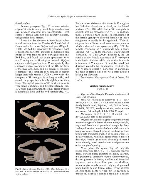
Fig. 2. Hiplyra elegans (Gravier, 1920). Male holotype, CL =<br />
9.90 mm, CB = 9.33 mm (ZMK CRU929). (A) dorsal surface; (B)<br />
ventral surface.<br />
For the male abdomen, the telson in H. elegans<br />
has 2 distinct elevations proximally on the lateral<br />
portions (Fig. 1D), while that of H. variegata is<br />
smooth, with no elevation (Fig. 7C). In addition,<br />
these 2 species have distinct morphologies of<br />
the female gonopore allowing females of these<br />
congeners to readily be distinguished. While H.<br />
elegans has a distinct oval membranous process<br />
which is directed anteroposteriorly (Fig. 1E), the<br />
female gonopore of H. variegata has a large<br />
opening (Fig. 7D) on the inner side of a prominent<br />
elevation. As Galil (2009) discussed, the 1st<br />
somite of the female abdomen of H. variegata<br />
is distinctly trilobate, while this somite is simple<br />
in females of H. elegans. It must be noted that<br />
drawings provided by Stephensen (1946: 88, fig.<br />
15F-K) clearly depict H. variegata, in particular,<br />
the male abdomen which shows a smooth telson<br />
lacking any elevation.<br />
Distribution: Madagascar, Gulf of Oman, Sri<br />
Lanka.<br />
Hiplyra ramli sp. nov.<br />
(Figs. 3, 4)<br />
Type locality: Al Aqah, Fujairah, east coast of<br />
UAE, Gulf of Oman.<br />
Material examined: Holotype 1 (SMF<br />
38466, CL = 7.4. mm, CB = 6.8 mm), Al Aqah, near<br />
Sandy Beach Hotel, Fujairah, UAE, Gulf of Oman,<br />
25°30'N, 56°22'E, sandy substrate, under stones<br />
and corals, 3-4 m depth, 4 July 1995, M. Apel.<br />
Paratypes: 6 , 9 (4 ovig.) (SMF<br />
38467), same data as for holotype.<br />
Diagnosis: Carapace slightly longer than wide;<br />
anterior margin of efferent channel nearly straight,<br />
separated from lateral granulated margin by deep<br />
U-shaped incision; somite 6 of male abdomen with<br />
triangular arrow-shaped process on distal portion;<br />
telson wide-triangular, swollen on basal portion; G1<br />
distally widened, with small apical process directed<br />
dorsally; female gonopore obliquely directed<br />
anterodorsally, small membranous oval process on<br />
outer margin of opening.<br />
Description: Carapace (Fig. 4A) slightly<br />
longer than wide (CL/CB = 1.1), distinctly convex;<br />
dorsal surface finely punctate medially, laterally,<br />
and posteriorly; carapace regions weakly defined,<br />
distinct grooves delimiting cardiac and intestinal<br />
regions; branchiocardiac grooves shallow;<br />
frontal region nearly smooth, slightly depressed<br />
immediately behind frontal ridge. Front slightly<br />
shorter than posterior margin of carapace,<br />
produced, slightly extended medially; shallow