216 Lin et al. – New Scleractinian Coral From Taiwan USA) instrument. Skeleton vouchers were deposited at the National Museum of Natural Science (NMNS), Taichung, Taiwan and at the Museum of Tropical Queensland (MTQ), Townsville, Australia. In the morphological description, the following abbreviations were used: CD, calicular diameter; GCD, great CD; Sx, septa of the x order; Px, pali of the x order; and H, height. Tissue samples preserved in CHAOS solution (Fukami 2004) were used for DNA extraction. Symbiodinium identification Following LaJeunesse (2002), denaturing gradient gel electrophoresis (DGGE) of the internal transcribed spacer (ITS)-2 region was performed to identify the Symbiodinium clade present in P. chaishanensis sp. nov. The ITS-2 region was amplified using primers ITS2 clamp and ITSintfor 2 developed by LaJeunesse and Trench (2000). A polymerase chain reaction (PCR) was performed with a touch-down cycle according to LaJeunesse (2002). PCR products were subjected to electrophoresis for 15-16 h on denaturing gradient gels (45%-80%) using a CBS Scientific System (Del Mar, CA, USA). Gels were stained with SYBR green (Molecular Probes, Eugene, OR, USA) for 20 min, and photographed for further analysis. Bands were excised from the gel and sent for direct sequencing. Resulting sequences were deposited in the NCBI database (with accession nos.: 180016-180021) Sequence analysis and phylogeny Forty mt16S rDNA and the cytochrome c oxidase subunit I (COI) sequences, including these 2 regions from the complete mt genome of P. chaishanensis sp. nov (Lin et al. 2011), were retrieved from GenBank. This dataset contained 11 robust and 4 complex scleractinian families. Phylogenetic analyses were performed using MEGA 4.0 (Tamura et al. 2007) for Neighborjoining (NJ) and MrBayes 3.1.2 (Huelsenbeck and Ronquist 2001) for Bayesian inference (BI). The most appropriate model of nucleotides was determined to be HKY+I using MrModeltest vers. 2.3 (Nylander 2004). The NJ analyses were performed with 500 replicates, and for the BI, 2 runs each of 10 6 generations were calculated for each marker with topologies saved every 100 generations. The 1st quarter of the saved topologies were discarded as burn-in, and the remaining ones were used to calculate posterior probabilities. Systematic description RESULTS Subclass Hexacorallia. Order Scleractinia Bourne, 1900. Suborder Caryophylliina Vaughan & Wells, 1943. Family Caryophylliidae Dana, 1846. Genus Polycyathus Duncan, 1876. Polycyathus chaishanensis sp. nov. Illustrations of the holotype are given in figures 3C, D, 4A-C; and illustrations of the paratype are given in figure 4D, E. Materials examined: Holotype: NMNS-6309- 001 (Taichung, Taiwan). Paratypes: NMNS-6309- 002, NMNS-6309-003 (Taichung, Taiwan), and MTQ G64703 (Queensland, Australia, 1 specimen). Type locality: 22°38'18''N, 120°15'19''E (Taiwan), 3 m in depth. Description: Small reptoid colonies formed by closely spaced cylindrical corallites arising from a common coenosteum or from stolons. Holotypic colony consisting of approximately 70 corallites. Extratentacular budding common; however, some corallites displaying intratentacular division. Calice circular to slightly elliptical. Largest corallite examined 3.65 × 3.73 mm in CD and 4.0 mm in H. Theca thick. Costae more prominent near calicular edge. All costae equal in width (about 0.21 mm wide), slightly convex, and bearing low, coarse granules. Intercostal striae deep and flat near calicular edge, becoming less distinct in direction of base. Coenosteum and theca white, but columellar elements usually light-brown pigmented. Vivid-red to dark brown sub-pellucid polyps considerably expanded above calicular edge; tentacles long, slender, with knobby end, and covered by small white verruca. Septa hexamerally arranged in 4 incomplete cycles, according to formula: S1 ≥ S2 > S3 > S4. Corallites < 2 mm in GCD with 12 or fewer septa, but larger corallites (up to 3.7 mm in GCD) with several pairs of S4 totaling up to 34 septa. S1 exsert (0.5-0.7 mm), with straight and almostvertical axial edges sometimes bearing small, cylindrical (0.24 mm in diameter) palus. S2 only slightly less exsert and equal or narrower than S1. S3 less exsert, thinner, and about 2/3 width of S2. Axial edges of S1-S2 dentate, those of S3 laciniated. S4 1/2-2/3 width of S3. Well-developed P3 (sometimes bilobated) present before S3. If
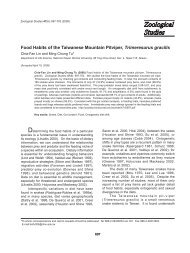
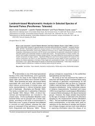
<strong>Zoological</strong> <strong>Studies</strong> 51(2): 213-221 (2012) 217 (A) 1 mm (B) (C) 1 mm 2 mm (D) (E) 500 μm 1 mm Fig. 4. (A) Calicular view of 1 corallite of the holotypic colony (NMNS-6309-001) undergoing extratentacular budding; (B) calicular view of 1 corallite of the holotypic colony (NMNS-6309-001) undergoing intratentacular budding; (C) calicular view of 1 corallite of the holotypic colony (NMNS-6309-001); (D) lateral view of a corallite from the paratype colony (NMNS-6309-002); (E) detail of columellar elements MTQ G64703.