annals 1-2.qxd - Centrum Zdrowia Dziecka
annals 1-2.qxd - Centrum Zdrowia Dziecka
annals 1-2.qxd - Centrum Zdrowia Dziecka

Create successful ePaper yourself
Turn your PDF publications into a flip-book with our unique Google optimized e-Paper software.
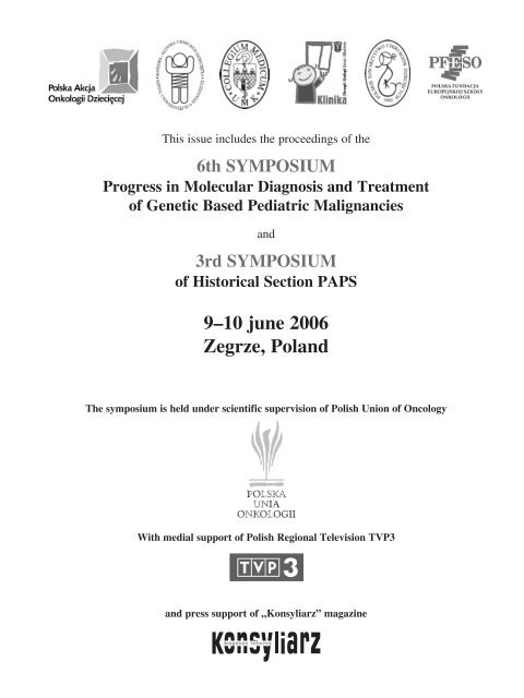
This issue includes the proceedings of the<br />
6th SYMPOSIUM<br />
Progress in Molecular Diagnosis and Treatment<br />
of Genetic Based Pediatric Malignancies<br />
and<br />
3rd SYMPOSIUM<br />
of Historical Section PAPS<br />
9–10 june 2006<br />
Zegrze, Poland<br />
The symposium is held under scientific supervision of Polish Union of Oncology<br />
With medial support of Polish Regional Television TVP3<br />
and press support of „Konsyliarz” magazine
This symposium is sponsored by
Annals of Diagnostic Paediatric Pathology<br />
Official Journal of the Polish Paediatric Pathology Society<br />
and Section of Oncological Surgery of Polish Association of Paediatric Surgeons<br />
EDITOR-IN-CHIEF<br />
CO-EDITORS<br />
ASSOCIATE EDITORS<br />
PRODUCTION EDITOR<br />
EDITORIAL OFFICE<br />
EDITORIAL BOARD<br />
B. M. WoŸniewicz, Warsaw<br />
B. Cukrowska, Warsaw<br />
A. I. Prokurat, Bydgoszcz<br />
J. Cielecka-Kuszyk, Warsaw A. Bysiek, Cracow<br />
E. Czarnowska, Warsaw P. Czauderna, Gdansk<br />
W. T. Dura, Warsaw J. Godziñski, Wroclaw<br />
M. Grajkowska, Warsaw J. Niedzielski, Lodz<br />
M. Liebhardt, Warsaw W. WoŸniak, Warsaw<br />
A. Wasiutyñski, Warsaw M. Wysocki, Bydgoszcz<br />
Lotos Poligrafia Ltd., Warsaw, www.drukarnia-lotos.pl<br />
Annals of Diagnostic Paediatric Pathology<br />
Department of Pathology<br />
The Children’s Memorial Health Institute<br />
Aleja Dzieci Polskich 20<br />
04 736 Warszawa, Poland<br />
Tel.: +48−22−815−19−72<br />
Fax: +48−22−815−19−75<br />
E−mail: b.cukrowska@czd.pl, b.wozniewicz@czd.pl<br />
J. P. Barbet, Paris J. Kobos, Lodz<br />
L. A. Boccon-Gibod, Paris G. Karpati, Montreal<br />
P. E. Campbell, Melbourne J. Las Heras, Santiago de Chile<br />
A. Chilarski, Lodz K. Madaliñski, Warsaw<br />
J. Czernik, Wroclaw D. M. F. Menezes, Rio de Janeiro<br />
E. Gilbert-Baarness, Tampa W. A. Newton, Jr., Johnstown<br />
A. A. Greco, New York B. Otte, Brussels<br />
M. D. Haust, London S. A. Pileri, Bologna<br />
A. Hinek, Toronto J. Plaschkes, Berne<br />
J. Huber, Utrecht F. Raafat, Birmingham<br />
C. G. Gopalakrishnan, Trivandrum S. W. Sadowinski, Mexico City<br />
S. Gogus, Ankara K. Sawicz-Birkowska, Wroclaw<br />
A. Jankowski, Poznan J. Stejskal, Prague<br />
P. Januszewicz, Warsaw Cz. Stoba, Gdansk<br />
B. Jarz¹b, Gliwice G. Thiene, Padova<br />
B. A. Kakulas, Perth S. Variend, Shieffield<br />
R. O. C. Kaschula, Rondebosch T. H. Wyszyñska, Warsaw<br />
W. Kawalec, Warsaw A. Zimmermann, Berne<br />
The journal is supported by the State Committee for Research.<br />
AIMS AND SCOPE<br />
The Annals of Diagnostic Paediatric Pathology is an international peer−reviewed journal. The focus of the journal is current progress in<br />
clinical paediatric pathology in both basic and clinical applications. Experimental studies and clinical trials are accepted for publication,<br />
as are case reports supported by literature review. The main policy of the Annals is to publish papers that present practical knowledge<br />
that can be applied by clinicians.<br />
© Copyright by Polish Paediatric Pathology Society, 2001<br />
ISSN 1427-4426
INSTRUCTIONS FOR AUTHORS<br />
General<br />
Send three copies of the manuscript written in<br />
English and three full sets of illustrations (two<br />
sets suitable for review and one for publication),<br />
with a cover letter, including the complete<br />
name, address, telephone and facsimile<br />
numbers, and e-mail address of the communicating<br />
author, and a statement that all authors<br />
have approved the manuscript, addressed to:<br />
Bogdan M. Wozniewicz, MD, PhD, ADPP Editor-in-Chief,<br />
Annals of Diagnostic Paediatric<br />
Pathology, Department of Pathology, The Children’s<br />
Memorial Health Institute, Aleja Dzieci<br />
Polskich 20, PL-04736 Warsaw, Poland; Telephone:<br />
(48)-22-815 1972, (48)-22-815 2732;<br />
(48)-22-815 1965; Fax: (48)-22-815 1973;<br />
e-mail: b.cukrowska@czd.pl<br />
Organization of Manuscript<br />
Manuscripts, including references should be<br />
typed on one side of paper with double spacing<br />
and broad margins. The pages should be numbered<br />
consecutively. The manuscript should be<br />
presented in the order indicated.<br />
Title page. The first page should contain the article’s<br />
title, the complete name of all authors<br />
(first name, middle initials, surname), the departmental<br />
and institutional affiliations and complete<br />
street or mailing addresses of all authors,<br />
the telephone and fax numbers and e-mail<br />
address for the corresponding author, and the<br />
city, state and country where the work was<br />
performed.<br />
Abstract and key words. An abstract of not more<br />
than 250 words should begin on page 2. It<br />
should be a factual, not descriptive, statement<br />
of study objectives, methods, principal results,<br />
and conclusions. Use Index Medicus terms<br />
from medical subject headings where possible.<br />
For the purpose of coding and subject indexing<br />
provide up to six key words in alphabetical<br />
order immediately following the abstract.<br />
Text. The text should be organized into an introductory<br />
section, including the background<br />
and purpose of the study, and then into sections<br />
titled “Methods”, “Results” and “Discussion”.<br />
In certain circumstances the nature of the<br />
report does not lend itself to this format. Underline<br />
any words or phrases to be emphasized by<br />
italics.<br />
Acknowledgments. Grant support and individuals<br />
who were of direct help in the reported work<br />
should be acknowledged by a brief statement<br />
on a separate page immediately following the<br />
text.<br />
References. References must be typed double<br />
spaced and listed in consecutive alphabetical<br />
and numerical order. All references must be cited<br />
in the text or tables. Once a reference has<br />
been cited, all subsequent citation should be to<br />
the original number. Unpublished data and personal<br />
communications are not acceptable references.<br />
All references must be verified against<br />
the original documents. Abbreviations of journals<br />
must conform to those in the list of Journals<br />
indexed in Index Medicus.<br />
Journals. References to journal articles should<br />
include surnames and initials of author(s) (list<br />
all authors if fewer than seven; if seven or more,<br />
list only the first three, followed by et al.),<br />
year, full title of article, abbreviated journal<br />
name, volume, and inclusive pagination:<br />
Dresdale DT, Schults M, Michtom RJ (1991)<br />
Primary pulmonary hypertension. I. Clinical and<br />
hemodynamic study. Am J Med 11: 686–705<br />
Books. References to a book should include<br />
surnames and initials of author(s), year, title of<br />
book, city, publisher and edition:<br />
Jones KL (1988) Smith’s recognizable patterns<br />
of human malformation, 4th edn. Saunders,<br />
Philadelphia<br />
Chapter in a book. References to a chapter in<br />
a book should include surnames and initials of<br />
authors), year, chapter title, editor(s), book title,<br />
publisher, city, volume (if applicable), section<br />
(if applicable), and inclusive pagination:<br />
Thurlbeck WM (1992) Respiratory system. In:<br />
Dimmick JE, Kalousek DK (eds) Developmental<br />
pathology of the embryo and fetus. Lippincott,<br />
Philadelphia, pp 437–449<br />
Papers in conference proceedings or abstracts.<br />
These references should include surnames and<br />
initials of author(s), year, title, title of conference,<br />
city, publisher, and inclusive pagination.<br />
Kjaer I (1994) The prenatal axial skeleton as<br />
marker of normal and pathological development<br />
of the human central nervous system. In:<br />
Alfred Benzon Symposium: Brain Lesions in<br />
the New-born. Munksgaard, Copenhagen, 37:<br />
124–138<br />
Polendo R, Lowe GN, McDougall S, Hahn TJ<br />
(1994) Mechanism of prostaglandin regulation<br />
of proteoglycan synthesis in rat chondrocytic<br />
cells (abstract). J Bone Miner Res 9: S187<br />
Tables. Each table should be typed double spaced<br />
on a separate sheet of paper, numbered consecutively<br />
with Arabic numbers, and contain<br />
a brief title at the top. All tables must be cited<br />
in the text.<br />
Figures. Legends for figures should be typed<br />
double spaced on a separate sheet and numbered<br />
consecutively with Arabic numbers corresponding<br />
to the figures. The figure legend should<br />
be as clear as possible and should fully<br />
describe the contents the figure. All figures<br />
must be cited in the text. Figures should be professionally<br />
drawn and photographed and submitted<br />
in triplicate as black and white, high<br />
quality glossy prints. Special attention should<br />
be paid to focus and contrast. Do not submit<br />
original artwork. Symbols, letters, arrows, and<br />
numbers must be of sufficient size and contrast<br />
to be clearly recognizable when the figure is<br />
reduced to publication size. Paste a label on the<br />
back of each figure, indicating its number, name<br />
of author, title of paper, and the desired<br />
orientation. The optimal size for a figure is<br />
8.6 × 10 cm (3.4 × 4 inch). If appropriate and<br />
acceptable these will be available for retrieval<br />
by readers in the online electronic version of<br />
the journal. A limited number of illustrations in<br />
full color will be considered, but the author<br />
may be required to bear the costs of part or all<br />
of their reproduction. Artwork to accepted manuscripts<br />
will not be returned unless a written<br />
request accompanies the page proofs.<br />
Electronic submission of final accepted manuscripts.<br />
Please send an electronic version of<br />
your manuscript with the final, accepted, double<br />
spaced hard copy. It is the author’s responsibility<br />
to ensure that the disk and hard copy<br />
match. Disks may be prepared on a PC in any<br />
version of Word or WordPerfect. Do not use<br />
desktop publishing software. Keep the document<br />
as simple as possible and avoid complex<br />
formatting. Follow the instructions for references<br />
very carefully. If incorrectly styled, the value<br />
of the diskette submission will be reduced.<br />
Abbreviations, spelling and units. Standard<br />
abbreviations and units listed in the Council of<br />
Biology Editors Style Manual: A Guide for<br />
Authors, Editors and Publishers in the Biological<br />
Sciences (5th edn, Bethesda, Md, Council<br />
of Biology Editors, 1983) should be used. All<br />
quantitative measurements should be in conventional<br />
metric units, followed when appropriate<br />
by the SI unit in parentheses.<br />
Permissions. Copyrighted material from other<br />
sources must be accompanied by written permission<br />
from both the publisher and author for<br />
reproduction. Any personal communication<br />
used in the text must be accompanied by a letter<br />
agreeing to be quoted as written.<br />
Review and action. Manuscripts will be examined<br />
by the editorial staff and sent to two reviewers.<br />
Authors will be notified of the disposition<br />
of their manuscript within 60 days.<br />
Case reports. Limit abstract to 150 words, use<br />
no more than three tables or figures and limit<br />
references to 15. Additional illustrations may<br />
be submitted for online storage and retrieval.<br />
Letters to the editor. Letters to the editor intended<br />
for publication should not exceed 800<br />
words in length. Up to three references and two<br />
illustrations may accompany the letter. Letters<br />
must include the name, departmental and institutional<br />
affiliation and complete mailing address<br />
of the author. The editorial board reserves<br />
the right to accept, reject, or excerpt letters<br />
without changing the views expressed by the<br />
author (s).<br />
Page Proofs and Offprints. All material accepted<br />
for publication is subjected to copyediting.<br />
Authors will receive page proofs of articules<br />
before publication and should answer all queries<br />
and carefully check all editorial changes<br />
at this point. Offprints may be ordered at cost<br />
price when the page proofs are returned.
Annals of Diagnostic Paediatric Pathology<br />
Official Journal of the Polish Paediatric Pathology Society<br />
and Section of Oncological Surgery of Polish Association of Paediatric Surgeons<br />
Volume 10 Number 1–2 Summer 2006<br />
CONTENTS<br />
Review<br />
paper<br />
Original<br />
papers<br />
Pathogenesis and clinical manifestation of celiac disease: analysis of atypical symptoms . . 7<br />
Bo¿ena Cukrowska, Barbara Burda-Muszyñska, Maria Zegad³o-Mylik, Jerzy Socha<br />
Polyoma BK virus nephropathy in children after kidney transplantation . . . . . . . . . . . . . . 13<br />
Przemys³aw Kluge, Ewa Œmirska, Sylwester Prokurat, Agnieszka Perkowska, Bo¿ena Cukrowska<br />
Primary malignant liver cell tumours in children – different treatment strategies . . . . . . . 17<br />
Andrzej Igor Prokurat, Ma³gorzata Chrupek, Roman KaŸmirczuk, Przemys³aw Kluge, Andrzej Koœciesza,<br />
Pawe³ Rajszys, El¿bieta Dzik, Arthur Zimmermann<br />
Utilization of internal sphincter in the reconstruction of anorectal malformations<br />
in girls . . . . . . . . . . . . . . . . . . . . . . . . . . . . . . . . . . . . . . . . . . . . . . . . . . . . . . . . . . . . . . . . . . . 23<br />
Andrzej Igor Prokurat, Ma³gorzata Chrupek, Roman KaŸmirczuk, Przemys³aw Kluge<br />
Comparison of histological changes in liver biopsy specimens in patients<br />
with biliary atresia of poor and good prognosis . . . . . . . . . . . . . . . . . . . . . . . . . . . . . . . . . . . 31<br />
Joanna Cielecka-Kuszyk, Piotr Czubkowski, Ludmi³a Bacewicz, Joanna Paw³owska, Irena Jankowska,<br />
Tamara Szymañska-Dêbiñska<br />
The impact of probiotic Lactobacillus casei and paracasei strains on cytokine profile<br />
in children with atopic dermatitis . . . . . . . . . . . . . . . . . . . . . . . . . . . . . . . . . . . . . . . . . . . . . . 37<br />
Ilona Rosiak, Urszula Witos³aw, Aldona Ceregra, El¿bieta Klewicka, Ilona Motyl, Zdzis³awa Libudzisz,<br />
Bo¿ena Cukrowska<br />
Case<br />
report<br />
Paraganglioma associated with neuroblastoma: rare composite tumor<br />
in a 16-year-old girl . . . . . . . . . . . . . . . . . . . . . . . . . . . . . . . . . . . . . . . . . . . . . . . . . . . . . . . . . 43<br />
Przemyslaw Galazka, Malgorzata Chrupek, Roman Kazmirczuk, Jaroslaw Peregud-Pogorzelski,<br />
Malgorzata Pacholska, Grzegorz Meder, Przemyslaw Kluge, Zdzislaw Skok, Krzysztof Kusza<br />
and Andrzej Igor Prokurat<br />
Program Case of the Symposium . . . . . . . . . . . . . . . . . . . . . . . . . . . . . . . . . . . . . . . . . . . . . . . . . . . . . . . . . . . . . . 49<br />
reports<br />
Abstracts . . . . . . . . . . . . . . . . . . . . . . . . . . . . . . . . . . . . . . . . . . . . . . . . . . . . . . . . . . . . . . . . . . . . . . . . . . . 51
SUBSCRIPTION<br />
Subscriptions: Published 2 numbers per year, subscriptions are sold on a calendar year basis by the<br />
volume number designated for that year. Payment by check in US dollars and credit cards (Visa card only).<br />
Professional researches subscription: 40 € (outside Europe) per year, 20 € (Europe) per year.<br />
Library and institutions subscriptions: 250 € (outside Europe) per year, 200 € (Europe) per year.<br />
I wish to pay my ADPP subscription G<br />
Name ____________________________________<br />
Europe<br />
Outside Europe<br />
Address __________________________________<br />
City ______________________________________<br />
Postal Code _______________________________<br />
Country ___________________________________<br />
Phone ____________________________________<br />
Fax ______________________________________<br />
E−mail ____________________________________<br />
Individual 20 € G 40 € G<br />
Institution 200 € G 250 € G<br />
Please indicate method of payment<br />
Visa G<br />
Name _______________________________<br />
Card Number _________________________<br />
Expiry Date ___________________________<br />
by check G<br />
_________________________________________<br />
Signature<br />
Pay to the order of<br />
Polish Paediatric Pathology Society<br />
Bank BPH<br />
Al. Jana Pawla II 23<br />
00−854 Warsaw<br />
account No 10601028−310000561979<br />
Please sign and return this form to:<br />
Annals of Diagnostic Paediatric Pathology<br />
Department of Pathology<br />
The Children’s Memorial Health Institute<br />
Aleja Dzieci Polskich 20<br />
04 736 Warszawa, Poland<br />
Tel.: +48−22−815−19−72<br />
Fax: +48−22−815−19−75<br />
E−mail: b.cukrowska@czd.pl<br />
b.wozniewicz@czd.pl
Annals of Diagnostic Paediatric Pathology 2006, 10(1–2):7–12<br />
© Copyright by Polish Paediatric Pathology Society Annals of<br />
Pathogenesis and clinical manifestation of celiac disease:<br />
analysis of atypical symptoms<br />
Bo¿ena Cukrowska 1 , Barbara Burda-Muszyñska 2 , Maria Zegad³o-Mylik 1 , Jerzy Socha 2<br />
Diagnostic<br />
Paediatric<br />
Pathology<br />
1<br />
Department of Pathology<br />
2<br />
Department of Gastrology, Hepatology and Immunology<br />
The Children’s Memorial Health Institute<br />
Warsaw, Poland<br />
Abstract<br />
Celiac disease is defined as enteropathy occurring after ingestion of gluten from cereals in genetically<br />
predisposed individuals. Recently, after introduction of sensitive and specific screening tests, the increase<br />
in prevalence of celiac disease has been observed. The celiac disease incidence of 1:100 – 1:300 is similar<br />
in Europe, United States, Australia and Asia. Still, a lot of celiac patients is undiagnosed. Difficulties in<br />
diagnosis are caused by the changes of clinical features of the disease into atypical and the occurrence of<br />
illness in older children and adults. In this review pathogenetic factors playing role in atypical celiac disease<br />
have been shown, intestinal and non-intestinal symptoms of disease, and risk groups, in which screening<br />
tests should be done, have been presented as well.<br />
Key words: celiac disease, gluten, screening tests, atypical symptoms, risk groups<br />
Introduction<br />
Celiac disease (CD) is an immune-mediated enteropathy caused<br />
by a permanent sensitivity to gluten, i.e. a protein present<br />
in cereals, occurring in genetically susceptible individuals,<br />
in whom ingestion of gluten induces typical changes in<br />
small intestine mucosa [16]. The development of reliable serological<br />
screening tests has dramatically increased our awareness<br />
of CD as a common condition. The prevalence of CD<br />
in a healthy population of Europe, United States, Australia<br />
and Asia varies between 1:100 – 1:300 [17, 24, 36, 37]. In<br />
Poland CD screening tests in healthy population have not been<br />
done, yet. Due to the fact that the prevalence of CD in Europe<br />
is so high, it is expected that even about 380 thousand<br />
of CD patients live in Polish country.<br />
CD has extremely changed since the moment Dutch<br />
physician Willem Dicke had described the clinical aspects of<br />
the disease [13]. Nowadays, so called classical type of CD<br />
with chronic diarrhea, abdominal distension, poor weight gain,<br />
starting under 2 years of age, occurs rather rare. CD appears<br />
in each age after the introduction of gluten to the diet, and<br />
in many older patients presents atypical non-gastrointestinal<br />
symptoms or has a silent form with minimal or no clinical<br />
symptoms [10]. Gastrointestinal symptoms of CD represent<br />
the tip of the celiac iceberg presented by Logan [32]. Because<br />
of atypical manifestations, many patients with CD remain<br />
undiagnosed or are treated symptomatically. In adults, CD is<br />
diagnosed on average over 10 years after the first symptoms<br />
[21]. The lack of proper treatment, i. e. a strict gluten free diet<br />
(GFD), increases the risk of life-threatening complications that<br />
are difficult to manage, such as cancers and malignant lymphomas<br />
[7]. There is also strong evidence that in unrecognized<br />
CD patients the frequency of other autoimmune diseases<br />
is higher than in general population [58].<br />
Pathogenesis<br />
CD is a highly genetically determined disease with the major<br />
genetic factor being the expression of specific HLA class<br />
II antigens. About 90–95% of CD patients express HLA-<br />
-DQ2 molecule, the rest of them are HLA-DQ8-positive<br />
[33]. These HLA-DQ receptors have the capacity to bind the<br />
Address for correspondence<br />
Bo¿ena Cukrowska, MD, PhD, DrSc<br />
Zak³ad Patologii<br />
Instytut „Pomnik-<strong>Centrum</strong> <strong>Zdrowia</strong> <strong>Dziecka</strong>”<br />
Aleja Dzieci Polskich 20<br />
04-734 Warszawa<br />
E-mail: ptpd@czd.waw.pl
8<br />
specific gliadin peptides that have been implicated in CD.<br />
HLA-DQ molecules present on the surface of antigen presenting<br />
cells (macrophages, dendritic cells), i.e. cells, which<br />
catch the gliadin peptides and present them in association<br />
with specific HLA-DQ receptors to T lymphocytes [43].<br />
Gluten contains peptides presenting extremely high<br />
affinity to DQ2 and DQ8 receptors [51]. These toxic peptides<br />
are characterized by high glutamine content, i.e. the amino<br />
acid which is a substrate for the enzyme – tissue transglutaminase<br />
(tTG). tTG catalyzes deamination of glutamine into<br />
glutamine acid. This reaction increases about 100-times<br />
the affinity of toxic peptides, and is essential for significant<br />
stimulation of gliadin-specific – T lymphocytes (Fig. 1). Activated<br />
gliadin-specific T lymphocytes produce pro-inflammatory<br />
cytokines responsible for histological changes in intestinal<br />
mucosa [10, 11].<br />
Fig. 1 Effector mechanisms of celiac disease – the activation of specific T<br />
lymphocytes<br />
Toxic peptides are present in wheat, barley and rye<br />
[9]. Corn, rise and, as recently has been shown, pure oats do<br />
not contain toxic peptides active in CD [23].<br />
The recent reports by Shan et al. shows that digestion<br />
of recombinant gliadin with gastric and pancreatic enzymes<br />
in vitro produces the highly stable 33-mer peptide that is rich<br />
in proline and glutamine, and which contains all known toxic<br />
peptides [51]. This 33-mer peptide is resistant to brush<br />
border enzymes, but it can be easily degestive by endopeptidases<br />
coming from intestinal bacteria [51].<br />
Findings showing that gut bacteria could deprive gliadin<br />
peptides of toxicity open a new therapeutic possibility<br />
with using probiotics, i.e. non-pathogenic bacteria regulating<br />
gut microflora ecosystem. It is not excluded that bacterial endopeptidases<br />
could be also used in preparation of non toxic<br />
cereals [12].<br />
HLA-DQ2 and -DQ8 are common in human population.<br />
This haplotype is present in 20–30% of health controls,<br />
suggesting that activation of pathological processes could be<br />
influenced by other genes. It seems that HLA class I related<br />
gene – the major histocompatibility complex class I chain-related<br />
gene A (MICA) is an attractive candidate for better un-<br />
derstanding of physiopathological mechanisms of CD [31].<br />
MICA molecule is mainly expressed on the intestinal epithelium<br />
and is recognized by T lymphocytes, CD8+ lymphocytes<br />
and natural killer cells. The binding of MICA to cells induces<br />
cytotoxic and inflammatory responses in the intestine.<br />
Lopez-Vazquez et al. found an association of the MICA A5.1<br />
allele with atypical form of CD. Up to 29% of patients with<br />
atypical CD expressed A5.1 allele in comparison to 13% of<br />
patients with typical CD and 7% of healthy population. The<br />
mechanism by which atypical manifestations are triggered is<br />
connected with the fact that MICA-A5.1 is responsible for secretion<br />
of soluble form of MICA molecule. This protein reacts<br />
with cytotoxic and pro-inflammatory lymphocytes and<br />
inhibits their activation in intestine by MICA present in gut<br />
epithelium.<br />
Clinical symptoms<br />
The classical clinical picture of childhood celiac disease consists<br />
of prolonged diarrhea with failure to thrive, abdominal<br />
distension, vomiting. Severe malnutrition and even cachexia<br />
can occur when diagnosis is delayed. This type of presentation<br />
has decreased in many European countries over the past<br />
few decades. The factors responsible for this change might be<br />
explained by the exclusion of gluten from the diet of babies<br />
and promotion of natural feeding [14]. Studies performed in<br />
United States on children with recognized CD showed that older<br />
age of CD onset and atypical manifestations were results<br />
of prolonged breast feeding [14]. On the other hand Swedish<br />
observed that breast feeding decreased occurrence of CD in<br />
children before 2 years of age, and high doses of gluten introduced<br />
to the baby's diet independent on the age without protection<br />
of breast milk increased the risk of CD [26]. Presented<br />
results open the discussion on the period of introduction<br />
of gluten to the infant diet. Experimental data show that oral<br />
tolerance is developed only in early ontogeny, thus too late<br />
administration of gluten to the diet could be responsible for<br />
gluten intolerance. It seems that low amounts of gluten in the<br />
diet before 6 months of life, but introduced during breast feeding<br />
could prevent the development of CD [25]. However,<br />
intake the gluten either before 3 month of life or under<br />
7 month of life increased the risk of CD [46].<br />
In Poland Szaflarska-Szczepanik observed the decreased<br />
frequency of classical form of CD in children starting<br />
from 1990 [55]. Children presented atypical symptoms, such<br />
as low weight, short stature, anemia, abdominal pain. Ludvigsson<br />
at al. reported similar tendency in children before<br />
2 years old, in whom in addition, irritability and muscle wasting<br />
were found [34].<br />
Recently, increased frequency of CD reaching 3,4%<br />
was found in patients with irritable bowel syndrome (IBS) [15,<br />
53]. These patients tend to experience diarrhea, vomiting, recurrent<br />
abdominal pain, constipation, nausea. Our studies also<br />
showed high frequency of CD in children with IBS (1:60) [4].<br />
Many symptomatic patients with newly diagnosed<br />
CD initially present with non-gastrointestinal manifestations<br />
of CD. Atypical symptoms of CD are presented in Table 1.
9<br />
Table 1<br />
Nongastrointestinal symptoms of celiac disease<br />
Iron and folic acid anaemia<br />
Short stature<br />
Pubertal delay<br />
Unexplained infertility, spontaneous abortions<br />
Epilepsy with intracranial calcifications<br />
Cerebellar ataxia<br />
Depression, irrtability<br />
Osteopenia /osteoporosis<br />
Arthritis<br />
Dental enemal hypoplasia<br />
Recurrent oral ulcers<br />
Unexplained hypertransaminaesemia<br />
Dermatitis herpetiformis<br />
The most common non-gastrointestinal manifestation<br />
of CD, especially in adults is iron and folic acid anemia.<br />
About 11% of adults with anemia resistant to oral iron or folate<br />
supplementation have CD [3, 41]. When patients represent<br />
unexplained iron deficiency anemia the frequency is lower<br />
ranging up to 8%. In children, short stature is the most<br />
common non-gastrointestinal symptom of CD. The incidence<br />
of CD in this group is 8–10% [56].<br />
Numerous studies confirm that disturbances in bone<br />
mineral density are often symptoms in celiac patients. Osteoporosis<br />
is one of the well-known complications of untreated<br />
CD [28, 44]. A GFD in children reverses bone density abnormalities<br />
and the beneficial effects of gluten withdrawal are<br />
persistent [42]. Contrary to pediatric cases, in adults affected<br />
by osteoporosis, a GFD does not reduce the risk of fractures<br />
[57]. In children dental enamel hypoplasia of permanent teeth<br />
could be a single manifestation of CD [1].<br />
A number of neurological and psychological manifestations,<br />
including epilepsy with intracranial calcifications,<br />
primary ataxia, autism, depression, headaches, have been reported<br />
in patients with CD, but the evidence for their association<br />
with CD in children is weak [65]. Our studies on frequency<br />
of CD at risk group of children did not confirm occurrence<br />
of CD in children with epilepsy and autism [4].<br />
There is some evidence for unexplained hypertransaminaesemia<br />
in untreated patients with CD. Up to 9% of<br />
adults with elevated serum transaminases present CD [61].<br />
In biopsies of liver obtained from these patients only nonspecific<br />
inflammatory changes were found, and tle level of enzymes<br />
appeared to normalize on a GFD [61]. Sjogren et al.<br />
found a nonspecific increase of anti-gliadin antibodies<br />
(AGA) in a numbers of liver diseases, such as alcoholic cirrhosis<br />
(20%), primary biliary cirrhosis (16%), primary sclerosing<br />
cholangitis (24%), hepatitis HCV (11%) [52]. However,<br />
the evidence of an increase of CD confirmed by histo-<br />
logical lesions was found only in patients with autoimmune<br />
hepatitis [59].<br />
Unexplained infertility could be also a syndrome of<br />
atypical CD [29]. Incidences of menstrual irregularities and<br />
spontaneous abortions appear more often in women with CD<br />
than in general populations [29]. In children with CD pubertal<br />
delay can occur.<br />
Other atypical syndromes of CD are recurrent oral<br />
ulcers and arthritis. Up to 3% of children with juvenile arthritis<br />
present CD [54].<br />
Nowadays, dermatitis herpetiformis is considered to<br />
be a cutaneous manifestation of gluten sensitivity [27]. Dermatitis<br />
herpetiformis is a severe itchy, blistering skin disease<br />
with abnormalities occurring on the elbows, knees and buttocks.<br />
In intestinal biopsy typical for CD histological lesions<br />
are found. The treatment with a GFD approves skin and intestinal<br />
abnormalities.<br />
Associated conditions<br />
CD is associated with a number of autoimmune and non autoagressive<br />
genetical diseases (Table 2). Autoimmune disorders<br />
occur ten times more frequently in adult patients with<br />
CD than in general populations. Up to 8% of patients with<br />
type 1 diabetes have the characteristic features of CD in<br />
small intestinal biopsy [8, 48]. This figure may be an underestimate,<br />
as serial screening of individuals with type 1 diabetes<br />
over a period of years has identified additional cases<br />
who initially had negative serological tests [8]. CD is also<br />
more often recognize in patients with autoimmune thyroiditis<br />
[54, 61], Sjogren's syndrome [35], cardiomyopathy [47],<br />
autoimmune myocarditis [18] and in autoagressive endocrynological<br />
disorders [45].<br />
The incidence of CD in individuals with Down syndrome<br />
is between 5% and 12% [20]. About one third of patients<br />
present atypical non-gastrointestinal syndromes. An increased<br />
prevalence of CD, which ranges from 4,1 to 8,1%<br />
Table 2<br />
Diseases associated with celiac disease<br />
Disease<br />
Percentage of association<br />
Diabetes mellitus type 1 1 5–8%<br />
Autoimmune thyroitidis 4–5%<br />
Sjogren’s syndrome 5%<br />
Autoimmune hepatitis 6,4%<br />
Juvenile arthritidis 6,6%<br />
Addison’s disease 7,9%<br />
Cardiomyopathies 2,1–5,76%<br />
Autoimmune myocarditis 4,4%<br />
Down syndrome 5–12%<br />
Turner syndrome 4–8%<br />
Wiliams syndrome 8,2%<br />
Selective IgA deficiency 2–8%
10<br />
has also been reported in Turner syndrome [2, 50] and in<br />
Wiliams syndrome (8, 2%) [19].<br />
Based on retrospective studies the frequency of selective<br />
immunoglobulin (Ig) A deficiency in CD was between<br />
1,7% and 7,7% [6].<br />
There is strong evidence that first-degree relatives of<br />
diagnosed CD cases are at increased risk for CD with a prevalence<br />
of 4% to 5% [22]. The frequency of CD is lower in<br />
second-degree relatives.<br />
Diagnosis<br />
Although serological tests have been improved during last<br />
years, still biopsy of small intestine is necessary for CD recognition.<br />
According criteria established by European Society<br />
of Gastrology, Hepatology and Nutrition in 1990, CD diagnosis<br />
is based upon histological evidence of typical small<br />
intestinal mucosal abnormalities and clinical improvement<br />
after introduction of gluten free diet [62]. CD affects the mucosa<br />
of the proximal small intestine, with damage gradually<br />
decreasing in severity towards the distal small intestine. Because<br />
the histological changes in CD may be patchy, it is recommended<br />
that multiple biopsy specimens should be obtained<br />
from the jejunum and examined according modified<br />
Marsh scale (Fig. 2) [38, 40].<br />
There is strong evidence that villous atrophy (Marsh<br />
grade III) is a characteristic histopathological feature of CD<br />
[22]. The presence of infiltrative changes with crypt hyperplasia<br />
(Marsh grade II) is compatible with CD but with less clear<br />
evidence. Diagnosis in these cases is dependent on positive<br />
serological tests. When serological tests are negative, other<br />
conditions for the intestinal changes should be considered.<br />
The presence of infiltrative intraepithelial lymphocytes alone<br />
(Marsh grade I) is not specific for CD. Concomitant positive<br />
serology increases the likehood of CD. In such cases it is recommended<br />
additional strategies, such as determination of<br />
the HLA antigens, repeat biopsy or a trial of treatment with<br />
a GFD and repeated measurement of antibodies [22].<br />
Thus, intestinal biopsy together with determination of<br />
specific antibodies in sera provides definitive CD diagnosis.<br />
As a GFD improved the intestinal lesions [64], it should be<br />
stressed that GF diet should not be introduced before planned<br />
biopsy.<br />
Serological tests are frequently used to identify individuals<br />
for whom the biopsy procedure. They are also useful<br />
for screening the population to determine the prevalence<br />
of CD and for monitoring of a GFD therapy [64].<br />
A comparison between several commercial available<br />
serological tests demonstrated that determination of anti-endomysial<br />
antibodies (EMA) by an indirect immunofluorescence<br />
technique (IIF) and anti-human recombinant tissue<br />
transglutaminase antibodies (hrtTG-Ab) by immunoenzymatic<br />
(ELISA) method are superior to anti-gliadin antibodies<br />
(AGA) and anti-guinea pig tissue transglutaminase antibodies<br />
(gTG-Ab) [63, 64]. The sensitivity and specificity<br />
of those tests range about 90%. Our studies showed that also<br />
anti-reticulin antibodies (ARA) determined by IIF are<br />
characterized by very high sensitivity (98%) and specificity<br />
(100%) [64].<br />
Thus, the autoantibodies produced against the same<br />
autoantigen, i.e. tissue transglutaminase [30], should be employed<br />
for diagnosis and screening of CD. Our observations<br />
confirm that in some patients EMA/ARA tests are negative,<br />
but hrtTG-Ab are present, so it seems that determination of<br />
autoantibodies by two tests performed by different methods<br />
are needed.<br />
Fig. 2 Histopathological lesions in celiac patients
11<br />
Screening tests<br />
Screening tests should be done at risk groups:<br />
! in first degree relatives of known CD cases,<br />
! in patients with diseases associated with CD (Table 1)<br />
! in patients with atypical syndromes (Table 2).<br />
Epidemiological studies have shown that screening<br />
tests at risk groups should be done in 2–3 years old children<br />
[5]. As the occurrence of CD at risk groups is increasing with<br />
age, it is recommended to repeat serological tests in the case<br />
of negative results.<br />
Some authors suggest that due to the high prevalence<br />
of CD and occurrence of atypical forms of CD, screening tests<br />
should be done not only at risk groups, but in the whole<br />
population (mass-screening) [39].<br />
Conclusions<br />
1. Celiac disease is a very common disorder with prevalence<br />
1:100 – 1:300.<br />
2. Most people with CD manifest atypical non-gastrointestinal<br />
syndromes or silent form of the disease.<br />
3. Screening tests should be done at least at-risk groups of<br />
patients.<br />
Acnowledgment<br />
The study was support by the projekt PBZ/KBN-<br />
097/P06/2003.<br />
References<br />
1. Aine L, Maki M, Colloin P, Keyrilainen<br />
O (1990) Dental enamel defects in celiac<br />
disease. J Oral Pathol Med 19:<br />
241–245<br />
2. Bonamico M, Paquino AM, Mariani P,<br />
et al (2002) Prevalence and clinical picture<br />
of celiac disease in Turner syndrome.<br />
J Clin Endocrinol Metab 87:<br />
5495–5498<br />
3. Bottaro G, Cataldo F, Rotolo N, Spina<br />
M, Corraza GR (1999) The clinical pattern<br />
of subclinical/silent celiac disease:<br />
an analysis on 1026 consecutive cases.<br />
Am J Gastroenterol 94:691–696<br />
4. Burda-Muszynska B, Oralewska B, Cukrowska<br />
B, et al (2006) Atypical celiac<br />
disease at risk groups of children. Pediat<br />
Wsp 2:99–102<br />
5. Castano L, Blarduni E, Ortiz L, et al<br />
(2004) Prospective population screening<br />
for celiac disease: high prevalence<br />
in the first 3 years of life. J Pediatr Gastroenterol<br />
Nutr 39:80–84<br />
6. Cataldo F, Marino V, Ventura A, Bottaro<br />
G, Corazza GR (1998) Prevalence<br />
and clinical features of selective immunoglobulin<br />
A deficiency in coeliac disease:<br />
an Italian multicentre study. Italian<br />
Society of Paediatric Gastroenterology<br />
and Hepatology (SIGEP) and “Club del<br />
Tenue” Working Groups on Coeliac Disease.<br />
Gut 42:362–365<br />
7. Catassi C, Bearzi I, Holmes GK (2005)<br />
Association of celiac disease and intestinal<br />
lymphomas and other cancers.<br />
Gastroenterology 128:S79–86<br />
8. Contreas G, Valletta E, Ulmi D, Cantoni<br />
S, Pinellli L (2004) Screening of celiac<br />
disease in North Italian children with type<br />
1 diabetes: limited usefulness of HLA-<br />
-DQ typing. Acta Paediatr 93:628–632<br />
9. Cornell H, Wieser H, Belitz HD (1992)<br />
Characterization of the gliadin-derived<br />
peptides which are biologically active<br />
in coeliac disease. Clin Chim Acta 213:<br />
37–50<br />
10. Cukrowska B (2003) Autoimmunological<br />
background of celiac disease. Nowa<br />
Ped 32:58–61 (in Polish)<br />
11. Cukrowska B (2005) Autoimmunological<br />
features of celiac disease. In: Autoimmunological<br />
diseases in children.<br />
J. Socha (Ed), PZWL, Warszawa, pp<br />
172–182 (in Polish)<br />
12. Di Cagno R, De Angelis M, Lavermicocca<br />
P, De Vincenzi M, Giovannimi<br />
C, Faccia M, Gobbetti M (2002) Proteolysis<br />
of sourdough lactic acid bacteria:<br />
effects on wheat flour protein fractions<br />
and gliadin peptides involved in human<br />
cereal intolerance. App Environ Microbiol<br />
68:623–633<br />
13. Dicke W, Weijers H, Van de Kamer J.<br />
Coeliac disease (1953) II. The presence<br />
in wheat of a factor having a deleterious<br />
effect in cases of coeliac disease.<br />
Acta Paediatr 42:34–42<br />
14. D’Amico MA, Holmes J, Stavropoulos<br />
SN, et al (2005) Presentation of pediatric<br />
celiac disease in the United States:<br />
prominent effect of breast feeding. Clin<br />
Pediatr 44:249–258<br />
15. El-Matary W, Spray Ch, Sandhau B<br />
(2004) Irritable bowel syndrome; the<br />
commonest cause of recurrent abdominal<br />
pain in children. Eur J Pediatr 163:<br />
584–588<br />
16. Farrel RJ, Kelly CP (2003) Celiac<br />
sprue. N Engl J Med 346:180–188<br />
17. Fasano A, Berti I, Gerarduzzi T, at al<br />
(2003) The iceberg cometh: establishing<br />
the prevalence of celiac disease in<br />
the United States and Finland. Arch Intern<br />
Med 163:286–292<br />
18. Frustaci A, Cuoco L, Chimenti C, at al<br />
(2002) Celiac disease associated with<br />
autoimmune myocarditis. Circulation<br />
105:2611–2618<br />
19. Giannoti A, Tiberio G, Castro M, at al<br />
(2001) Coeliac disease in Wiliams syndrome.<br />
J Med Genet 38:767–678<br />
20. Goldacre MJ, Wotton CJ, Seagroatt V,<br />
Zeates D (2004) Cancers and immune<br />
related diseases associated with Down’s<br />
syndrome: a record linkage study. Arch<br />
Dis Child 89:1014–1017<br />
21. Green PH (2005) The many faces of<br />
celiac disease: clinical presentation of<br />
celiac disease in the adult population.<br />
Gastroenterology 128:S74–78<br />
22. Hill ID, Dirks MH, Liptak GS, at al<br />
(2005) Guideline for the diagnosis and<br />
treatment of celiac disease in children:<br />
recommendation of the North American<br />
Society for Pediatric Gastroenerology,<br />
Hepatology and Nutrition. J. Pediatr<br />
Gastroenerol Nutr 40:1–19<br />
23. Hogberg L, Laurin P, Falth-Magnusson<br />
K, at al (2004) Oats to children with<br />
newly diagnosed celiac disease: a randomized<br />
double blind study. Gut 53:<br />
649–654<br />
24. Hovell CJ, Collet JA, Vautier GI (2001)<br />
High prevalence of celiac disease in<br />
a population-based study from Western<br />
Australia: a case for screening Med J<br />
Aust 175:247–250<br />
25. Ivarsson A (2005) The Swedish epidemic<br />
of coeliac disease explored using<br />
an epidemiological approach – some<br />
lessons to be learnt. Best Prac Res Clin<br />
Gastroenterol 19:425–440<br />
26. Ivarsson A, Hernell O, Stenlud H, Persson<br />
LA (2002) Breast-feeding protects<br />
against celiac disease. Am J Clin Nutr<br />
75:14–21
12<br />
27. Karpati S (2004) Dermatitis herpetiformis:<br />
close to unravelling a disease.<br />
J Dermatol Sci 34:83–90<br />
28. Kempainnen T, Kroger H, Janatuinen E<br />
(1999) Osteoporosis in adult patients<br />
with celiac disease. Bone 24:249–255<br />
29. Kotze LMS (2002) Gynecologic and<br />
obstetric findings related to nutritional<br />
status and adherence to a gluten-free<br />
diet in Brazilian patients with celiac disease.<br />
J Clin Gastroenterol 38:567–574<br />
30. Korponay-Szabo IR, Laurila K, Szondy<br />
Z, at al (2003) Missing endomysial and<br />
reticulin binding of coeliac antibodies<br />
in transglutaminase 2 knockout tissues.<br />
Gut 52:199–204<br />
31. Lopez-Vasquez A, Rodrigo L, Fuentes<br />
D (2002) MICA-A5.1 allele is associated<br />
with atypical forms of celiac disease<br />
of HLA-DQ2-negative patients. Immunogenetics<br />
53:989–991<br />
32. Maki M, Colin P (1997) Coeliac disease.<br />
Lancet 349:1755–1759<br />
33. Louka AS, Sollid LM (2003) HLA in<br />
celiac diseases: unraveling the complex<br />
genetics of a complex disorder. Tissue<br />
Antigens 61:105–117<br />
34. Ludvigsson JF, Ansved P, Falth-Magnusson<br />
K (2004) Symptoms and signs<br />
have changed in Swedish children with<br />
coeliac disease. J Pediatr Gastroenterol<br />
Nutr 38:181–186<br />
35. Luft LM, Barr SG, Martin LO, Chan<br />
EK, Fritzler MJ (2003) Autoantibodies<br />
to tissue transglutaminase in Sjogren's<br />
syndrome and related rheumatic diseases.<br />
J Rheumatol 30:2613–2619<br />
36. Maki M, Mustalahti K, Kokkonen J<br />
(2003) Prevalence of celiac disease<br />
among children in Finland. N Engl J<br />
Med 348:2517–2524<br />
37. Malekzadeh R, Sachdev A, Ali AF<br />
(2005) Coeliac disease in developing<br />
countries: Middle East, India and North<br />
Africa. Best Prac Res Clin Gastroenterol<br />
19:351–358<br />
38. Marsh MN (1992) Gluten, major histocompatibility<br />
complex and the small intestine.<br />
Gastroenterology 102:330–354<br />
39. Mearin ML, Ivarsson A, Dickez<br />
W (2005) Coeliac disease: is time for<br />
mass screening. Best Prac res Clin Gastroenterol<br />
19:441–452<br />
40. Mino-Kenudson N, Brown I, Lauwers<br />
GY (2005) Histopathological diagnosis<br />
of gluten sensitive enteropathy. Curr<br />
Diagn Pathol 11:274–283<br />
41. Mody RJ, Brown PI, Wechsler DS<br />
(2003) Refractory iron deficiency anemia<br />
as the primary clinical manifestation<br />
of celiac diseases. J Pediatr Hematol<br />
Oncol 25:169–172<br />
42. Mora S, Barera G, Beccio S, at al<br />
(2001) A prospective, longitudinal study<br />
of a long-term effect of treatment on<br />
bone density in children with celiac disease.<br />
J Pediatr 139:516–521<br />
43. Mowat A (2003) Coeliac disease –<br />
a meeting point for genetics, immunology,<br />
and protein chemistry. Lancet<br />
361:1290–1292<br />
44. Mustalahti K, CollinP, Sievanen H,<br />
Salmi J, Maki M (1999) Osteopenia in<br />
patients with clinically silent coeliac disease<br />
warrants screening. Lancet 354:<br />
744–745<br />
45. Myhre AG, Aarsetoy H, Undlien DE,<br />
Hovdenak N, Aknes L, Husebye ES<br />
(2003) High frequency of celiac disease<br />
among patients with autoimmune adrenocortical<br />
failure. Scan J Gastroenterol<br />
38:511–515<br />
46. Norris JM, Barriga K, Hoffenberg EJ, at<br />
al (2005) Risk of celiac disease autoimmunity<br />
and timing of gluten introduction<br />
in the diet of infants at increased<br />
risk of disease. JAMA 293:2410–2412<br />
47. Not T, Faleschini E, Tommasini A<br />
(2003) Celiac disease in patients with<br />
sporadic and inherited cardiomyopathies<br />
and in their relatives. Eur Heart J<br />
24:1455–1461<br />
48. Prazny M, Skrha J, Limanova Z, at al<br />
(2005) Screening of associated autoimmunity<br />
in type 1 diabetes mellitus with<br />
respect to diabetes control. Physiol Res<br />
54:41–48<br />
49. Rujner J, Gregorek H, Lewartowska A, at<br />
al (2002) Antibodies against tissue transglutaminase<br />
and endomysium in patients<br />
with untreated celiac disease. Ped Pol<br />
2002; LXXVII:487–493 (in Polish)<br />
50. Rujner J, Wiœniewski A, WoŸniewicz B,<br />
Witas HW, M³ynarski W (1999) Celiac<br />
disease in girl with Turner syndrome.<br />
Pediat Pol LXXIV:181–183 (in Polish)<br />
51. Shan L, Molberg O, Parrot I, at al<br />
(2002) Structural basis for gluten intolerance<br />
in celiac sprue. Science 297:<br />
2275–2279<br />
52. Sjoberg K, Lindgren S, Eriksson S (1997)<br />
Frequent occurrence of non-specific gliadin<br />
antibodies in chronic liver disease.<br />
Scand Jgastroenterol 32: 1162–1167<br />
53. Spiegel BM, DeRosa VP, Gralnek IM,<br />
at al (2004) Testing for celiac sprue in<br />
irritable bowel syndrome with predominant<br />
diarrhea: a cost -effectiveness analysis.<br />
Gastroenterology 126:1721–1732<br />
54. Stagi S, Giani T, Simonini G, Falcini F<br />
(2005) Thyroid function, autoimmune<br />
thyroiditis and coeliac disease in juvenile<br />
idiopathic arthritis. Rheumatology<br />
44:517–520<br />
55. Szaflarska – Szczepanik A (2003) Chainging<br />
of clinical features of celiac disease<br />
in children. Pediat Pol LXXVIII:<br />
45–51 (in Polish)<br />
56. Turner L, Hasanoglu H, Aybay C<br />
(2001) Endomysium antibodies in the<br />
diagnosis of celiac disease in short-statured<br />
children with no gastrointestinal<br />
symptoms. Pediatr Int 43:71–73<br />
57. Vasques H, Mazure R, Gonzalez D, at<br />
al (2000) Risk of fractures in celiac disease<br />
patients: a cross-sectional, case-<br />
-control study. 95:183–189<br />
58. Ventura A, Magazzu G, Greco L (1999)<br />
Duration of exposure to gluten and risk<br />
for autoimmune disorders in patients<br />
with celiac disease. Gastroeneterology<br />
117:297–303<br />
59. Villalta D, Givolami D, Bidoli E, Bizzaro<br />
N, Tampoia M, Yozzoli R (2005)<br />
High prevalence of celiac disease in autoimmune<br />
hepatitis detected by anti-tissue<br />
transglutaminase autoantibodies. J<br />
Clin Lab Anal 19:6–10<br />
60. Volta U, De Franceschi L, Lari F, Molinaro<br />
N, Zoli M, Bianchi FB (1998) Coeliac<br />
disease hidden by cryptogenic hypertransaminesaemia.<br />
Lancet 352: 26–29<br />
61. Volta U, Ravaglia G, Granito A, at al<br />
(2001) Coeliac disease in patients with<br />
autoimmune thyroiditis. Digestion 64:<br />
61–65<br />
62. Walker-Smith JA, Guandalini S,<br />
Schmitz J, Shmerling DH, Visakorpi JK<br />
(1990) Revised criteria for diagnosis of<br />
celiac disease. Arch. Dis Child 65:<br />
909–911<br />
63. Zegad³o-Mylik A, Cukrowska B, Syczewska<br />
M, at al (2003) Autoantibodies<br />
to tissue transglutaminase in children<br />
with celiac disease: comparison of guinea<br />
pig and human transglutaminase antigens.<br />
Ann Diagn Paediatr Pathol 4:29–32<br />
64. Zegad³o-Mylik A, Cukrowska B, Gregorek<br />
H (2006) Autoantibodies to tissue<br />
transglutaminase, reticulin, endomysium<br />
and anti-gliadin antibodies in diagnosis<br />
and monitoring of celiac disease<br />
in children. Proceedings of pediatrics<br />
(in press)<br />
65. Zelnik N, Pacht A, Obeid R, Lerner A<br />
(2004) Range of neurologic disorders in<br />
patients with celiac disease. Pediatrics<br />
113(6):1672–1676
Annals of Diagnostic Paediatric Pathology 2006, 10(1–2):13–15<br />
© Copyright by Polish Paediatric Pathology Society Annals of<br />
Polyoma BK virus nephropathy in children after kidney<br />
transplantation<br />
Przemys³aw Kluge 1 , Ewa Œmirska 2 , Sylwester Prokurat 2 , Agnieszka Perkowska 3 ,<br />
Bo¿ena Cukrowska 1<br />
Diagnostic<br />
Paediatric<br />
Pathology<br />
1<br />
Department of Pathology<br />
2<br />
Department of Nephrology, Kidney Transplantation and Arterial Hypertension<br />
The Children's Memorial Health Institute<br />
Warsaw, Poland<br />
3<br />
Institute of Transplantology Medical University<br />
Warsaw, Poland<br />
Abstract<br />
Polyoma BK virus nephropathy in transplanted kidney may lead to premature graft failure in up to 10% of<br />
adult patients. In children there are only very few data from the literature on this problem. We examined<br />
retrospectively the material (kidney biopsies and nephrectomy specimens) collected at the Department of<br />
Pathology, the Children’s Memorial Health Institute from 1997–2004. Two out of 69 patients developed<br />
Polyoma BK virus associated nephropathy (2,9%). In one of them virus caused injury resulted in graft lost.<br />
Key words: children, kidney transplantation, nephropathy, polyoma BK virus<br />
Introduction<br />
Polyomavirus hominis type 1, known also as BK virus represents<br />
a family of DNA viruses. BK virus genome shares a homology<br />
with other viruses of the family: JC virus and SV40<br />
virus [3]. This homology has important implications for some<br />
diagnostic procedures. Primary BK virus infection occurs<br />
during childhood and about 75% of adults are seropositive.<br />
The humoral response results in production of antibodies in<br />
various classes of immunoglobulins. In a host with normal<br />
immunity the infection has a clinically indolent course,<br />
which is subclinical or with slight flu-like symptoms [3].<br />
After primary infection virus particles persist mainly in epithelial<br />
cells of urinary tract including kidneys.<br />
The clinical course is different in immunocompromised<br />
patients [1, 3]. In both inherited and secondary immune<br />
dysfunctions previously „silent” virus undergoes reactivation.<br />
In some patients this results in an injury of infected organs,<br />
viremia and sometimes generalized disease. In practice<br />
BK virus caused organ injury is mainly observed in transplanted<br />
kidneys. In up to 10% of adult patients after kidney<br />
replacement Polyoma BK virus associated nephropathy<br />
(PAN) occurs, sometimes leading to graft failure [1, 3]. The<br />
prevalence of PAN seems to become more and more frequent,<br />
probably because of using modern, potent immunosuppressive<br />
drugs.<br />
According to data from the literature PAN was diagnosed<br />
6–270 weeks after kidney transplantation (KT) – average<br />
44 weeks. In retrospective studies PAN was associated<br />
with graft failure in 10–100% patients after 12–240 weeks of<br />
follow up [3]. In morphological picture kidney injury consist<br />
of necroinflammatory damage of tubules with occurrence of<br />
characteristic nuclear inclusions containing virus particles<br />
[1–4]. This damage, in some patients, subsequently results in<br />
irreversible scarring of renal parenchyma and organ lost.<br />
To classify a morphological pattern of PAN a 3-step<br />
system is proposed [1, 3]. In stage (or pattern) A there is only<br />
focal involvement of tubular medullar epithelium. Nuclear<br />
inclusion and inflammatory cells are not numerous. In this<br />
stage a false-negative diagnosis is probable because of sam-<br />
Address for correspondence<br />
Przemys³aw Kluge, MD Fax: + 48 22 815 19 75<br />
Department of Pathology<br />
The Children’s Memorial Health Institute<br />
Aleja Dzieci Polskich 20<br />
04-730 Warsaw, Poland
14<br />
pling error, particularly in kidney core biopsies. In stage B<br />
a pattern of necroinflammatory injury is evident occurring<br />
multifocally or diffusely. Nuclear inclusions are numerous,<br />
they may be observed in tubular as well as Bowman capsule<br />
epithelial cells. The inflammatory infiltrates include lymphocytes,<br />
plasma cells and polymorphonuclears. The differential<br />
diagnosis in such cases may sometimes be difficult. It<br />
includes acute cellular rejection, other viral infections (particularly<br />
CMV virus and Adenovirus) as well as tubular cells<br />
nuclear pleomorphism occurring during regeneration after<br />
acute tubular necrosis or recovery from acute cellular rejection.<br />
In difficult cases immunomorphology should be used<br />
with SV40 antibody. This antibody, originally developed for<br />
SV40 virus, is useful because of above mentioned homology<br />
of BK and SV40 viruses. „In situ” hybridization also may<br />
be used for more precise diagnosis. In stage C diffuse, chronic<br />
injury is seen with fibrosis and scarring of kidney parenchyma.<br />
In this stage virus specific changes may be very focal<br />
and again difficult to observe.<br />
When PAN is diagnosed antiviral therapy should be<br />
considered, but there are no specific drugs against BK virus.<br />
In some cases therapy with cidofovir is helpful [3].<br />
Little is known about PAN in children after kidney<br />
transplantation but probably it occurs comparable to adults<br />
[5, 6]. The aim of our study is to examine the prevalence of<br />
PAN after KT in our, pediatric group of patients.<br />
function injury (creatynine level was 5 mg%) and severe<br />
pneumonia. As the patient did not improved after therapy<br />
with antibiotics, the immunosuppression was stopped and nephrectomy<br />
was decided. Macroscopically the kidney was enlarged<br />
(12 × 7 × 6 cm) and firmed. Microscopically there were<br />
changes in kidney cortex and medulla (Fig. 1). Numerous<br />
nuclear inclusions of characteristic morphology were found<br />
in tubular cells as well as in epithelium of Bowman capsule<br />
of numerous glomeruli. The infected cells were also seen in<br />
tubular lumina. Extensive inflammatory infiltrates were observed.<br />
They consisted of lymphocytes, plasma cells and polymorphonuclears.<br />
Interstitial oedema was also found. No<br />
necrotic foci were observed. There were no morphological<br />
signs of cellular vascular rejection (no arteritis). The number<br />
of inflammatory cells (lymphocytes) not exceeded 4 per 10<br />
tubular epithelium cells (pattern t1 according to Banff classification).<br />
Reaction with SV40 antibody was strongly positive<br />
in infected cells (Fig. 2). The pattern found was consistent<br />
with stage B changes. It was found that in this patient<br />
core kidney biopsy was performed 3 months earlier. Origi-<br />
Material and methods<br />
Patients<br />
Retrospectively, 102 microscopic slides collected at the Department<br />
of Pathology, the Children’s Memorial Health Institute.<br />
Material including core biopsies and nephrectomy<br />
specimens were obtained from 69 KT pediatric patients. All<br />
biopsies were performed in 1997–2004 because of transplanted<br />
organ injury. There were no protocol biopsies. In all patients<br />
calcineurin inhibitors have been used as basic immunosuppression.<br />
PAN diagnosis<br />
PAN was primarily diagnosed with light microscopy from<br />
HE slides. The diagnosis was confirmed by the immunohistochemical<br />
method using SV40 T Ag antibody (Calbiochem).<br />
Anti-mouse IgG labeled with biotin was used as the<br />
secondary antibody (Vector). The reaction was visualized by<br />
Vectastatin ABC kit (Vector) and AEC substrate chromogen<br />
(Dakocytomation).<br />
Fig. 1 PAN – Stage B pattern in transplanted kidney. Numerous nuclear<br />
inclusions in tubular epithelium as well as inflammatory infiltrates. HE 200×<br />
Results<br />
PAN was microscopically diagnosed in 2 out of 69 patients<br />
(2,9%).<br />
Case 1<br />
In the first patient (6 year old girl) the diagnosis was established<br />
from the nephrectomy specimen. The operation was<br />
performed 12 months after KT in the patient with allograft<br />
Fig. 2 PAN in transplanted kidney. Numerous cells are positive vith SV40<br />
antibody. 100×
15<br />
Discussion<br />
Fig. 3 PAN – Stage A pattern in transplanted kidney. Only single tubuli<br />
contain virus nuclear inclusions. HE 100×<br />
nally, only borderline changes (according to Banff classification)<br />
were diagnosed. In second review of slides PAN in<br />
stage A was diagnosed. In those slides there were only a few<br />
foci of infected tubular cells in the medullar part of biopsy<br />
specimen (Fig 3). Finally, after nephrectomy the patient improved,<br />
no specific antiviral drugs were used, dialysis program<br />
has been started.<br />
Case 2<br />
In the second patient (8 years old girl) core kidney biopsy<br />
was performed because of transplanted organ dysfunction 18<br />
months post KT (creatynine level was 4,6 mg%). Microscopically<br />
extensive changes consistent with PAN stage B were<br />
found. Nuclear inclusions were found in tubular epithelium,<br />
there were also prominent exfoliations of infected cells.<br />
Inflammatory infiltrates consist of lymphocytes and plasma<br />
cells focally forming t1-type pattern according to Banff classification.<br />
No features of cellular vascular rejection were observed.<br />
After diagnosis of PAN antiviral therapy with cidofovir<br />
was administered. It resulted in slowly improving renal<br />
function (finally, creatynine level was 2,4 mg%).<br />
In the analyzed group of pediatric patients PAN was observed<br />
in 2/69 (2,9%). This result is in line with studies concerning<br />
adult patients. The prevalence of PAN is 1–7%<br />
according to retrospective studies [3]. PAN was diagnosed<br />
on average 44 weeks post-transplantation including a peak<br />
around the fist 24 weeks, i.e. similar as in our patients. Persistence<br />
of PAN was associated with irreversible allograft failure<br />
in 10–100%. In our both patients clinical course was rather<br />
severe with evident injury of transplanted kidney function.<br />
In one patient kidney injury was so severe that,<br />
particularly in the context of severe pneumonia, the only reasonable<br />
treatment was nephrectomy after stopping of immunosuppression.<br />
Morphological findings confirmed very<br />
extensive damage of the kidney parenchyma. It is possible,<br />
that in this patient earlier proper diagnosis would be helpful.<br />
In the first biopsy of the patient the PAN pattern was observed,<br />
but morphological changes are discrete and difficult to<br />
observe.<br />
In the second patient the main problem in microscopical<br />
diagnosis was to determine the etiology of injury as<br />
well as excluding of the acute cellular rejection. PAN diagnosis<br />
was preferable because of identification of infected<br />
cells. It should be mentioned however that coexistence of<br />
PAN and „true” acute cellular rejection could exist. The final<br />
result of treatment (antiviral therapy, no anti-rejection<br />
therapy) confirmed BK virus etiology as main factor of kidney<br />
injury.<br />
In summary we can conclude that transplanted kidney<br />
biopsy should to be considered as the useful method of PAN<br />
diagnosis, but it should be stressed that visible organ injury<br />
during PAN occurs rather late in a course o BK virus caused<br />
disease. Other methods, for example, serial viral load measurements<br />
may be very helpful.<br />
Acknowledgment<br />
The study was supported by internal grant from the Children’s<br />
Memorial Health Institute.<br />
References<br />
1. Drachenberg CB, Hirsch HH, Ramos E,<br />
Papadimitriou JC (2005) Polyomavirus<br />
disease in renal transplantation. Review<br />
of pathological findings and diagnostic<br />
methods. Hum Pathol 36:1245–1255<br />
2. Drachenberg CB, Papadimitriou JC,<br />
Hirsch HH, et al (2004) Histological<br />
pattern of Polyomavirus nephropathy:<br />
correlation with graft outcome and viral<br />
load. Am J Transplant 4:2082–2092<br />
3. Hirsch HH, Steiger J (2003) Polyomavirus<br />
BK. Lancet Infect Dis 3:611–623<br />
4. Singh HK, Nickleit V (2004) Kidney<br />
disease caused by viral infections. Curr<br />
Diag Pathol 10:11–21<br />
5. Smith JM, McDonald RA, Finn LS,<br />
Healey PJ, Davis CL, Limaye AP<br />
(2004) Polyomavirus nephropathy in<br />
pediatric kidney transplat recipients.<br />
Am J Transplant 4:2109–2117<br />
6. Vats A (2004) BK virus-associated<br />
transplant nephropathy: need for increased<br />
Awareness in children. Pediatr<br />
Transplant 8:421–425
Annals of Diagnostic Paediatric Pathology 2006, 10(1–2):17–22<br />
© Copyright by Polish Paediatric Pathology Society Annals of<br />
Primary malignant liver cell tumours in children –<br />
different treatment strategies<br />
Andrzej Igor Prokurat 1,2 ,Ma³gorzata Chrupek 1,2 , Roman KaŸmirczuk 3,4 , Przemys³aw Kluge 5 ,<br />
Andrzej Koœciesza 6 , Pawe³ Rajszys 6 , El¿bieta Dzik 6 , Arthur Zimmermann 7<br />
Diagnostic<br />
Paediatric<br />
Pathology<br />
1<br />
Department of Paediatric Surgery<br />
3<br />
Department of Anaestesiology and Intensive Care Unit<br />
L. Rydygier Collegium Medicum<br />
N. Copernicus University in Toruń, Poland<br />
2<br />
Department of Paediatric Surgery and Organ Transplantation<br />
4<br />
Department of Anaestesiology and Intensive Care Unit<br />
5<br />
Department of Pathology<br />
6<br />
Department of Radiology<br />
The Children's Memorial Health Institute in Warsaw, Poland<br />
7<br />
Department of Surgical Pathology<br />
Institute of Pathology of the University in Berne, Switzerland<br />
Abstract<br />
Address for correspondence<br />
Malignant liver cell tumours in children still present a real diagnstic and therapeutic challenge. The histology<br />
of these tumours is often not clearly defined, and the differentiation between hepatoblastoma (HBL) and<br />
hepatocellular carcinoma (HCC) is sometimes difficult. Intermediate forms between HBL and HCC called<br />
„Transitional Liver Cell Tumours” (TLCT) can also be observed. The introduction of complex treatments<br />
(chemotherapy and operation) has led to an increase in the 3-year survival rate of patients with HBL of over<br />
70% but not improved the survival rate of children with HCC and TLCT, which is still not higher than 40%.<br />
The most important factors affecting the final results of treatment in children with malignant liver cell<br />
tumours still are microscopic radical operations, the presence of extrahepatic spread of disease, and distant<br />
metastases. The aim of this study is the analysis of the results of treatment in children with malignant liver<br />
cell tumours using 3 different treatment strategies: typical anatomcal liver resection, atypical borderline non<br />
anatomical liver resection for giant tumours and liver transplantation. The results of treatment of 59 children<br />
with primary malignant liver cell tumours treated during last 20 years have been analysed; the patients<br />
consisted of 29 patients with HBL (children up to 4 years of age), 10 patients with TLCT (children over<br />
5 years of age) and 20 patients with HCC. Liver resection (typical or atypical) was performed in 39 children<br />
and liver transplantation (primary or secondary) was performed in 8 children. Type and size of tumour,<br />
response of tumour to chemotherapy, the possibility of radical resection, type of operation and its microscopic<br />
radicality and final results of complex treatment against the morphology of the tumour were analysed in all<br />
patients. Clear differences in the treatment response are indicated, depending on the histology of the tumour.<br />
Very good results with a complex treatment have been confirmed in the group of small children with HBL,<br />
except for patients with an unfavourable histology (anaplasia or SCUD-small cell undifferentiated HBL),<br />
patients with TLCT and HCC demonstrated unsatisfactory results after complex treatment. Detailed<br />
histological analysis also demonstrated difficulties in achieving microscopic radicality in the operative<br />
treatment of patients with TLCT and HCC as well as in children with HBL in cases the tumour size exceeded<br />
10 cm in diameter. In conclusions, it is suggested that it worth verifying the indications for traditional surgical<br />
treatment and adapting the treatment to the histology of tumour as well as broadening the indications for<br />
liver transplantation as a primary operation, which may guarantee microscopic surgical radicality.<br />
Key words: liver tumours in children, liver resections, liver transplantation<br />
Prof. Andrzej I. Prokurat Phone: +48 52 585 40 15<br />
Department of Paediatric Surgery L.Rydygier Collegium Medicum Fax: +48 52 585 40 95<br />
N. Copernicus University in Toruñ, Poland E-mail: aprokurat@cm.umk.pl<br />
ul. M. Sk³odowskiej-Curie 9<br />
04-736 Bydgoszcz, Poland
18<br />
Introduction<br />
Primary malignant liver cell tumours are the most common<br />
hepatic tumours in children. They account to 70% of all hepatic<br />
lesions encountered in paediatric age group [20, 25].<br />
These tumours represent a heterogeneous group, whereby hepatoblastoma<br />
(HBL) tend to occur predominantly in younger<br />
children and hepatocellular carcinoma (HCC) in older children<br />
and adolescents. The rare intermediate tumours „Transitional<br />
Liver Cell Tumours” (TLCT) are on the borderline<br />
between HBL and HCC: they have intermediate clinical and<br />
histological features, their biological behaviour is not completely<br />
known and the treatment strategy has not yet been defined<br />
[16, 18]. The histological picture of liver cell tumours<br />
is often unclear, which makes the differentiation between<br />
HBL, HCC and TLCT sometimes difficult [18]. This results<br />
in difficulties in selection and standardisation of the best treatment<br />
strategies, aggravated by the high percentage of giant<br />
tumours or multifocal lesions [14, 15], the unpredictable response<br />
to chemotherapy [15, 18] and big differences in biological<br />
behaviour between HBL, HCC and TLCT [15–18].<br />
Advances in liver surgery [3, 4], the determination of risk<br />
factors, and standardisation of the protocols of chemotherapy<br />
have significantly improved the results of treatment of<br />
HBL in children [5, 19]. However this has not affected the<br />
treatment results for HCC and TLCT, where – despite the application<br />
of different treatment strategies – results still remain<br />
poor [7, 15–18].<br />
The aim of the study is to analyse the treatment results<br />
of children with malignant liver cell tumours using three different<br />
treatment strategies: typical anatomical liver resection,<br />
atypical borderline non anatomical liver resection for giant<br />
tumours and liver transplantation. The most reliable surgical<br />
procedures for different types of tumours are discussed.<br />
Materials and methods<br />
Retrospective analysis of the methods and results of surgical<br />
treatment in 59 patients with malignant liver cell tumours treated<br />
during last 20 years in the Children’s memorial Health<br />
Institute in Warsaw was carried out. The patients consisted<br />
of 29 children with HBL, 10 children with TLCT, and 20<br />
children with HCC. The age of the children with HBL ranged<br />
from 1 month to 4 years (mean 1.3 years), of children<br />
with TLCT from 5 to 17 years (mean 10 years), and of children<br />
with HCC from 2 to 20 years (mean 12.2 years). The final<br />
diagnosis was based on surgical biopsies before treatment,<br />
the resected specimen after surgery or post mortem<br />
protocols, together with previously described clinical and histological<br />
criteria [16, 18, 20, 25]. Before initiating therapy<br />
(chemotherapy, surgery) all patients underwent radiological<br />
examination with imaging techniques (angio-CT scan, USG).<br />
Further monitoring of tumour response to chemotherapy was<br />
carried out with the same scanning procedure. Initial chemotherapy<br />
was introduced prior to surgery in the majority of patients.<br />
Before 1989, chemotherapy for HBL and TLCT was<br />
given on an individual basis (mainly VCA) and after 1990<br />
was switched to PLADO, while for HCC chemotherapy consisted<br />
mainly of VCAF or PLADO. Radical surgical treatment,<br />
either resection of the tumour with part of the liver or<br />
orthotopic liver transplantation (OLT), was undertaken in 47<br />
of 59 children according to previously described standards<br />
[12, 14]. Partial liver resection (typical or atypical) together<br />
with resection of the tumour was performed in 39 children<br />
and OLT (primary or secondary) in 8 patients. Typical liver<br />
resections were undertaken when the localisation of tumour<br />
allowed resection within the planes of the segmental division<br />
of the liver as proposed by Couinaud [4, 6]. In cases of atypical<br />
resection due to the location or size of the tumours, the<br />
planes of resection differed from the anatomical division of<br />
the liver. Primary OLT was undertaken when traditional liver<br />
resection was impossible, whereas secondary OLT was<br />
done in cases of local tumour recurrence after previous liver<br />
surgery. Follow-up ranged from 1 to 20 years. Retrospective<br />
analysis investigated the type and size of the tumour, the<br />
response of chemotherapy, the possibility of radical tumour<br />
resection, biliary complications, microscopic radicality of<br />
surgery, and the final results after complex treatment.<br />
Results<br />
Nature and size of malignant liver cell tumours<br />
„Resectability” of a tumor was determined by analyzing the<br />
nature and size of the lesion. All hepatic lesions were divided<br />
into three categories depending on the localization and<br />
type of the tumor: lesions up to 5 cm, up to 10 cm, and over<br />
10 cm or multifocal (Table 1).<br />
Table 1<br />
Diameter of malignant liver cell tumours before treatment<br />
Initial diameter of tumour before treatment<br />
Up to 5 cm Up to 10 cm Over 10 cm<br />
or multifocal<br />
HBL n=29 – 8 (27,5%) 21 (72,5%)<br />
TLCT n=10 – 2 (20%) 8 (80%)<br />
HCC n=20 6 (30%) 3 (15%) 11 (55%)<br />
In cases of HBL and TLCT most lesions (about 70-<br />
80%) were very advanced (with diameter greater than 10 cm<br />
or multifocal) (Fig. 1). In children with HCC advanced lesions<br />
were noted in about 50% cases, and lesions of up to<br />
5 cm in 30% of patients. Despite the high percentage of very<br />
advanced lesions in patients with HBL and TLCT, after<br />
initial chemotherapy about 80% of tumours were found to be<br />
resectable (table 2). In cases of HCC, despite the higher percentage<br />
of relatively small lesions, only 35% of tumors were<br />
assessed as traditionally suitable for resection and 65% of<br />
tumors were assessed as not resectable according to traditional<br />
criteria.
microscopic radicality of surgery (Table 4). Complete microscopic<br />
radicality was documented in 70% of children with<br />
HBL. Detailed histologic examination in cases of TLCT and<br />
HCC indicated that complete microscopic radicality in these<br />
tumours is very difficult to achieve. Traditional liver resections<br />
performed in children with TLCT and HCC showed incomplete<br />
microscopic radicality in 75% and 71% of patients,<br />
respectively. Microscopic radicality and incidence of postoperative<br />
biliary complications depended on the type of performed<br />
resection (Table 5).<br />
19<br />
Table 4<br />
Fig. 1 Liver CT of 17-year-old girl. Giant tumour (TLCT) in the right liver<br />
(seg V, VI, VIII)<br />
Table 2<br />
Resectability of malignant liver cell tumours in children<br />
Resectability of tumour<br />
resectable not respectable<br />
HBL n=29 23 (79%) 6 (21%)<br />
TLCT n=10 8 (80%) 2 (20%)<br />
HCC n=20 7 (35%) 13 (65%)<br />
Treatment of malignant liver cell tumours<br />
Obvious differences depending on the histology of the tumour<br />
were demonstrated in the response of the tumours to treatment<br />
(Table 3). A good response after initial chemotherapy<br />
was noted in 56% of children with HBL, as opposed to<br />
only 11% of patients in whom no response was noted. In cases<br />
of TLCT and HCC the majority of patients (80% and<br />
91% of cases, respectively) showed no response to initial<br />
chemotherapy.<br />
Clear differences between the types of malignant liver<br />
cell tumours have been demonstrated with respect to the<br />
Table 3<br />
Response to initial treatment of malignant liver cell tumours<br />
Reduction of tumour after chemotherapy<br />
Up to 50% Up to 25% No reduction<br />
HBL* 15 (56%) 9 (33%) 3 (11%)<br />
TLCT – 2 (20%) 8 (80%)<br />
HCC* – 1 (9%) 10 (91%)<br />
* patients after OLT were excluded<br />
Microscopic radicality after resection of malignant liver cell<br />
tumours<br />
Table 5<br />
Microscopic radicality<br />
radical not radical<br />
HBL* n=23 16 (70%) 7 (30%)<br />
TLCT n=8 2 (25%) 6 (75%)<br />
HCC* n=7 2 (29%) 5 (71%)<br />
* patients after OLT were excluded<br />
Microscopic radicality and biliary complications and the type<br />
of resection<br />
Microscopic<br />
Biliary<br />
radicality complications<br />
radical not radical absent present<br />
typical 17 (71%) 7 (29%) 23 (96%) 1 (4%)<br />
resections<br />
n=24<br />
atypical 4 (27%) 11 (73%) 10 (67%) 5 (33%)<br />
resections<br />
n=15<br />
In cases of typical liver resections microscopic radicality<br />
was documented in 71% of patients and postoperative<br />
biliary complications occurred only in 4% of children. In<br />
contrast, in atypical liver resections a lack of microscopic radicality<br />
was noted in 73% of patients with postoperative biliary<br />
complications observed in 33% of children.<br />
Final results of treatment of malignant<br />
liver cell tumours<br />
Analysis of the treatment results indicated very satisfactory<br />
final results in children with HBL and unsatisfactory results<br />
in patients with TLCT and HCC (Table 6). Out of the whole<br />
group of children with HBL 63% are alive without recurrence<br />
of disease at follow-up of 1 year and longer, and in the<br />
group of patients after surgery the survival rate is 74 %.
20<br />
Table 6<br />
Microscopic radicality and biliary complications and the type<br />
of resection<br />
Whole group Operated group<br />
n=53 n=39<br />
died alive died alive<br />
HBL* 10 (37%) 17 (63%) 6 (26%) 17 (74%)<br />
TLCT 7 (70%) 3 (30%) 6 (67%) 3 (33%)<br />
HCC* 13 (81%) 3 (19%) 4 (57%) 3 (43%)<br />
* patients after primary OLT were excluded /HBL-2, HCC-4/<br />
In contrast, in the groups of patients with TLCT and<br />
HCC 30% and 19% of patients, respectively are alive without<br />
recurrence of disease at follow-up of 1 year and longer and<br />
in the groups of patients having undergone surgery the survival<br />
rates are only 33% and 43% respectively.<br />
Liver transplantation<br />
Primary OLT was done in 2 children with HBL and in 4 children<br />
with HCC, while secondary OLT was performed in<br />
1 patient with TLCT and in 1 patient with HCC (Table 7).<br />
Table 7<br />
Liver transplantations in patients with malignant liver cell tumours<br />
Type of liver transplantation<br />
Primary OLT<br />
HBL n=2 N=2 –<br />
1pat. – died after 6 mo. (PTLD)<br />
1pat. – alive (NAD 1y.after OLT)<br />
TLCT n=1 – N=1 died 1,5 y. (metastases)<br />
HCC n=5 N=4 N=1 alive (NAD 1y. after OLT)<br />
2pat. – alive (NAD 2y. after OLT)<br />
2pat. – died (PNF, infection)<br />
PTLD – post transplant lymphoproliferative disease, PNF-primary graft non function.<br />
Three of 6 children after primary OLT are alive without<br />
recurrence of disease with a follow-up longer than<br />
1 year and the remaining 3 children died due to reasons not<br />
connected with malignancy. The only death after OLT due<br />
to dissemination of malignancy was noted in a patient with<br />
TLCT and a history of unsuccessful attempts at traditional radical<br />
surgery.<br />
Discussion<br />
Primary malignant liver cell tumours, despite their relatively<br />
low incidence in children, represent a real diagnostic and therapeutic<br />
challenge due to their not completely known biology<br />
and wide spectrum of morphology. Hepatoblastoma and<br />
hepatocellular carcinoma, located on the opposite sides of the<br />
spectrum, account together for over 90 % of primary malignant<br />
hepatic tumours in children [11]. Malignant hepatic tumours<br />
contribute to about 70% of all primary hepatic tumours,<br />
and about 0.5–2% of all malignant diseases in children<br />
[9, 23]. The incidence of malignant liver tumours is higher in<br />
the first 2–3 years of age and in children over 10 years of age.<br />
The first age peak is dominated by HBL and the second by<br />
HCC. Incidence and age peaks on the other hand are unknown<br />
for TLCT. It seems, however, that the age distribution for<br />
TLCT is similar to that of HCC in children [16, 18]. Data<br />
from the presented series seem also to confirm this statement.<br />
Causes of malignant liver cell tumours are not well<br />
known. Genetic, environmental, and viral factors have all been<br />
considered as contributing factors. The best explanation<br />
for the development of HBL is the theory of interrupted organogenesis,<br />
an inappropriate continuation of proliferation of<br />
persistent embryonic tissue which transforms further into<br />
embryonal tumour [11]. In contrast to HBL, it has been proven<br />
that transmission and incorporation of the DNA of HBV<br />
or HCV onto human genetic material as well as the effect of<br />
metabolic factors generating liver cirrhosis serve as oncogenic<br />
factors for HCC [9]. The etiology of TLCT is less known.<br />
According to the hypothesis proposed by Zimmermann [18],<br />
these tumours arise from the cellular lines developing on the<br />
transitional ways between blastemic<br />
tumours and adult-type<br />
tumours, which would explain<br />
the atypical biological behaviour<br />
of these lesions [16]. The different<br />
etiology of HBL, TLCT,<br />
Secondary OLT<br />
and HCC, as well as their different<br />
clinical picture and biological<br />
behaviour seems to require<br />
different treatment strategies.<br />
The introduction of new radiological<br />
imaging techniques has<br />
increased the accuracy with<br />
which the localisation and<br />
extension of the tumour within<br />
the liver can be assessed, allowing<br />
monitoring of chemotherapy<br />
and detailed planning of<br />
the extent of surgery. Modern diagnostic instruments do not<br />
allow, however, the prediction of biological behaviour. This<br />
seems to be the most serious difficulty standing in the way<br />
of prompt therapeutic decision, in addition to the fact that<br />
apart from HCC these are not well standardised [7, 15–19].<br />
Despite the introduction of chemotherapy, the most<br />
important factor in deciding on the final treatment of malignant<br />
liver cell tumours is still the „resectability” of the tumour.<br />
The microscopic radicality of surgery is the critically<br />
important factor [24]. This is closely related to the nature of<br />
the tumour and its response to initial chemotherapy, which<br />
substantially influences the possibility of radical surgery [10,<br />
14, 16, 19]. Striking differences in initial tumour size, response<br />
to initial chemotherapy and resectability between
21<br />
HBL, TLCT, and HCC were noted in the presented series.<br />
Whereas the vast majority of patient with HBL and TLCT<br />
presented with over 10 cm or multifocal giant tumours in<br />
children with HCC these features were noted in only about<br />
50% of children. At the same time only children with HBL<br />
demonstrated a satisfactory rate (about 50%) of patients with<br />
good response to initial chemotherapy. These data explain<br />
the high number of children, up to 80% of patients with HBL,<br />
in whom tumours liable for resections with the use of traditional<br />
hepatic surgery could be found. Similar data have been<br />
also reported by others [10, 11]. It appears that this effect<br />
is related to the high sensitivity to chemotherapy of HBL, resulting<br />
not only in a reduction of tumour mass but also in an<br />
increase of tumour density, reduction of vascularity and increase<br />
of the tumour’s „resistance” to surgical manipulation<br />
[10]. This together with the limited propensity of HBL to<br />
form micro metastases in healthy liver tissue, permits microscopically<br />
radical surgery to be carried out in the majority of<br />
patients during traditional liver surgery [11, 16]. In contrast<br />
to this HCC, compared to HBL, presented a clearly higher<br />
percentage of multifocal lesions, a bad response to chemotherapy,<br />
and more aggressive biological behaviour what results<br />
in a clearly lower percentage of resectable lesions. In<br />
the presented series the percentage of resectable lesions in<br />
patients with HCC did not exceed 39% of children. In the<br />
presented series a very low (lower than 30% of patients) percentage<br />
of tumours which demonstrated microscopic radicality<br />
after traditional hepatic surgery was noted. Similar data<br />
have been published also by others [7, 10, 15, 17, 19, 23].<br />
The potential for resectability seems to be also limited by liver<br />
cirrhosis associated with HCC in a majority of patients<br />
and additionally affecting the possibility of traditional liver<br />
surgery [7, 10, 15, 17, 19, 23, 25]. Fibrolamellar HCC remains<br />
the only subtype of HCC in which the potential radicality<br />
of traditional hepatic surgery has been claimed to be higher<br />
[9, 15, 17]. However, this has been contradicted by<br />
others [7, 22, 23]. In the presented series the only microscopically<br />
radical surgery in HCC was achieved in FL-HCC.<br />
In cases of TLCT, in contrast to HBL and HCC, despite<br />
tumours of considerable size with a bad response to initial<br />
chemotherapy, a high percentage of resectable lesions was<br />
noted. In the presented series however, due to the giant size<br />
of the tumours, surgery was connected with many technical<br />
difficulties (atypical resections) and resulted in a high percentage<br />
of patients in whom microscopic radicality was impossible,<br />
determining the final outcome of treatment. Unclear<br />
etiology and biological behaviour of TLCT as well as the<br />
small group of analysed patients does not, however, permit<br />
a definitive assessment of the resectability of these tumours.<br />
Analysis of the microscopic radicality of surgery and<br />
the incidence of biliary complications indicated their clear<br />
dependence on the type of surgical resection. In the presented<br />
series typical resections, despite the type of tumour,<br />
which were possible with smaller tumours and performed in<br />
agreement with the planes of liver division [3, 4, 6], had<br />
a high percentage of microscopic radicality and a low percentage<br />
of postoperative biliary complications. Atypical resections<br />
on the other hand, performed in cases of giant marginally<br />
resectable tumours or in cases with liver cirrhosis, were<br />
connected with a low percentage of microscopically radical<br />
operations and a significant incidence (30% of patients)<br />
of postoperative biliary complications. These data, confirmed<br />
by others [7, 14, 16-18], cast some doubt on the arguments<br />
in favour of performing atypical liver resections, especially<br />
in HCC and TLCT cases with aggressive biological behaviour.<br />
For these cases alternative therapeutic strategies are<br />
needed other than traditional hepatic surgery.<br />
The assessment of the final results of treatment in the<br />
presented series of children with malignant liver cell tumours<br />
showed clear differences between HBL and HCC or<br />
TLCT cases. Very good results in small children with HBL,<br />
except in cases with an unfavourable histology (anaplasia or<br />
small cell undifferentiated SCUD) have been confirmed,<br />
while really unsatisfactory results after the treatment of patients<br />
with TLCT and HCC were noted. The results of treatment<br />
in patients with HBL, similar to other published series<br />
[5, 9, 11, 19, 23, 24], seems to confirm already accepted therapeutic<br />
strategies for HBL.<br />
It also appears that the reason for the large discrepancy<br />
between the results of treatment achieved in HBL and<br />
HCC or TLCT is a result of the different biological nature of<br />
these tumours, the lack of sensitivity to chemotherapy of these<br />
tumours, and the limited possibilities of achieving microscopic<br />
radicality during traditional hepatic surgery. These<br />
differences are also the basis to look into new treatment strategies<br />
which might be used to treat children with malignant<br />
liver cell tumours, particularly HCC or TLCT and patients<br />
with HBL presenting with very large tumours.<br />
In the last decade the interest of paediatric oncologists<br />
in a more extensive incorporation of orthotopic liver<br />
transplantation in the treatment of malignant liver cell tumours<br />
has increased. However, the role of OLT as a routine treatment<br />
strategy for these tumours in children is still a matter<br />
of debate, due to limited experience and relatively small series<br />
of patients [1, 7, 8, 12, 15, 22, 23]. Because of that the<br />
main indications for OLT in paediatric oncology remain non<br />
resectable tumours [1, 2, 8, 9, 13, 15, 17, 19, 21–23].<br />
In the presented series of patients with malignant liver<br />
cell tumours OLT was carried out in 8 patients. Particularly<br />
encouraging results were achieve in the group of patients with<br />
primary OLT, of whom 3 patients are alive without evidence<br />
of disease and the deaths of the remaining 3 children were due<br />
to reasons not connected with malignancy. These results indicate<br />
potential possibilities to expand the indications for<br />
OLT as a routine alternative treatment to traditional hepatic<br />
surgery, for patients with TLCT and HCC as well as for a limited<br />
group of patients with HBL with marginally resectable<br />
tumours. The use of OLT in these cases allows the problem<br />
of doubtful surgical microscopic radicality to be excluded<br />
which is a weak point of traditional liver surgery.<br />
The encouraging results of the use of OLT in paediatric<br />
liver malignancies (mainly not resectable HBL), which<br />
have already been published [1, 2, 8, 13, 21, 22] and which<br />
documents at least two years’ survival by 50–70% of patients,
22<br />
are in agreement with the results achieved in the presented series.<br />
However the role of secondary OLT in patients with recurrence<br />
of tumour after primary treatment by traditional hepatic<br />
resection remains unclear. It seems that the indications<br />
for OLT after primary unsuccessful traditional liver surgery<br />
may be limited due to the high incidence of recurrence of malignancy<br />
after OLT [21, 22]. In the presented series, out of<br />
two patients who underwent secondary OLT, one patient with<br />
TLCT died because of disseminated malignancy, and the follow-up<br />
of another patient with FL-HCC, because of typical<br />
tumour recurrences even after several years [9, 11, 23], is not<br />
long enough to allow any prognostic conclusions.<br />
Results of the presented series together with the results<br />
of other already published series seem to document the<br />
need to differentiate between treatment strategies for malignant<br />
liver cell tumours. This differentiation must consider<br />
not only the size and location of the lesion but also the tumour’s<br />
nature and type, which may lead to different biological<br />
behaviour determining the long-term results. The use of<br />
traditional liver surgery (hepatectomy procedures) seems to<br />
be appropriate for the treatment of smaller lesions of all types<br />
and in the majority of radiologically documented, evidently<br />
resectable HBL cases, with the predictable possibility<br />
of microscopically radical surgery. In cases of giant tumours<br />
and majority of HCC and TLCT cases, the indications for<br />
liver transplantation as a primary surgical treatment should<br />
be considered. Such a strategy based on the biological behaviour<br />
of different malignant liver cell tumour will increase<br />
the possibility of full surgical microscopic radicality significantly<br />
determining the final treatment outcome.<br />
References<br />
1. Achilleos O, Buist L, Kelly D, et al<br />
(1996) Unresectable hepatic tumours in<br />
childhood and the role of liver transplanatation.<br />
J Pediatr Surg 31:1563–1567<br />
2. AL-Qabandi, Jenkinson HC, Buckels<br />
JA, et al (1999) Orthotopic liver transplantation<br />
for unresectable hepatoblastoma:<br />
A single center’s experience, J<br />
Pediatr Surg 34:1261–1264<br />
3. Bismuth H, Houssin D, Castaing D<br />
(1982) Major and minor segmentectomies<br />
“reglees” in liver surgery. World J<br />
Surg 6:10–24<br />
4. Bismuth H (1982) Surgical anatomy<br />
and anatomical surgery of the liver.<br />
World J Surg 6:3–6<br />
5. Brown J, Perilongo G, Shafford E, et al<br />
(2000) Pretreatment prognostic factors<br />
for children with hepatoblastoma-results<br />
from the International Society of<br />
Paediatric Oncology (SIOP) Study SIO-<br />
PEL1. Eur J Cancer 36:1418–1425<br />
6. Couinaud C (1957) Le foie. Etudes anatomiques<br />
et chirurgicales. Paris, Masson<br />
7. Czauderna P (2002) Hepatocellular<br />
carcinoma in children-treatment perspectives<br />
on the basis of international<br />
SIOPEL 1 trial experience. Ann Diagn<br />
Paed Pathol 6:51–55<br />
8. Dover N, Smith L (2000) Liver transplantation<br />
for malignant liver tumors in<br />
children. Med Pediatr Oncol 34:<br />
136–140<br />
9. Greenberg M, Filler M (1997) Hepatic<br />
tumors. In: Pizzo P and Poplack D<br />
(eds): Principles and Practice of Pediatric<br />
Oncology, Lippincott-Raven Publishers,<br />
Philadelphia<br />
10. Gulglielmi M, Perilongo G, Cecchetto<br />
G, et al. (1993) Rationale and results of<br />
the International Society of Pediatric<br />
Oncology (SIOP) Italian pilot study on<br />
children with hepatoma: Surgical resection<br />
d’ emblee or after or after primary<br />
chemotherapy J Surg Oncol<br />
3:122–126<br />
11. Howard ER, Heaton ND (1991) Benign<br />
and malignant tumours, w Howard ER<br />
(ed): Surgery of Liver Disease in Children,<br />
Oxford, Great Britain, Butterworth<br />
12. Kaliciñski P, Kamiñski A, Paw³owska<br />
J, Prokurat A et al. (2000): Liver transplanatation<br />
in children – The Children’s<br />
Memorial Health Institute experience<br />
Surg Childh Intern 8:135–141<br />
13. Proceedings of International Research<br />
Workshop on Pediatric Liver Tumours<br />
(1999) “Into the Year 2000”. Berno<br />
Switzerland, 18-20 March<br />
14. Prokurat A, Chrupek M, Polnik D, Kaliciñski<br />
P (1999) Use of fibrin glue in<br />
the prevention of biliary and haemorrhagic<br />
complication after liver resection<br />
in children. Surg Childh Internat 7:<br />
26–31<br />
15. Prokurat A, Chrupek M, Kluge P, et al.<br />
(2000) Treatment of hepatocellular carcinoma<br />
in children – 16 years experience<br />
in a single center. Surg Childh Intern<br />
8:196–202<br />
16. Prokurat A, Zimmermann A, Kluge P,<br />
et al. (2000) Hepatoblastoma of young<br />
children and adolescents-the same or<br />
different entity. Surg Childh Intern 3:<br />
190–195<br />
17. Prokurat A, Kluge P, Chrupek M, Koœciesza<br />
A (2002) Possibilities and limitations<br />
in the treatment of heparocellular<br />
carcinoma in children. Ann Diagn<br />
Paed Pathol 6:51–55<br />
18. Prokurat A, Kluge P, Kosciesza A, Perek<br />
D, Kappeler A, Zimmermann A<br />
(2002) Transitional liver cell tumours<br />
(TLCT) in older children and adolescents:<br />
A novel group of aggressive hepatic<br />
tumours expressing beta-catenin,<br />
Med Ped Oncol 39:510–518<br />
19. S. I. O. P. International Society of<br />
Paediatric Oncology (1990–1999)<br />
Materials of Liver Tumour Studies,<br />
SIOPEL 1 (1990–1994), SIOPEL 2<br />
(1995–1998), SIOPEL 3 (1999)<br />
20. Schmidt D., Harms D., Lang W. (1985)<br />
Primary malignant hepatic tumours in<br />
childhood. Virchows Arch 407:387–405<br />
21. Superina R, Bilik R (1996) Results of<br />
liver transplantation in children with<br />
unresectale liver tumours. J Pediatr<br />
Surg 31:835–839<br />
22. Tagge E, Tagge D, Reyes J (1992) Resection<br />
including transplantation for<br />
hepatoblastoma and hepatocellular carcinoma:<br />
impact on suevival. J Pediatr<br />
Surg 27:292–296<br />
23. Tagge E, Tagge D (1997) Hepatoblastoma<br />
and hepatocellular carcinoma. In:<br />
Oldham K, Colombani P and Foglia R<br />
(eds) Surgery of Infants and Children,<br />
Lippincott-Raven Publishers, Philadelphia<br />
24. von Schweinitz D, Hecker HJ, Harms<br />
D, et al. (1995) Complete resection before<br />
development of drug resistance is<br />
essential for survival from advanced hepatoblastoma-a<br />
report from the German<br />
Cooperative Pediatric Liver Tumour<br />
Study HB 89. J Pediatr Surg 30:<br />
845–852<br />
25. Weinberg A., Finegold M. (1983) Primary<br />
hepatic tumours of childhood.<br />
Hum Pathol 4:512–537
Annals of Diagnostic Paediatric Pathology 2006, 10(1–2):23–29<br />
© Copyright by Polish Paediatric Pathology Society Annals of<br />
Utilization of internal sphincter in the reconstruction<br />
of anorectal malformations in girls<br />
Andrzej Igor Prokurat 1,2 , Ma³gorzata Chrupek 1,2 , Roman KaŸmirczuk 3,4 ,<br />
Przemys³aw Kluge 5<br />
Diagnostic<br />
Paediatric<br />
Pathology<br />
1<br />
Department of Paediatric Surgery<br />
3<br />
Department of Anaestesiology and Intensive Care Unit<br />
L. Rydygier Collegium Medicum<br />
N. Copernicus University in Toruń, Poland<br />
2<br />
Department of Paediatric Surgery and Organ Transplantation<br />
4<br />
Department of Anaestesiology and Intensive Care Unit<br />
5<br />
Department of Pathology<br />
The Children's Memorial Health Institute in Warsaw, Poland<br />
Abstract<br />
Recto-cutaneous or recto-vestibular fistulas are the most common types of anorectal malformations in girls.<br />
Among the different methods of reconstruction of these defects the most frequently recommended are<br />
posterior sagittal anorectoplasty (PSARP) as described by Peòa and anterior sagittal anorectoplasty<br />
(ASARP) as described by Mollard; both of these procedures exist in several modifications.<br />
The aim of the study is to compare the results after surgical treatment of girls with anorectal malformations<br />
and external fistula using both approaches. The results in 55 girls with anorectal malformations and external<br />
fistula (recto-cutaneous, recto-vestibular, recto-vaginal, and recto-cloacal) were analysed. In 28 girls<br />
posterior sagittal anorectoplasty (PSARP) as described by Peòa with resection of the end of fistula was<br />
performed. In 27 girls anterior sagittal anorectoplasty (ASRP), a modification of Okada operation with<br />
preservation of the external fistula, was performed. In both groups of the defects congenital associated<br />
anomalies were noted. Reconstructive surgery in all girls operated on with ASARP procedure was preceded<br />
by a daily dilatation of the fistula to obtain an appropriate diameter size as well as manometric studies of<br />
the fistula to assess the quality of internal sphincter muscle. After reconstruction of ARMs in both groups<br />
the postoperative results of continence were clinically assessed with the use of 4 clinical methods and<br />
anorectal manometry.<br />
In the group having undergone ASARP operation the presence of the internal sphincter was confirmed<br />
before and after reconstruction of defects. In the group having undergone PSARP operation the presence<br />
of an internal sphincter was confirmed after operation only in 3 children. The best functional results as<br />
demonstrated by both clinical and manometric studies occurred in the group of girls operated on with<br />
ASARP technique in whom the whole external fistula was preserved.<br />
In conclusion, the results achieved demonstrated the need of internal sphincter saving procedures by<br />
utilizing the whole fistula during reconstruction of anorectal malformations to improve the results of<br />
postoperative continence.<br />
Key words: Anorectal malformations; internal sphincter; manometry<br />
Address for correspondence<br />
Prof. Andrzej I. Prokurat Phone: +48 52 585 40 15<br />
Department of Paediatric Surgery L.Rydygier Collegium Medicum Fax: +48 52 585 40 95<br />
N. Copernicus University in Toruñ, Poland E-mail: aprokurat@cm.umk.pl<br />
ul. M. Sk³odowskiej-Curie 9<br />
04-736 Bydgoszcz, Poland
24<br />
Introduction<br />
Recto-cutaneous or recto-vestibular fistulas are the most<br />
common types of anorectal malformations in girls. Among<br />
the different methods of reconstruction of these defects the<br />
most frequently recommended are posterior sagittal anorectoplasty<br />
(PSARP) as described by Peňa [20] and anterior sagittal<br />
anorectoplasty (ASARP) as described by Mollard [16],<br />
and known in several modifications [12, 27, 28]. The introduction<br />
of the modern concept of function of the external<br />
sphincter based on anatomical and physiological studies [2]<br />
and operative procedures, known as PSARP, for the reconstruction<br />
of anorectal malformations (ARM) [19] has been<br />
a breakthrough in the treatment of these anomalies and resulted<br />
in a significant improvement in postoperative continence<br />
[20]. This concept however does not include the possibility<br />
of utilization of the internal anal sphincter (IAS), documented<br />
in all fistulas in an experimental model of ARM [13].<br />
ASARP, understood as an operative procedure analogous to<br />
PSARP, makes use of the whole fistula as an embryologic<br />
form of ectopic anus [11] together with the IAS [9]. This<br />
operative procedure also described as an „internal sphincter<br />
saving procedure” has been shown to improve postoperative<br />
continence [1]. However, better functional results after utilization<br />
of IAS in reconstructive procedures for ARM have been<br />
denied by some authors due to recto-urogenital / recto-cutaneous<br />
innervation defects [15]. The aim of the study is to<br />
assess the possibilities of utilizing the IAS during reconstructive<br />
procedures for ARM in girls with external fistula operated<br />
on with PSARP and ASARP procedures.<br />
Materials and methods<br />
The records of 55 girls treated in the CMHI in Warsaw for<br />
anorectal malformations with external fistula (recto-cutaneous,<br />
recto-vestibular, recto-vaginal, and recto-cloacal) between<br />
1987 and 1999 were retrospectively analysed. Reconstructive<br />
procedures for ARM were performed in all patients.<br />
28 consecutively treated girls (group 1) were treated using<br />
posterior sagittal anorectoplasty (PSARP) as described by<br />
Peňa [19] with resection of the end of fistula. In the next 27<br />
consecutively treated girls (group 2), modified anterior sagittal<br />
anorectoplasty (ASARP), a further modification of Okada's<br />
operation [23] with utilization of the whole external fistula,<br />
was performed. None of the operated girls were excluded<br />
from the study. In both groups the type o defect (type of<br />
fistula) and associated congenital anomalies were assessed.<br />
The group of girls operated on using the ASARP procedure<br />
underwent several dilatations of the fistula prior to reconstructive<br />
surgery until a diameter of 10 H was achieved. In<br />
this group manometric studies of the fistula were performed<br />
before the operation to assess the quality of the internal<br />
sphincter muscle. In both groups of patients, after the standard<br />
course of dilatation and at least 6 months after reconstruction<br />
of defects or colostomy closure, postoperative continence<br />
was clinically assessed with the use of 4 clinical scales:<br />
Kelly [8], Holschneider and Rintala [26], Kiesewetter<br />
and Chang [10], and Peňa [21]. The assessment of continence<br />
was finally completed with anorectal manometric studies.<br />
Dantec 5500 was used for the manometric studies,<br />
with continuous 30 ml/h flow, together with Polyvinyl Zinetics<br />
Medical PMCA4-A multichannel catheters with a latex<br />
balloon and 3-cm distance between the orifices [23, 24]. Manometric<br />
studies were performed in the lithotomy position<br />
without sedation. During the studies the following parameters<br />
were evaluated:<br />
– Length of high pressure zone (HPZ) in mm obtained during<br />
profilometry<br />
– Maximal anal resting pressure (MARP) in cm H 2 O obtained<br />
during profilometry<br />
– Recto-anal relaxation reflex (RARR), assessed by % of<br />
decrease of MARP after insufflations of air into a latex<br />
balloon located in rectum. RARR was described as full<br />
(F) when at least a 50% decrease of MARP was recorded<br />
and partial (P) when only 25–50% decrease of MARP<br />
was achieved<br />
– Voluntary anal squeeze pressure (VASP), assessed by %<br />
of increase of MARP during voluntary anal squeezing of<br />
the external anal sphincter (EAS). VASP was recorded as<br />
positive when the achieved pressure reached the level of<br />
at least 2 × MARP<br />
– Rectal sensation to balloon distension (RSBD) in ml was<br />
the lowest volume of insufflated air which lead to filling<br />
of the rectal distension.<br />
Results of manometric studies in both groups were<br />
compared to the results of studies obtained in the same way<br />
in age-matched controls, each consisting of 10 patients without<br />
ARM which served as a standard for mannometric values.<br />
Results<br />
Assessment of the type of fistula and associated<br />
congenital anomalies<br />
In both groups, ARM with recto-cutaneous or recto-vestibular<br />
fistula was found in 86% of patients (Table 1). Vaginal and<br />
cloacal defects had the lowest incidence in both groups. Embryological<br />
and anatomical similarities, which mandated a nearly<br />
identical surgical approach, were the reason that both<br />
groups of girls with vestibular and vaginal fistulas were analysed<br />
together in further studies. The most common type of<br />
ARM in group 1 was recto-vestibular/vaginal fistula and in<br />
group 2 recto-cutaneous fistula. There was no significant statistical<br />
difference between both groups with respect to the type<br />
of ARM. In each group chi 2 test indicated a significantly<br />
lower incidence (p
25<br />
Table 1<br />
Incidence of different types of defects<br />
Type of ARM – type of fistula (n) % of patients Statistical p-value<br />
group 1<br />
test<br />
group 2<br />
Recto-cutaneous (9) 33%<br />
(12) 45% Fisher NS<br />
Recto-vestibular or vaginal (15) 53%<br />
(11) 41% Fisher NS<br />
Cloacal (4) 14%<br />
(4) 14% Fisher NS<br />
Table 2<br />
Incidence of associated congenital anomalies<br />
Associated congenital anomalies (n) % ACA (% tot) Statistical p-value<br />
group 1<br />
test<br />
group 2<br />
Genito-urinary defects (13) 87% (46%)<br />
(12) 86% (45%) Fisher NS<br />
Skeletal defects (12) 80% (43%)<br />
(8) 57% (30%) Fisher NS<br />
Alimentary tract defects (4) 27% (14%)<br />
(4) 29% (15%) Fisher NS<br />
Cardiovascular defects (3) 20% (11%)<br />
(5) 36% (19%) Fisher NS<br />
CUN defects (2) 13% (7%)<br />
(3) 22% (11%) Fisher NS<br />
% ACA – % of children in the group with associated congenital anomalies;<br />
% tot – % of children with congenital anomaly in the whole group.<br />
test indicated a significantly higher incidence (p
26<br />
Table 3<br />
Incidence of different types of defects<br />
Type of ARM – type of fistula (n) % of pat. with ACA Statistical p-value<br />
group 1<br />
test<br />
group 2<br />
Total (15) 54%<br />
(14) 52% Fisher NS<br />
Recto-cutaneous (–) –<br />
(2) 17% Fisher NS<br />
Recto-vestibular or vaginal (11) 73%<br />
(8) 73% Fisher NS<br />
Cloacal (4) 100%<br />
(4) 100% Fisher NS<br />
Table 4<br />
Manometric studies of the fistula before reconstruction of ARM in group 2 and anorectal manometry in control group<br />
Group 2 before Control group Statistical p-value<br />
reconstruction<br />
test<br />
(fistula)<br />
(anus)<br />
n=27 n =10<br />
Mean age SD (years) 32,29 ± 22,30 25,50 ± 15,50 t-Student NS<br />
HPZ (mm) mean value SD 19,55 ± 3,93 20,40 ± 3,09 t-Student NS<br />
MARP (cm H 2 O) mean value SD 63,33 ± 16,87 75,0 ± 12,69 t-Student NS<br />
Positive RARR (n) %pac (27) 100% (10) 100% t-Student NS<br />
Type of RARR* (n) %<br />
F RARR /P RARR (19) 70% / (8) 30% (10) 100% / (0) – Fisher NS<br />
HPZ, high pressure zone; MARP, maximal anal resting pressure; RARR, recto-anal relaxation reflex;<br />
*F, number (n) and % of patients with full RARR; P, number (n) and % of patients with partial RARR.<br />
(p=0.08) and the Kiesewetter/Chang score showed no statistically<br />
significant difference.<br />
The manometric studies indicated statistically significant<br />
better medium results in group 2 after ASARP for all<br />
studied manometric parameters. The most significant statistical<br />
difference between both groups (p
27<br />
Table 5<br />
Clinical score and results of manometric studies in girls after PSARP and ASARP<br />
Group 1 Group 2 Statistical p-value<br />
after PSARP after ASARP test<br />
n=28 n=27<br />
Mean age SD (years) 13,96 ± 3,70 7,22 ± 2,43 t-Student < 0,00001<br />
Kelly score mean value SD 4,10 ± 2,06 5,07 ± 1,10 t-Student < 0,05<br />
Holschneider/Rintala 10,03 ± 3,27 11,48 ± 2,63 t-Student NS<br />
score mean value SD<br />
(p=0,08)<br />
Medium value SD (1,32 ± 0,72) (1,59 ± 0,57)<br />
(after transformation of scale) t-Student NS<br />
Kiesewetter/Chang* score 46% / 39%/ 14% 63% / 33% / 4%<br />
(p=0,1)<br />
%G / %P / %B<br />
Medium value SD (1,25 ± 0,80) (1,63 ± 0,56)<br />
(after transformation of scale)<br />
met. Pena**<br />
t-Student < 0,05<br />
%F / %P / %IN 46% / 33% / 21% 66% / 30% / 4%<br />
Constipation (2 and 3 deg.) (9) 32% (10) 37% Fisher NS<br />
(n) % pat.<br />
HPZ (mm) mean value SD 23,78 ± 8,07 27,77 ± 4,12 t-Student < 0,05<br />
MARP (cm H 2 O) mean value SD 48,92 ± 25,72 74,44 ± 26,21 t-Student < 0,001<br />
RSBD (ml) mean value SD 40,71 ± 27,07 53,70 ± 38,24 t-Student NS<br />
Positive RARR (n) % pac (3) 11% (26) 96% Fisher < 0,01<br />
Positive VASP ■ (n) % pac (14) 50% (26) 96% Fisher < 0,01<br />
*G, good continence; P, medium continence; B, bad continence (after author of scale); **F, full continence; P. partial continence; IN,<br />
incontinence (after author of scale); HPZ, high pressure zone; MARP, maximal anal resting pressure; RSBD, rectal sensation to balloon<br />
distension; RARR, recto-anal relaxation reflex; VASP, voluntary anal squeeze pressure; positive VASP ■ – pressure reaching the level of<br />
at least 2×MARP; (n), number of patients.<br />
Table 6<br />
Manometric studies in the age-matched controls<br />
Control group for PSARP<br />
Control group for ASARP<br />
n=10 n=10<br />
Mean age SD (years) 13.00 ± 1,33 7,50 ± 2,01<br />
HPZ (mm) mean value SD 31,20 ± 3,79 27,60 ± 4,19<br />
MARP (cm H 2 O) mean value SD 93,00 ± 10,59 78,0 ± 14,75<br />
RSBD (ml) mean value SD 20,00 ± 6,66 32,0 ± 7,88<br />
Positive RARR (n) % pat (10) 100% (10) 100%<br />
Positive VASP* (n) % pat (10) 100% (10) 100%<br />
HPZ, high pressure zone; MARP, maximal anal resting pressure; RSBD, rectal sensation to balloon distension; RARR, recto-anal<br />
relaxation reflex; VASP, voluntary anal squeeze pressure; Positive VASP* – pressure reaching the level of at least 2×MARP; (n), number<br />
of patients.
28<br />
value of MARP between manometric studies of the fistula,<br />
the same studies undertaken in group 2 after reconstruction<br />
of ARM and healthy controls.<br />
The results of manometric studies of patients from<br />
group 2 indicated that the manometric parameters describing<br />
the proper function of IAS, both before and after reconstruction<br />
of ARM, did not differ significantly in this group from<br />
the normal values of healthy controls (table 4–6). The study<br />
revealed also that in both groups (group 1 and 2) there were<br />
no significant statistical differences in RSBD or the incidence<br />
of postoperative constipation (Table 5).<br />
Discussion<br />
Significant improvement have been achieved during the last<br />
two decades in the treatment of anorectal malformations.<br />
Both the knowledge of the anatomy, physiology, and pathogenesis<br />
of ARM and the methods of surgical correction and<br />
assessment of results have radically changed [9, 12, 19–21].<br />
The introduction and popularization of a treatment concept<br />
described by Alberto Peňa [2, 19] and the acceptance of the<br />
role of the internal anal sphincter (IAS) in the mechanism of<br />
continence [13] have had an important influence on improving<br />
the results of surgical corrections of ARM. Scientific<br />
documentation of improvements in the results of treatment<br />
still remains a problem due to differences in classifications<br />
applied in the past and the present ant the subjectivity of clinical<br />
methods for assessing continence. Not only the preservation<br />
of the external sphincter (EAS) but also the utilization<br />
of the internal sphincter (IAS), which are both still<br />
a matter of debate, seem to be important.<br />
ARMs with an external fistula in female patients, occurring<br />
in about 90% of all girls with ARM [20, 21], offer<br />
a classic opportunity to study the development of IAS and the<br />
influence of its preservation during surgery on postoperative<br />
continence. In the presented series ARMs with recto-cutaneous<br />
or recto-vestibular fistula were recorded in 86% of patients.<br />
These results are similar to data published by Peňa<br />
[21], who found these fistulas to be the most common ARM<br />
occurring in girls. In the presented series, in contrast to older<br />
studies [4], the most common ARM was recto-vestibular<br />
fistula, occurring in 40% of patients of both groups. This probably<br />
resulted from differences in classification and difficulties<br />
in the detailed clinical assessment of the perineum. Such<br />
differences in classification and difficulties in assessment<br />
may be responsible for the differences between data from published<br />
series on the incidence of associated congenital anomalies.<br />
In this series associated congenital anomalies were<br />
noted in more than 50 % of girls with ARM and external fistula.<br />
A high incidence of associated anomalies points to the<br />
need of active screening, also in the group of patients with<br />
„few defects”, in whom the incidence of associated anomalies<br />
was underestimated in the past [3, 18]. In these studies<br />
associated congenital anomalies were recorded in 17% of<br />
girls with recto-cutaneous fistula and in 73% of girls with<br />
recto-vestibular/vaginal fistula. Heinen [5] also stressed the<br />
possibility of understanding the occurrence of congenital<br />
CUN anomalies, which both in his series and in the presented<br />
series were equal, occurring in 10% of girls with ARM.<br />
Underestimation of these defects could be connected with the<br />
problem of a broad application of modern imaging techniques<br />
(CT, NMR) in diagnostic schedules in patients with ARM<br />
[7, 14].<br />
The use of clinical scales for the assessment of continence<br />
indicated clear differences in postoperative continence<br />
between both study groups. In group 1, compared to age-<br />
-matched controls, the results of postoperative continence, as<br />
assessed by all four clinical scales, were distinctly worse<br />
with results achieving statistical difference. The most significant<br />
statistical difference in the assessment of continence<br />
was noted when using the Holschneider/Rintala scale, which<br />
appears to be the most sensitive scale for all methods. In group<br />
2, clinical assessment of postoperative continence with<br />
three of the four methods did not reveal significant statistical<br />
differences compared to healthy controls. The only statistical<br />
differences were noted when using the Holschneider/Rintala<br />
scale, which indicated even delicate disturbances<br />
of continence between both study groups, and could be<br />
explained by utilization in group 2 of the whole length of the<br />
fistula containing the IAS, and the use of ASARP (in comparison<br />
with PSARP) allowing a limitation in the length of<br />
separation of the fistula during reconstruction of ARM [1].<br />
Manometric studies for the assessment of continence also indicated<br />
better results in postoperative continence in group<br />
2 after the ASARP procedure. A direct comparison of all manometric<br />
parameters indicated statistically significant better<br />
results in group 2. This together with the fact, that no statistical<br />
differences were noted between the manometric studies<br />
in group 2 and the manometric results obtained in healthy<br />
controls, is a strong argument for the use of the ASARP procedure<br />
as a surgical technique for the reconstruction of ARM<br />
in girls.<br />
The presence and function of the internal anal sphincter<br />
in the fistula in patients with ARM, as well as its utilization<br />
during reconstruction and its influence on the quality of<br />
postoperative continence are still a matter of debate. In the<br />
presented series manometric studies were used for the assessment<br />
of the quality and function of IAS in girls with ARM<br />
and external fistula. The lack of differences in the results of<br />
manometric studies of the fistula in girls with ARM and the<br />
result in age-matched healthy controls, clearly point to the<br />
functional presence of IAS in the fistula. These results together<br />
with the evidence of recto-anal relaxation reflex in the<br />
fistula in all girls before surgery for ARM and the evidence<br />
of RARR in 96 % of patients from group 2 after reconstruction<br />
of defects not only proves the presence of IAS but also<br />
its full reflexive ability. The evidence of RARR in 3 patients<br />
after PSARP procedure was probably connected with remnants<br />
of the IAS remaining after resection of the end of the<br />
fistula, while evidence of partial RARR in the fistula of girls<br />
with ARM prior to reconstruction could be explained by partial<br />
maldevelopment of the IAS and the presence of a common<br />
vagina and fistula wall resulting in disturbances in relaxation<br />
of IAS, as has been noted in other studies [23].
29<br />
The interpretation of the results achieved allows the<br />
conclusion to be drawn that an embryologically well developed<br />
and functionally efficient IAS is always present in the<br />
external fistula in girls with ARM despite the type of defect.<br />
These results also confirm the results of embryologic experimental<br />
studies by Kluth and Lambrecht [11, 13] and of studies<br />
by Nievelstein in human embryos [17], which considered<br />
the fistula in ARM to be part of the ectopy of the primitive<br />
anal canal, providing the basis for the utilization of the<br />
whole fistula during reconstructive ARM procedures.<br />
The results of histologic studies by Meier-Ruge [15]<br />
and Holschneider [6] remain in opposition to this suggestion.<br />
Disturbances in the innervation of the distal part of the fistula,<br />
similar to typical aganglionosis or Hirschsprung disease,<br />
hypoganglionosis, dysganglionosis, or intestinal neuronal<br />
dysplasia [15] are claimed to be responsible for the high incidence<br />
of constipation connected with preservation of the fistula<br />
during reconstruction of ARM [6]. These observations,<br />
however, are not confirmed by others [22, 25] and in the presented<br />
series the incidence of postoperative constipation after<br />
reconstruction of ARM was not different in patients, irrespective<br />
of whether the procedures had utilized or not utilized<br />
the whole external fistula. Although the controversy respecting<br />
ARM procedures with potentially hypoplastic, yet<br />
functionally completely efficient internal sphincters is still<br />
there and will generate further detailed studies, the better results<br />
of continence achieved here in the group in whom an<br />
„internal sphincter saving procedure” was carried out suggests<br />
that this procedure should be the procedure of choice for<br />
all girls with anorectal malformations.<br />
References<br />
1. Chen CC, Lin CL, Lu WT, et al (1998)<br />
Anorectal function and endopelvic dissection<br />
in patients with repaired imperforate<br />
anus. Pediatr Surg Int 13:<br />
133–137<br />
2. deVries PA, Pena A (1982) Posterior<br />
sagittal anorectoplasty. J Pediatr Surg<br />
17:638–643<br />
3. Hassink E, Rieu P, Hamel B (1996) Additional<br />
congenital defects in anorectal<br />
malformations. Eur J Pediatr brak volume<br />
155–447<br />
4. Hecker WC, Holschneider AM, Kraeft<br />
H, et al. (1980) Complications, lethality<br />
and long-term results after surgery of<br />
anorectal atresia. Z Kinderchir 29:<br />
238–244<br />
5. Heinen FL (1997) The surgical treatment<br />
of low anal defects and vestibular<br />
fistulas. Semin Periatr Surg 6:204–216<br />
6. Holschneider AM, Ure BM, Pfrommer<br />
W, et al (1996) Innervation patterns of<br />
the rectal pouch and fistula in anorectal<br />
malformations: A preliminary report.<br />
J Pediatr Surg 31:357–362<br />
7. Kakizaki H, Nonomura K, Asano Y, et<br />
al (1994) Preexisting neurogenic voiding<br />
dysfunction in children with imperforate<br />
anus: Problems in management.<br />
J Urol 151:1041–1044<br />
8. Kelly JH (1972) The clinical and radiological<br />
assessment of anal continence in<br />
childhood. Aust NZ J Surg 42:62–63<br />
9. Kerremans RPJ, Penninckx FMA, Beckers<br />
JPV (1974) Functional evaluation<br />
of ectopic anus and its surgical consequences.<br />
Am J Dis Child 128:811–814<br />
10. Kiesewetter WB, Chang JHT (1977)<br />
Imperforate anus: a five to thirty year<br />
follow up perspective. Prog Pediatr<br />
Surg 10:11–120<br />
11. Kluth D, Lambrecht W (1997) Current<br />
concepts in the embryology of anorectal<br />
malformations. Seminars in Pediatric<br />
Surgery 6:180–186<br />
12. Laberge JM (1997) The anterior sagittal<br />
approach to the treatment of anorectal<br />
malformations: Evolution of Mollard<br />
aproach. Semin Periatr Surg 6:<br />
196–203<br />
13. Lambrecht W, Lierse W (1987) The internal<br />
sphincter in anorectal malformations:<br />
morphologic investigations in<br />
neonatal pigs. J Pediatr Surg 22:<br />
1160–1168<br />
14. Levitt MA, Patel M, Rodriguez G, Gaylin<br />
DS, Pena A (1997) The tethered spinal<br />
cord in patients with anorectal malformations.<br />
J Pediatr Surg 32:462–468<br />
15. Meier-Ruge WA, Holschneider AM<br />
(2000) Histopatologic observations of<br />
anorectal abnormalities in anal atresia.<br />
Pediatr Surg Int 16:2–7<br />
16. Mollard P, Marechal JM, Jaubert de Beaujeu<br />
(1975) Le reperege de la sangle<br />
du releveur au cours du traitement des<br />
imperforations ano-rectales hautes. Ann<br />
Chir Inf 16:461–468<br />
17. Nievelstein RA, van der Werf JF, Verbeek<br />
FJ, et al (1998) Normal and abnormal<br />
embryonic development of the anorectum<br />
in human embryos. Teratology<br />
57:70–78<br />
18. Parrot TS (1977) Urologic implications<br />
of imperforate anus. Urology 10:<br />
407–413<br />
19. Peňa A, de Vries PA (1982) Posterior<br />
sagittal anorectoplasty: Important technical<br />
considerations and new applications<br />
J Pediatr Surg 17:796–811<br />
20. Peňa A (1997) Advances in anorectal<br />
malformations. Semin Pediatr Surg 6:<br />
165–169<br />
21. Peňa A (1995) Anorectal malformations.<br />
Semin Pediatr Surg 4:35–47<br />
22. Pilmane M, Zabunova M, Mozgis D,<br />
Engelis A (2001) Neuropeptide-containing<br />
innervation in fistulae of the anorectal<br />
region. Surg Childh Intern 9:<br />
80–86<br />
23. Prokurat A, Chrupek M, Kamiñski<br />
W (1998) The internal sphincter muscle<br />
in fistula – manometric studies of<br />
external visible fistula in girls with low<br />
and intermediate anorectal malformation.<br />
Surg Childh Intern 6:148–152<br />
24. Prokurat A, Chrupek M, Polnik D<br />
(2000) Constipation after reconstruction<br />
of anorectal malformations in children.<br />
Surg Childh Intern 3:217–221<br />
25. Rintala R, Lindhal H, Sariola H et al.<br />
(1990) The rectourogenital connection<br />
in anorectal malformations is an ectopic<br />
anal canal. J Pediart Surg 25:665–668<br />
26. Rintala R, Mildh L, Lindahl H (1992)<br />
Fecal continence and quality of life in<br />
adult patients with an operated low anorectal<br />
malformations. J Pediatr Surg 27:<br />
902–905<br />
27. Sigalet DL, Laberge JM, Adolph VR, et<br />
al (1996) The anterior sagittal approach<br />
for high imperforate anus: A simplification<br />
of Mollard approach. J Pediatr Surg<br />
31:625–629<br />
28. Yazbeck S, Luks F, St-Vil D (1992)<br />
Anterior perineal approach and threeflap<br />
anoplasty for imperforate anus:<br />
Optimal reconstruction with minimal<br />
destruction. J Pediatr Surg 27:190–195
Annals of Diagnostic Paediatric Pathology 2006, 10(1–2):31–35<br />
© Copyright by Polish Paediatric Pathology Society Annals of<br />
The impact of probiotic Lactobacillus casei and paracasei<br />
strains on cytokine profile in children with atopic dermatitis<br />
Ilona Rosiak 1 , Urszula Witos³aw 2 , Aldona Ceregra 3 , El¿bieta Klewicka 4 ,<br />
Ilona Motyl 4 , Zdzis³awa Libudzisz 4 , Bo¿ena Cukrowska 1<br />
Diagnostic<br />
Paediatric<br />
Pathology<br />
1<br />
Department of Pathology<br />
3<br />
Department of Paediatrics<br />
The Children's Memorial Health Institute, Warsaw, Poland<br />
2<br />
Warsaw Agricultural University<br />
Warsaw, Poland<br />
4<br />
Institute of Fermentation Technology and Microbiology<br />
Technical University of Lodz, Lodz, Poland<br />
Abstract<br />
Probiotic bacteria have been shown to improve the severity of allergic disease such as atopic dermatitis.<br />
We presented novel Lactobacillus casei and paracasei strains which improved the clinical status of children<br />
with cow milk protein allergy. The aim of this study was to evaluate the impact of those novel Lactobacillus<br />
strains on the level of interleukin (IL)-5, IL-10, IL-12, tumor necrosis factor (TNF) -alpha, interferon (IFN)<br />
-gamma and transforming growth factor (TGF)-beta1 in blood cell cultures of infants with atopic dermatitis.<br />
The study included 30 children with cow milk protein allergy at the mean age 10 months. Blood cells<br />
were incubated with the mixture of three selected Lactobacillus strains and polyclonal activator<br />
phytohemagglutinin (PHA). The level of cytokines was measured in culture supernatants by<br />
immunoenzymatic assays. The levels of TGF-beta, TNF-alpha. IL-12 and IFN-gamma were markedly<br />
higher after incubation with probiotic bacteria as compared with PHA. Only differences in IFN-gamma<br />
level did not achieve statistical significance. In contrast, IL-10 and IL-5 were produced in significantly<br />
lower amounts when cells were activated by the mixture of Lactobacilllus strains than by PHA. Our results<br />
show that novel probiotic Lactobacillus casei and paracasei strains can affect the immune system by<br />
induction of both regulatory and proinflammatory cytokine production and by inhibition of pro-allergic<br />
cytokine profile.<br />
Key words: allergy, atopic dermatitis, Lactobacillus, probiotics<br />
Introduction<br />
Probiotics are defined as viable microorganisms that exhibit<br />
beneficial health effects when consumed in adequate amount<br />
[5]. Most current probiotics are lactic acid bacteria, especially<br />
Lactobacillus and Bifidobacterium species. It is known clinical<br />
fact that Lactobacillus rhamnosus GG can be successfully<br />
used for the treatment of atopic dermatitis [6]. Probiotics<br />
(Lactobacillus rhamnosus strain GG and Escherichia<br />
coli strain O83) protect also from the development of allergic<br />
diseases when they are administrated to atopic mothers<br />
or newborns [7, 8, 10].<br />
Recently, we observed that novel probiotic Lactobacillus<br />
strains genetically identified as L. casei and L. paracasei<br />
significantly decreased the severity of atopic dermatitis<br />
in infants with cow milk allergy [4]. Clinical examinations<br />
showed the significant improvement of atopic dermatitis<br />
symptoms measured by SCORAD index in children rece-<br />
Address for correspondence<br />
Bo¿ena Cukrowska, MD, PhD Fax: +48 22 815 19 75<br />
Department of Pathology<br />
E-mail: b.cukrowska@czd.pl<br />
The Children’s Memorial Health Institute<br />
Aleja Dzieci Polskich 20<br />
04-736 Warsaw, Poland
32<br />
iving probiotics as compared with the placebo group. Immunological<br />
examinations showed a decrease in level of IgE in<br />
children receiving probiotics as compared with placebo group.<br />
In contrast to clinical and immunological findings Lactobacillus<br />
application did not significantly change the composition<br />
of gut microflora. These results suggest that beneficial<br />
impact on allergy development is dependent on direct<br />
activation of the immune system by probiotics.<br />
It is supposed that lactobacilli contribute to the activation<br />
of regulatory T-helper cell immune response and to<br />
the maintaince of T-helper-1 (Th1)/Th2 cytokine homeostasis<br />
[3, 18]. They were shown to enhance production of pro-<br />
-inflammatory Th1 cytokines and/or to reduce Th2 dominance<br />
in allergic patients [12, 13].<br />
The aim of this study was to evaluate the impact of<br />
novel probiotic Lactobacillus casei and paracasei strains<br />
on the level of pro-inflammatoy (IL-12, IFN-gamma,<br />
TNF-a-lpha), proallergic (IL-5) and regulatory (TGF-β 1 ,<br />
IL-10) cytokines in blood cell cultures obtained from allergic<br />
infants.<br />
Material and methods<br />
Patients’ characteristics<br />
The study included 30 children aged 3 months – 15 months<br />
(mean age 10 months) with atopic dermatitis caused by cow<br />
milk allergy. The severity of changes measured by SCORAD<br />
index was 32,5. The infants participated in the study with the<br />
informed consent of their parents and the study was approved<br />
by the Ethics Committee of the Children’s Memorial Health<br />
Institute.<br />
Genetic identification of Lactobacillus strains<br />
Lactobacillus bacteria were isolated from human (infant) and<br />
animal (rat) alimentary tract and from fermented milk beverages,<br />
and than collected at Collection of Industrial Microorganisms<br />
of the Institute Fermentation Technology and Microbiology<br />
£OCK 105 of Technical University, Lodz. Finally,<br />
twenty four Lactobacillus strains were identified by analysis<br />
of 16S ribosomal RNA (rRNA) sequences according Salama<br />
et al. [16]. Total DNA was extracted from bacteria as previously<br />
described by Tailiez et al. [17]. After PCR amplification<br />
of 16S rRNA gene, sequencing with universal 343 F and 1406<br />
GR primers was done using BigDye Terminator Cycle Sequencing<br />
(Applied Biosystem, USA). Sequences were read in<br />
ABI 377 apparatus (Applied Biosystem, USA) and analysed<br />
by computer comparison with BLAST software.<br />
Three Lactobacillus strains (£OCK 0900, 0908,<br />
0919) presenting the highest antagonism against pathogens,<br />
the highest adherence to Caco-2 epithelial cell line, and the<br />
highest viability in low pH and high bile salt concentration<br />
[1] were chosen for analyses of cytokine activation in blood<br />
cultures.<br />
Blood cultures<br />
The cell cultures were prepared from the whole blood. Heparinized<br />
blood was diluted (1:5) with RPMI-1640 medium<br />
(Sigma) without fetal calf serum with antibiotics (Sigma).<br />
Blood sample (150 µl) and equal quantity of the mixture of<br />
heat inactivated probiotic Lactobacillus casei and paracasei<br />
in final concentration 2,5 × 10 6 /mL was added to a microtitter<br />
well. As a positive and negative control polyclonal activator<br />
phytohemagglutinin (PHA) (Difco Laboratories, Detroit,<br />
USA) in final concentration 10 µg/mL and alone cultivated<br />
medium were used respectively. The cultures were<br />
incubated at 37 o C in humid air containing 5% CO 2 for 72<br />
hours. After this time supernatants were collected and frozen<br />
at –70 o C until analysis.<br />
Cytokine determination<br />
The cytokine level was determined in supernatants using<br />
R&D System Kits according to the instructions of the manufacturer.<br />
Briefly, 50 µl of supernatants was added to<br />
a microtitter well coated with the specific monoclonal antibody<br />
and left for 24 hour in 4 o C. After incubation the<br />
wells were washed and 100 µl of detection antibody was<br />
added to each well and the plates were incubated for 2 hours<br />
in room temperature. Then the wells were washed again<br />
and 100 µl of streptavidin was added for 20 minutes. Finally,<br />
the plates were washed and 50 µl of solution containing<br />
o–phenylenediamine dihydrochloride (Sigma) was<br />
added. The reaction was stopped with acid sulphuric and<br />
the plates were read on micro-ELISA reader at 450. The<br />
amounts of cytokines were calculated from the standard curve.<br />
The results were expressed in pg/mL as arithmetical<br />
means.<br />
Statistical analysis<br />
The results were statistically analyzed using nonparametric<br />
Mann-Whitney test. P
33<br />
Fig. 2 The level of pro-allergic IL-5 in blood cultures of allergic infants<br />
Fig. 1 The level of pro-inflammatory Th 1 cytokines in blood cultures of<br />
allergic infants<br />
Fig. 3 The level of regulatory cytokines in blood cultures of allergic infants<br />
Discussion<br />
Allergy was recently described as being the result of a deficit<br />
of bacterial stimulation during childhood [21]. The administration<br />
of probiotic bacteria, mainly Lactobacillus rhamnosus<br />
GG into breast-feeding mothers and to newborn babies<br />
led to a high inhibition (50%) of the risk of atopic dermatitis<br />
in children [7, 8]. Different Lactobacillus and Bifidobacterium<br />
strains have been shown to be useful in the treatment<br />
of atopic dermatitis in children [6, 15, 19, 20].<br />
Our group presented novel probiotic L. casei and L.<br />
paracasei strains, which in infants significantly reduced the<br />
severity of atopic dermatitis [4]. The results of the present<br />
study show that novel selected probiotic strains are potent in-
34<br />
ducers of pro-inflammatory cytokines and TGF-beta1 in allergic<br />
children. In contrast, they do not trigger pro-allergic<br />
cytokines produced by type 2 helper cells.<br />
It is believed that probiotics exert the beneficial effects<br />
in allergic patients by regulation of Th1/Th2 cytokine<br />
profile. Allergic diseases are characterized by expression of<br />
a type 2 cytokine profile. Indeed, IL-4, IL-5, IL-13 and IL-9<br />
are involved in the initiation and maintenance of the allergic<br />
reactions [11]. Thus, probiotic bacteria resulting in reduction<br />
of Th2 cytokines release and enhance of Th1 cytokine production<br />
can exert beneficial effects in allergic patients.<br />
The presented probiotic strains activated IL-12, TNFalpha<br />
and IFN-gamma production in higher amounts than<br />
known polyclonal activator PHA. Simultaneously IL-5 secretion<br />
was not detected. Our results correlate with studies<br />
performed with known probiotic strains. Pochard et al. [12]<br />
showed that Lactobacillus bacteria including L. rhamnosus<br />
GG inhibited production of IL-4 and IL-5 in blood mononuclear<br />
cells isolated from allergic patients, and this effect was<br />
dependent on enhanced production of IL-12 and IFN-gamma.<br />
Among the soluble factors that influence the Th1/Th2<br />
balance, IL-12 is a key cytokine responsible for initiating and<br />
maintaining IFN-gamma-producing Th1 cells. A wide diversity<br />
of microbial components trigger this cytokine by reactions<br />
with pattern recognition receptors, such as Toll-like receptors<br />
(TLR), which in human are widely expressed in gut<br />
epithelium, intestinal dendritic cells, lymphocytes [14].<br />
On the other hand, it is necessary to stress that continuous<br />
activation of immune response with the production of<br />
inflammatory cytokine could be devastating to the host. Therefore,<br />
probiotic strains should control and prevent an inappropriate<br />
and dangerous activation of inflammatory cascades<br />
and should augment development of tolerance towards allergens.<br />
We demonstrated that our selected probiotic strains induced<br />
production of TGF-beta1 and in lower amounts IL-10,<br />
i.e. the regulatory cytokine playing role in maintaining the balance<br />
between Th1/Th2 responses and in induction of oral tolerance<br />
[9]. TGF-beta blocks the differentiation of Th1 and<br />
Th2 cells via inhibition of the transcriptional factors required<br />
for expression of INF-gamma and IL-4. IL-10 can also inhibit<br />
both IL-12 synthesis and Th1 differentiation, but in some<br />
mouse model IL-10 produced by lung dendritic cells appeared<br />
to be critical for Th2 responses to inhaled antigens [2].<br />
Concluding our study showed that novel probiotic<br />
Lactobacillus casei and paracasei strains can affect the immune<br />
system by induction of both regulatory and proinflammatory<br />
cytokine production and by inhibition of pro-allergic<br />
cytokine profile.<br />
Acknowledgment<br />
This study is supported by the State Committee for Research<br />
(project 2P05E 067 26) and Institute Danone.<br />
References<br />
1. Ceregra A, Kozakova H, Czarnowska<br />
E, at al (2005) Effect of gastrointestinal<br />
application of probiotic Lactobacillus<br />
casei/paracasei strains on bacteria translocation<br />
and cytokine production. Folia<br />
Histochem Cytobiol 43(Supl 1):28<br />
2. Constant SL, Brogdon JL, Piggott<br />
DADA, Herrick CA, Visintin I, Ruddle<br />
NH, Bottomly K (2002) resident lung<br />
antigen-presenting cells have the capacity<br />
to promote Th2 cell differentiation<br />
in situ. J Clin Invest 110:1441–1448<br />
3. Cukrowska B (2002) The role of the intestinal<br />
microflora in the development<br />
of allergies: the modulation of the immune<br />
system by probiotics. Ann Diag<br />
Paediatr Pathol 6:89–96<br />
4. Cukrowska B, Piontek E, Najberg E, et<br />
al (2005) The effect of new probiotic<br />
Lactobacillus casei and casei/paracasei<br />
strains on gut ecosystem and clinical<br />
symptoms of children with atopic dermatitis.<br />
Allegy Clin Immunol Intern<br />
(Supl 1):1645<br />
5. Isolauri E (2001) Probiotics in human disease.<br />
Am J Clin Nutr 73:1142S–1146S<br />
6. Isolauri E, Matikainen S, Vuopio-Varkila<br />
J, et al (2000) Probiotics in the management<br />
of atopic eczema. Clin Exp<br />
Allergy, 2000, 30, 1604–1610<br />
7. Kalliomaki M, Salminen S, Arvilommi<br />
H, Koro P, Koskinen P, Isolauri E<br />
(2001) Probiotics in primary prevention<br />
of atopic disease: a randomized placebo-<br />
-controlled trial. Lancet 357:1076–1079<br />
8. Kalliomaki M, Salminen S, Poussa T,<br />
Arvilommi H, Isolauri R (2003) Probiotics<br />
and prevention of atopic disease:<br />
4-year follow-up of a randomized placebo-controlled<br />
trial. Lancet 361:<br />
1869–1871<br />
9. Levings MK, Bacchetta R, Schultz U,<br />
Roncarolo MG (2002) The role of Il-10<br />
and TGF-beta in the differentiation and<br />
effecto function of T egulatory cells. Int<br />
Arch Allergy Immunol 129:263–276<br />
10. Lodinova-Zadnikova R, Cukrowska B,<br />
Tlaskalova-Hogenova H (2003) Oral<br />
administration of probiotic Escherichia<br />
coli after birth reduces frequency of<br />
allergies and repeated infections later in<br />
life (after 10 and 20 years). In Arch<br />
Allergy Immunol 131: 209-211<br />
11. Novak N, Bieber T, Leung DYM<br />
(2003) Immune mechanisms leading to<br />
atopic dermatitis. J Allergy Clin Immunol<br />
112:S128–139<br />
12. Pochard P, Gosset P, Grangette C, et al<br />
(2002) Lactic acid bacteria inhibit TH2<br />
cytokine production by mononuclear<br />
cells from allergic patients. J Allergy<br />
Clin Immunol 32:563–570<br />
13. Pohjavuori E, Viljanen M, Korpela R<br />
(2004) Lactobacillus GG effect in increasing<br />
IFN-gamma production in infants<br />
with cow’s milk allergy. J Allergy<br />
Clin Immunol 114:131–136<br />
14. Rakoff-Nahoum S, Paglino J, Eslami-<br />
-Varzaneh F, Edberg S, Medzhitov R<br />
(2004) Recognition of commensal microflora<br />
by toll-like receptors is required<br />
for intestinal homeostasis. Cell<br />
118:229–241<br />
15. Rosenfeld V, Benfeldt E, Nielsen SD,<br />
et al (2003) Effects of probiotic Lactobacillus<br />
strains in children with atopic<br />
dermatitis. J Allergy Clin Immunol<br />
111:389–395
16. Salama M, Sandine W, Giovannoni S<br />
(1991) Development and application of<br />
oligonucleotide probes for identification<br />
of Lactococcus lactis subsp. cremoris.<br />
Appl Environ Microbiol 57(5):<br />
1313–1318<br />
17. Tailliez P, Tremblay J, Ehrlich SD,<br />
Chopin A (1998) Molecular diversity<br />
and relationship with in Lactococcus<br />
lactis, as revealed by randomly amplified<br />
polymorphic DNA (RAPD). System<br />
Appl Microbiol 21:530–538<br />
18. Tlaskalova-Hogenova H, Stepankova<br />
R, Hudcovic T, et al (2004) Commensal<br />
bacteria (normal mikroflora), mucosal<br />
immunity and chronic inflammatory<br />
and autoimmune diseases. Immunol<br />
Lett 93:97–10<br />
19. Viljanen M, Savilahti E, Haahtela T, et<br />
al (2005) Probiotics in the treatment of<br />
atopic eczema/dermatitis syndrome in<br />
infants: a double-blind placebo-controlled<br />
trial. Allergy 60:494–500<br />
35<br />
20. Westen S, Halbert A, Richmond P, Prescott<br />
SL (2005) Effects of probiotics on<br />
atopic dermatitis: a randomized controlled<br />
trial. Arch Dis Child 90:892–897<br />
21. Wold AE (1998) The hygiene hypothesis<br />
revised: is the rising frequency of allergy<br />
due to changes in intestinal flora<br />
Allergy 53(Supl):20–25
Annals of Diagnostic Paediatric Pathology 2006, 10(1–2):37–42<br />
© Copyright by Polish Paediatric Pathology Society Annals of<br />
Comparison of histological changes in liver biopsy specimens<br />
in patients with biliary atresia of poor and good prognosis<br />
Joanna Cielecka-Kuszyk 1 , Piotr Czubkowski 2 , Ludmi³a Bacewicz 3 ,<br />
Joanna Paw³owska 2 , Irena Jankowska 2 , Tamara Szymañska-Dêbiñska 1<br />
Diagnostic<br />
Paediatric<br />
Pathology<br />
1<br />
Department of Pathology<br />
2<br />
Department of Gastroenterology, Hepatology and Immunology<br />
3<br />
Department of Pediatric Surgery and Organ Transplantation<br />
The Children’s Memorial Health Institute, Warsaw, Poland<br />
Warsaw, Poland<br />
Abstract<br />
In order to compare intrahepatic histological changes in patients with biliary atresia of poor and good<br />
prognosis, we have examined retrospectively 29 liver biopsy specimens taken during hepatoportoenterostomy<br />
modo Kasai. We divided the material into two groups: 12 biopsy specimens were qualified to<br />
the first – unfavorable group with poor prognosis, because patients died or needed liver transplantation<br />
within 2 years after Kasai procedure and 17 biopsy specimens were qualified to the second – favorable<br />
group with good prognosis, because children have survived over 5 years with native liver. Presence of<br />
prominent giant cell transformation, lobular inflammation and features of ductal plate malformation differed<br />
in these two groups. Lobular cholestasis in zone 2 and 3 was of statistical significance in patients with poor<br />
prognosis.<br />
Key words: biliary atresia, hepatoportoenterostomy, ductal plate malformation, giant cells transformation,<br />
lobular inflammation, cholestasis<br />
Introduction<br />
Biliary atresia (BA) is the commonest cause of infantile cholestasis<br />
and leading indication for liver transplantation in children.<br />
[1]. Progressive fibro-inflammatory changes lead to irreversible<br />
cholangiopathy with complete obliteration of extra<br />
and intrahepatic bile ducts, though basic pathomechnisms of<br />
disease remain unclear. BA could also result from a developmental<br />
aberration occurring between 11 and 13 weeks gestation<br />
[13]. Destructive character of BA leads to death in the<br />
first 2 years of life if untreated [4]. The only chance to limit<br />
disease progression is to establish bile flow by Kasai hepatoportoenterostomy<br />
with best results if procedure is performed<br />
before 8 weeks of age. [11]. Introducing liver transplantation<br />
(LTx) in children opened new era in outcome of BA treatment,<br />
reaching overall 5 year survival in almost 80% of patients.<br />
LTx timing is an important factor influencing outcome<br />
so it is crucial to distinguish early prognostic factors. Characteristic<br />
histopathological changes in liver include cholestasis,<br />
inflammation, fibrosis and ductular proliferation. Prognostic<br />
value of liver histopathology is believed to be crucial but so<br />
far no firm criteria were established [4].<br />
The present study is based on a histological analysis<br />
of the intrahepatic histopathological changes in patients with<br />
poor and good prognosis of the disease. The aim of the study<br />
was to compare the stage of liver fibrosis, inflammation,<br />
ductular proliferation, giant cell transformation and cholestasis<br />
in surgical specimens taken during hepatoportoenterostomy<br />
modo Kasai from patients who had good and poor prognosis.<br />
Address for correspondence<br />
Jaonna Cielecka-Kuszyk Phone: +48 22 815 19 60<br />
Department of Pathology<br />
E-mail: j.kuszyk@czd.pl<br />
The Children’s Memorial Health Institute<br />
Al. Dzieci Polskich 20<br />
04-730 Warsaw, Poland
38<br />
Patients, material and methods<br />
The Children's Memorial Health Institute is the referral hospital<br />
for children with biliary atresia. From children hospitalized<br />
between May 1991 and April 2001 who underwent<br />
Kasai hepatoportoenterostomy at age between 30 and 70<br />
days (average 55 days) we distinguished two groups according<br />
to clinical outcome after operation: 12 liver biopsy<br />
specimens were qualified to the first – unfavorable group<br />
with poor prognosis, because patients, 16% male and 74%<br />
female, died or needed liver transplantation within 2 years<br />
after Kasai procedure (Table 1A) and 17 other liver biopsy<br />
specimens were qualified to the second – favorable group<br />
with good prognosis, because patients, 23% male and 77%<br />
female, survived over 5 years with no complications (Table<br />
1B). The liver wedge biopsies were collected during<br />
Kasai procedure. The research was carried out in accordance<br />
with the recommendations of the Bioethic Commission<br />
in the hospital.<br />
Histological examination<br />
All liver biopsy specimens were fixed in 10% neutral buffered<br />
formalin and embedded in paraffin. Sections displaying<br />
at least 10 portal tracts were routinely stained by Hematoxylin-Eosin,<br />
Periodic Acid-Schiff method, with and without<br />
diastase, Gomori silver stain, Azan method. In the course of<br />
evaluation histological activity of the disease, each sample<br />
was described using Ludwig classification for fibrosis and inflammation<br />
[2]. Liver fibrosis was assessed as follows: grade<br />
1 mild fibrosis: expansion of fibrous tissue in the portal<br />
tract; grade 2 moderate fibrosis with portal to portal bridging;<br />
grade 3 severe fibrosis with portal to portal bridging; grade<br />
4 cirrhosis with a reconstruction of hepatic lobules. Inflammatory<br />
changes were classified as grade 1 minimal inflammation;<br />
grade 2 mild inflammation; grade 3 moderate inflammation;<br />
grade 4 severe inflammation. The following categories<br />
of lesions were investigated: piecemeal necrosis,<br />
lobular inflammation, microabscesses, focal necrosis, portal<br />
and lobular inflammation, giant cell transformation, cholestasis,<br />
cholangitis, bile duct dilatation, cholangiolitis. These<br />
histological criteria were assessed as follows [7] – grade<br />
0 absent; 1 minimally present; 2 moderately present; 3 severe<br />
present.<br />
Immunohistochemical study<br />
The ductal and ductular phenotype was examined on paraffin<br />
embedded tissue using monoclonal antibodies directed<br />
against Cytokeratin 7 (OV-TL 1:50 Dako) and 19 (RCK 108<br />
1:100 Dako) and SMA by means of EnVision system<br />
DAKO. This method allowed to determine changes specific<br />
for ductal plate malformation by the positive staining on bile<br />
ductules epithelia and abnormal ductal structures. Immunologic<br />
staining for SMA was performed by an immunoperoxidase<br />
method allowed to demonstrate the lobular and portal<br />
architecture of the liver. One negative control was used<br />
from the liver tissue obtained from a child with autoimmune<br />
hepatitis.<br />
Table 1<br />
Characteristics of patients<br />
A. Group I: The first unfavorable group, patients died<br />
or needed liver transplantation within 2 years after Kasai<br />
procedure<br />
No patients Age at Kasai Survival/LTx<br />
procedure (days) (ages)<br />
1 F 50 0,4<br />
2 M 64 0,6<br />
3 M 60 0,6<br />
4 F 68 0,7<br />
5 F 49 0,7<br />
6 F 60 0,7<br />
7 F 46 0,9<br />
8 F 68 1,1<br />
9 F 55 1,2<br />
10 F 39 1,8<br />
11 F 47 2,7<br />
12 F 31 4,0<br />
B. Group II: The second favorable group, patients survived<br />
over 5 years with native liver after Kasai procedure<br />
No patients Age at Kasai Survival/LTx<br />
procedure (days) (ages)<br />
1 M 52 5,7<br />
2 M 60 6,1<br />
3 F 61 6,1<br />
4 F 48 6,2<br />
5 F 68 6,3<br />
6 F 38 8,1<br />
7 F 42 8,4<br />
8 F 63 8,8<br />
9 F 63 10,0<br />
10 F 55 10,0<br />
11 F 65 10,3<br />
12 M 63 11,0<br />
13 F 39 11,6<br />
14 F 48 12,0<br />
15 F 55 12,7<br />
16 F 69 16,9<br />
17 M 58 17,9<br />
Statistical analysis<br />
Chi-square Independence test and Kolmogorov-Smirnov test<br />
(significance level 0,05) was applied for the purpose of comparison<br />
the various histopathological intrahepatic changes<br />
with the age at Kasai procedure and length of survival period<br />
or time to the liver transplantation after operation.
39<br />
Results<br />
Morphological changes and cytokeratin expression<br />
in the liver<br />
The frequency of histological changes is demonstrated in Table<br />
2. We have found fibrosis, inflammatory changes in the<br />
portal tracts and lobules, cholestasis in zone 3, bile ductular<br />
proliferation with abnormal structures (concentric, tubular,<br />
reduplicated), cholangiolitis in all cases, but the intensity of<br />
these changes was different (Table 3).<br />
Table 2<br />
Histological changes in the first group of patients with poor<br />
prognosis and in the second group of patients with good<br />
prognosis<br />
No of cases (%) No of cases (%)<br />
Group I Group II<br />
Cholestasis<br />
Zone 1 100% 100%<br />
Zone 2 83% 41%<br />
Zone 3 83% 35%<br />
Ductules 66% 29%<br />
Ducts 83% 88%<br />
Dilatation of bile ducts 0% 0%<br />
Cholangitis 33% 41%<br />
Cholangiolitis 66% 41%<br />
Bile ductular proliferation 100% 100%<br />
Piecemeal necrosis 66% 64%<br />
Giant cell transformation 66% 29%<br />
Foci of hematopoesis 66% 41%<br />
Abnormal structures<br />
(concentric, tubular,<br />
reduplicated) 100% 100%<br />
Ductal plate malformation 66% 35%<br />
Fibrosis<br />
Grade 1 0% 0%<br />
Grade 2 33% 35%<br />
Grade 3 66% 64%<br />
Grade 4 0% 0%<br />
Table 3<br />
Comparison of significant histological parameters in the first<br />
group with poor prognosis and the second group with good<br />
prognosis<br />
Z3 cholestasis in zone 3, Ch cholangiolitis, GCT giant cell transformation,<br />
DPM ductal plate malformation<br />
The degree of fibrosis was similar in both groups at<br />
the time of Kasai procedure: we found moderate fibrosis of<br />
portal tracts and fibrous portal to portal septa in 4 infants<br />
from the first unfavorable group, in 6 infants from the second<br />
favorable group and severe fibrosis with septal bridging in<br />
8 infants from the first unfavorable group, in 11 infants from<br />
the second favorable group (Fig. 1C). Cirrhosis was absent<br />
at the time of Kasai procedure.<br />
Mild inflammatory changes with the presence of diffuse<br />
polymorphous infiltration consisted of granulocytes<br />
and lymphocytes in the portal tracts and fibrous septa were<br />
observed in all cases, but piecemeal necrosis was not a constant<br />
feature. In contrast, lobular inflammation was more<br />
prominent in the first unfavorable group. Prominent inflammation<br />
with massive infiltration of granulocytes and lymphocytes<br />
was seen only in two cases from the first unfavorable<br />
group, aged 49 days and 60 days at the time of Kasai<br />
procedure. They had also features of destructive cholangitis.<br />
We observed active necro-inflammatory lesions of the<br />
bile ducts and concentric fibrosis around their lumina. One<br />
of these patients died 7 months after Kasai procedure and<br />
the second one needed liver transplantation 8 months after<br />
Kasai procedure.<br />
In summary, cholangitis was seen in 4 and cholangiolitis<br />
in 8 patients from the first group as slight inflammation<br />
and infiltration of epithelial bile duct cells with few<br />
lymphocytes (periductal and intraepithelial infiltration). We<br />
found similar changes in 7 patients from the second favorable<br />
group. We didn`t observe any paucity of intrahepatic<br />
ducts.<br />
Bile ductular proliferation, seen in all cases in both<br />
groups was present at the limiting plate, in periportal zones<br />
and in the center of portal tracts, often as curved, tubular,<br />
concentric structures, and was not always associated with the<br />
inflammation. The degree varied between cases of the same<br />
group but was not dependent on other factors.<br />
Ductal plate malformation, persistence of fetal biliary<br />
structures, seen in 8 cases (66%) from the first unfavorable<br />
group and 6 cases (41%) from the second favorable group<br />
was characterized by an increased number of bile ducts at<br />
the limiting plate, without connective tissue between the lobule,<br />
irregular contours of the ductal lumina, lack of the centrally<br />
located bile duct and lack of a portal venule. These<br />
structures were curved, concentric, sometimes around a fibrous<br />
core in the portal tract (Fig. 1D). Their distribution was<br />
irregular, they were not present in all portal tracts. The<br />
expression of CK7 and CK19 was useful to identify features<br />
of ductal plate malformation, especially when inflammatory<br />
infiltrates were abundant (Fig. 1D).<br />
Hepatocytic giant cells were observed in 8 infants<br />
from the first unfavorable group (66%) and in 5 infants from<br />
the second favorable group (29%) of examined material. The<br />
number of nuclei ranged from 8 till 15 nuclei, usually they<br />
had bile pigment accumulation in the center forming often intralobular<br />
rosettes. Many well-defined rosettes were partially<br />
lined by giant cells (Fig. 1B).
40<br />
A<br />
D<br />
B<br />
C<br />
Fig. 1 Demonstration of histological changes in biliary atresia<br />
A. Cholestasis in zone 3, focus of hematopoesis (H&E)<br />
B. Giant cell transformation (H&E)<br />
C. Severe fibrosis (H&E)<br />
D. Features of ductal plate malformation CK7 and CK 19 (EnVision)<br />
Discussion<br />
The distribution of cholestasis was significantly different<br />
in both groups (Fig. 1A). Cholestasis in zone 1 was seen<br />
in all cases, but regular in zone 2 and 3 in the first group<br />
and was statistically different from the second group. Chi-<br />
-square Independence test and Kolmogorov-Smirnov test<br />
performed on liver specimens from patients with the first<br />
unfavorable group and the second favorable group (significance<br />
level 0.05) showed dependency between cholestasis in<br />
zone 2 and 3.<br />
There are two clinical forms of biliary atresia: embryonic and<br />
perinatal. The embryonic type occurs in 35% of patients, is<br />
characterised by early onset of neonatal cholestasis without<br />
jaundice free period and ductal plate remnants are often present<br />
in the liver. The perinatal type occurs in 65% of patients,<br />
has a later onset of cholestasis and jaundice free period may<br />
be present after physiological jaundice. The proposed prognostic<br />
factors after Kasai procedure include age at operation,<br />
ductal size at porta hepatic, grade of liver fibrosis, ductal plate<br />
malformation and ductular proliferation [7, 9, 16]. Our<br />
work based on the retrospective selection of patients at the same<br />
age during Kasai procedure, but different survival with native<br />
liver. The grade of fibrosis was similar in both groups and<br />
we didn`t observe any cirrhotic changes, and age of the patients<br />
ranged from 31 to 69 days. Some authors believe that<br />
hepatic fibrosis, rather than age, is an important prognostic<br />
factor [14]. In our material, two infants who underwent Kasai<br />
hepatoportoenterostomy at the age of 31 and 39 days, pro-
41<br />
gressed quickly to liver failure and liver transplantation after<br />
4 and 1,8 years respectively. In the initial biopsy they presented<br />
similar changes like other patients from the first, unfavorable<br />
group, but lobular inflammation, giant cell transformation,<br />
ductal plate malformation and cholestasis in zone 3 were<br />
exceptionally severe. It doesn`t mean, as pointed in the<br />
literature, that outcome of early hepatic portoenterostomy for<br />
biliary atresia is worse, but it may reflect a difference in the<br />
pathogenesis of the disease – fetal and perinatal type, phenotype<br />
for different disease processes [15].<br />
Ductal plate malformation may indicate an early onset<br />
of disease and can be seen in cases, when injury of bile<br />
ducts occurs early in fetal life with destruction and without<br />
selection of immature bile ducts. Ductal plate is formed around<br />
9 weeks of gestation and from 12 weeks of gestation begins<br />
the remodeling with incorporation into the connective<br />
tissue surrounding portal vein branches. The process of remodeling<br />
leads until term and by 4 weeks of life bile structures<br />
are similar to those of adult bile ducts. That is why the<br />
presence of ductal plate malformation in neonates is of pathological<br />
significance. The differential diagnosis of the persistence<br />
of fetal structures includes marginal ductular proliferation<br />
in neonatal cholestasis. In the literature it is pointed,<br />
that, when diagnosis is made on the basis of surgical and radiological<br />
findings, retrospective examination of the liver<br />
showed presence of ductal plate malformation in 21% – 85%.<br />
Other conditions of BA represent injury in a later stage with<br />
destruction of fully formed extraheaptic and intrahepatic bile<br />
ducts [3, 5, 12]. It is connected with abnormality of mesenchymal<br />
tissue around primitive bile ducts and disturbance<br />
in the epithelial-mesenchymal interactions [13]. In our<br />
material, ductal plate malformation was seen in 8 infants<br />
(66%) in the first unfavorable group and in 6 infants (35%)<br />
in the second favorable group. It was not consistent with degree<br />
of fibrosis. Presence of ductal plate malformation may<br />
play role as an indicator of prognosis.<br />
Ductular proliferation, reported by some authors as<br />
predictor of poor prognosis [16] was observed in our material<br />
in all patients in both two groups and was rather associated<br />
with cholestasis. Ductal proliferation was more intensive<br />
when ductal plate malformation was present. Epithelial cells<br />
forming bile ducts expressed CK7 and CK19 in addition to<br />
CK8 and CK 18 which was also positive in hepatocytes. Former<br />
immunohistochemical assessments showed that the<br />
number of CK7 positive cells in initial biopsy is significantly<br />
higher in a poor bile drainage group [8]. We didn`t demonstrate<br />
differences of the expression of CK7 and CK19<br />
because of lack of morphometric study.<br />
Predominance of giant cells in the biopsy specimens<br />
of patients with poor prognosis was well documented in our<br />
material. These giant cells are not regenerative/proliferative<br />
cells, they can be considered as a fusion of rosette forming<br />
hepatocytes and they are not associated with viral infection<br />
[10]. In the present study we have demonstrated that bile duct<br />
proliferation, cholestasis, cholangiolitis are almost invariably<br />
present at the time of surgical biopsy during Kasai procedure,<br />
indicating that neonatal reaction to cholestasis is similar<br />
to adult liver.<br />
In summary, presence of prominent giant cell transformation,<br />
lobular inflammation and features of ductal plate<br />
malformation differed in these two groups. Lobular cholestasis<br />
in zone 2 and 3 was of statistical significance in patients<br />
with poor prognosis.<br />
Acknowledgment<br />
Research was supported by Grant Nr PB 695/PO5/2001/21<br />
and PB 0722/P05/2004/27.<br />
References<br />
1. Balistreri WF, Grand R, Hoofnagle JH,<br />
et al (1996) Biliary atresia: Current concepts<br />
and research directions. Summary<br />
of a symposium. Hepatology 23:<br />
1682–1689<br />
2. Batts K, Ludwig J (1995) Chronic hepatitis.<br />
An update on terminology and<br />
reporting. Am J Surg Pathol 19:<br />
1409–1417<br />
3. Chardot C, Carton M, Spire-Bendelac<br />
N, et al (2001) Is the Kasai operation<br />
still indicated in children older then<br />
3 months diagnosed with biliary atresia<br />
J Pediatr 138:224–229<br />
4. Chardot C, Carton M, Spire-Bendelac<br />
N, et al (1999) Prognosis of biliary atresia<br />
in the era of liver transplantation:<br />
French national study from 1986 to<br />
1996. Hepatology 30(3):606–611<br />
5. Cielecka-Kuszyk J, Paw³owska J, Dura<br />
WT, Jankowska I, Socha P, Czarnowska<br />
E, Szymczak M (1999) Immunopathology<br />
of biliary atresia: clinical and<br />
pathological study of 7 infants with biliary<br />
cirrhosis. Ped Develop Pathol 2:<br />
293–298<br />
6. Consolato S, Sven A, Kahl P, Herwart<br />
F (2000) The remodeling of the primitive<br />
human biliary system. Early Hum<br />
Dev 58:167–178<br />
7. Kenneth S, Azarow S, James Philips M,<br />
Sandler A, Hagerstrand I, Superina R<br />
(1997) Biliary atresia: should all patients<br />
undergo a portoentrostomy J Pediatr<br />
Surg 32(2):168–174<br />
8. Kinugasa Y, Nakashima Y, Matsuo S,<br />
Shono K, Suita S, Sucishi K (1999) Bile<br />
duct proliferation as a prognostic factor<br />
in biliary atresia: an immunohistochemical<br />
assessment. J Pediatr Surg 34<br />
(11):1715–1720<br />
9. Knisely A (2002) Paediatic biliary tract<br />
disease. Curr Diagn Pathol 8:152–159<br />
10. Koukoulis G, Mieli-Vergani G, Portmann<br />
B (1999) Infantile liver giant<br />
cells: immunohistochemical study of<br />
their proliferative stage and possible<br />
mechanisms of formation. Ped Develop<br />
Pathol 2:353–359<br />
11. Paw³owska J (2000) Dziecko z niedro¿-<br />
noœci¹ dróg ¿ó³ciowych u progu XXI<br />
wieku. Pediatria Polska LXXV (2):<br />
99–110 (in Polish)
42<br />
12. Raweily E, Gibson A, Burt A (1990)<br />
Abnormalities of intrahepatic bile ducts<br />
in intrahepatic biliary atresia. Histopathol<br />
17:521–527<br />
13. Tan C, Driver M, Howard R, Moscoso<br />
G (1994) Extrahepatic biliary atresia:<br />
a first trimester event Clues from light<br />
microscopy and immunohistochemistry.<br />
J Ped Surg 29:808–814<br />
14. Weerasoriya V, White F, Shepherd R<br />
(2004) Hepatic fibrosis and survival in<br />
biliary atresia. J Pediatr 144:123–125<br />
15. Volpert D, White F, Finegold M, Molleston<br />
J, De Baum M, Perlmutter D<br />
(2001) Outcome of early hepatic portoenterostomy<br />
for biliary atresia. J Pediatr<br />
Gastroenterol Nutr 32(3):265–268<br />
16. Yee Low, Vijaylaxmy Vijayan, Carolyn<br />
Tan (2001) The prognostic value of<br />
ductal plate malformation and other histologic<br />
parameters in biliary atresia.<br />
An immunohistochemical study. J Pediatr<br />
139:320–322
Annals of Diagnostic Paediatric Pathology 2006, 10(1–2):43–46<br />
© Copyright by Polish Paediatric Pathology Society Annals of<br />
Paraganglioma associated with neuroblastoma:<br />
rare composite tumor in a 16-year-old girl<br />
Przemyslaw Galazka 1 , Malgorzata Chrupek 1 , Roman Kazmirczuk 2 ,<br />
Jaroslaw Peregud-Pogorzelski 3 , Malgorzata Pacholska 1 , Grzegorz Meder 4 ,<br />
Przemyslaw Kluge 5 , Zdzislaw Skok 6 , Krzysztof Kusza 2 and Andrzej Igor Prokurat 1<br />
Diagnostic<br />
Paediatric<br />
Pathology<br />
1<br />
Department of Paediatric Surgery<br />
2<br />
Department of Anaesthesiology and Intensive Care<br />
4<br />
Department of Radiology<br />
6 Department of Pathology<br />
Nicolaus Copernicus University,<br />
Collegium Medicum in Bydgoszcz, Poland<br />
3<br />
Department of Oncology<br />
I Clinic of Pediatric Disorders,<br />
Pomeranian Medical University in Szczecin, Poland<br />
5<br />
Department of Pathology<br />
Children’s Memorial Health Institute in Warsaw, Poland<br />
Abstract<br />
Composite pheochromocytomas are very rare and account for only 3% of both adrenal and extraadrenal<br />
pheochromocytomas. We report herein a case of 16-year-old girl with tumor of the retroperitoneal space.<br />
In her history she had episode of circulatory-respiratory collapse with pulmonary oedema during induction<br />
to general anaesthesia. Diagnosis from microscopic examination was unclear: paraganglioma in specimens<br />
obtained by core needle biopsy; neuroblastoma in material from open tumor biopsy. Preoperative imaging<br />
and scintigraphic studies as well as laboratory tests did not allowed to distinguish between paraganglioma<br />
and neuroblastoma. Preoperative studies have not shown any metastasis; the tumor size did not decrease<br />
after 4 courses of chemotherapy for neuroblastoma. After preoperative preparation with phenoxybenzamine<br />
patient underwent surgical removal of the tumor. Histopathologic study revealed paraganglioma with<br />
extensive areas of fibrosis. Patient remains disease free in 2 years follow up. In case of composite<br />
pheochromocytomas/paragangliomas an adequate preoperative diagnosis is not always possible using<br />
clinical, laboratory and imaging studies. In particular cases, even histopathologic examination of the biopsy<br />
specimen did not allow to establish firm diagnosis.<br />
Key words: composite paraganglioma, pheochromocytoma-neuroblastoma<br />
Introduction<br />
Composite pheochromocytomas are very rare and account<br />
for only 3% of both adrenal and extraadrenal pheochromocytomas<br />
[1]. These tumors contain a ganglioneuroma or less<br />
frequently a ganglioneuroblastoma component. Children<br />
with composite pheochromocytomas according to the literature<br />
tended to be older than patients with classical pheochromocytoma,<br />
and the tumors were mainly functional (catecholamine<br />
secreting) [5]. Some of these cases were associated<br />
with neurofibromatosis and multiple endocrine neoplasia<br />
[1, 8]. Extraadrenal paraganglioma is rare in the pediatric po-<br />
Address for correspondence<br />
Przemyslaw Galazka Phone: +48 052 585 40 15<br />
Department of Pediatric Surgery Fax. +48 052 585 40 95<br />
Ul. Sklodowskiej-Curie 9<br />
E-mail: kikchirdz@cm.umk.pl<br />
85-094 Bydgoszcz, Poland
44<br />
pulation and occur mostly in older children and adolescents<br />
[12]. We report herein a rare case of 16-year-old girl with<br />
a composite tumor (paraganglioma with neuroblastoma<br />
component) located in the retroperitoneal space.<br />
Case report<br />
A 16-year-old girl was referred to the Department of Oncology<br />
in Szczecin, Poland for the treatment of the retroperitoneal<br />
tumor. She had 2-year history of periodic, recurrent abdominal<br />
pain managed with spasmolythic agents. She did not<br />
have any additional complaints. On physical examination she<br />
was in good general condition, antropometrically presenting<br />
short height, and slightly disturbed body proportions (long<br />
body corpse and short extremities) similar to her mother. Her<br />
blood pressure and glycemia was normal, she did not have<br />
any episodes of vomiting, hypertension, or tachycardia. On<br />
physical examination there was an ill- defined, slightly tender,<br />
non-movable mass over the left side of her abdomen.<br />
Abdominal ultrasonography revealed left paraspinal mass,<br />
with intensive vascularity and mixed echogenicity. Fine needle<br />
aspiration biopsy detected atypical cells. Contrast enhanced<br />
computed tomography of the abdomen revealed a left<br />
retroperitoneal tumor (65 mm × 46 mm × 90 mm) below the<br />
pancreatic tail, attached to aorta and reaching aortic bifurcation.<br />
The mass was well defined, good vascularized, a pressure<br />
effect on the left illiopsoas muscle and lower pole of the<br />
left kidney was seen. The tumor displaced left renal, splenic<br />
and lower mesenteric vessels. There was no extension in the<br />
vertebral foramina (Fig. 1A, Fig. 1B).<br />
During induction to general anaesthesia, she had an<br />
episode of circulatory-respiratory collapse with pulmonary<br />
oedema. Using epidural analgesia, core needle biopsy of the<br />
tumor was performed, and the specimen was delivered for<br />
hematoxylin/eosin staining as well as for immunohistochemical<br />
studies. These studies were performed with commercially<br />
available antibodies (S-100 protein, chromogranin, sy-<br />
naptophysin, MIB-1, NSE and vimentin) and according to<br />
standard protocols. Pathological studies of the tumor revealed<br />
paraganglioma.<br />
In further diagnostic evaluation Metaiodobenzyl guanidine<br />
I 131 scintigraphy (MIBG) revealed uptake only<br />
within the tumor in left mid-abdomen with no metastasis.<br />
Laboratory data included urine catecholamine metabolites<br />
revealed increased level of vanilmandelic acid (VMA)<br />
(15,2 – 16,1 mg/24 h, N: 0,41 – 3,74). Based on these findings,<br />
pheochromocytoma or neuroblastoma was suspected.<br />
Open tumor biopsy was performed and histological evaluation<br />
revealed neuroblastoma (Fig. 2A, Fig. 2B). N-myc was<br />
not amplified in the molecular study of the tumor sample.<br />
Treatment with combination chemotherapy was executed<br />
(PACE protocol for III stage neuroblastoma: 4 preoperative<br />
cycles of vincristine, doxorubicin, cyclophosphamide and<br />
cisplatin). Tumor response for oncological treatment was<br />
judged as negative. Tumor size remained unchanged; whereas<br />
significant decrease of previously elevated level of<br />
VMA in 24-hour urine collection was noted (8,6 mg/24 h vs<br />
15,2 mg/24 h).<br />
Patient was referred to the Department of Pediatric<br />
Surgery, Collegium Medicum in Bydgoszcz, for the surgical<br />
treatment. After 3-week preoperative preparation with phenoxybenzamine<br />
patient underwent surgery. Transperitoneal<br />
approach was used for the complete removal of the tumor.<br />
Macroscopically tumor displayed hemorrhagic areas. Histopathologic<br />
study revealed paraganglioma with extensive areas<br />
of fibrosis (Fig. 3A, Fig. 3B). There was no perioperative<br />
and postoperative complications. In 2 years follow up patient<br />
remains free of disease.<br />
Discussion<br />
Composite pheochromocytomas – ganglioneuro (blasto)<br />
mas are rare composite tumors of both adrenal and extraadrenal<br />
pheochromocytomas [6]. Other non-pheochromo-<br />
A<br />
B<br />
Fig. 1<br />
A. Abdominal computed tomography revealed left retroperitoneal tumor. Preoperative study, after 4 courses of chemotherapy for neuroblastoma.<br />
B. 3-D reconstruction of a contrast enhanced abdominal CT scans provided good orientation in tumor vascularity and preoperative anatomical circumstances.
45<br />
A<br />
B<br />
Fig. 2<br />
A. Neuroblastoma. Specimen obtained by open tumor biopsy. Nests of small, primitive cells distributed between connective tissue are seen (hematoxylin and<br />
eosin, original magnification × 125).<br />
B. Histopathological examination of the specimen obtained by open tumor biopsy showed neuroblastoma: neuroblasts with hyperchromatic nuclei in smallfiber<br />
stroma consisted of unmyelinized neurons are seen (hematoxylin and eosin, original magnification × 225).<br />
A<br />
B<br />
Fig. 3<br />
A. Pheochromocytoma. Histopathological study of the resected tumor<br />
revealed extensive areas of fibrosis without features of necrosis (hematoxylin<br />
and eosin, original magnification × 125).<br />
B. Histopathological examination of the resected tumor showed<br />
pheochromocytoma where chief cells are large with a dark granular cytoplasm<br />
and vesicular nuclei (hematoxylin and eosin, original magnification × 225).<br />
cytoma components have been also reported [2,3,7,10].<br />
Such type of pheochromocytoma is designated „composite”<br />
or „mixed”, depending whether the pheochromocytoma<br />
and non-pheochromocytoma components show the same<br />
embryonic origin [4]. Pediatric composite pheochromocytomas<br />
associated with neuroblastomatous elements are very<br />
rare [8]. According to the fact that these tumors exhibit<br />
areas of divergent differentiation, the diagnosis of can be<br />
complicated. Yoshimi et al described pathological features<br />
of composite pheochromocytoma-ganglioneuroma. Histologically,<br />
the two components of this tumor were minimally<br />
admixed with abrupt transition [13]. In other reported case,<br />
Pytel et al have described intimately admixed areas of<br />
ganglioneuroma and paraganglioma and this histomorphological<br />
finding allowed to proper diagnose composite paraganglioma<br />
[9]. One can conclude that histology of composite<br />
tumors may vary in each case. According to literature<br />
pheochromocytoma component in composite tumors is usually<br />
predominant [5]. To avoid the misdiagnosis of concomitant<br />
histological pattern in composite tumor, it is essential<br />
to adequately sample the tumor specimen. In case of<br />
composite tumors, the most challenging situation for pathologist,<br />
is when multiple histological patterns are present in<br />
the same tumor, and when there is limited amount of tissue<br />
available for evaluation (such as core needle biopsy). For<br />
these reasons authors reevaluated microscopic specimens<br />
obtained from the tumor by open biopsy (Fig. 2A, Fig. 2B).<br />
Surprisingly, one of three independent pathologists (all<br />
from reference centers) recognized in examined specimens<br />
only paraganglioma, but not with neuroblastoma components<br />
as was done by the others. This could illustrate problems<br />
in equivocal judgement of histomorphological studies<br />
in case of rare tumors when multiple histological patterns<br />
are present in the same tumor.<br />
Further problems which are arising is that because of<br />
the rarity, adjuvant therapy standards and final outcomes for
46<br />
composite pheochromocytomas / paragangliomas with neuroblastoma<br />
component yet has not been well established. Nakagawara<br />
et al suggested that the metastatic pattern is defined<br />
by the major component of tumor; one can ask the question<br />
if the tumor would not metastasize to the other organs,<br />
even if the foci of neuroblastoma are small [8]. In our case,<br />
although preoperative evaluation did not reveal any signs of<br />
metastatic disease, the suspicion of neuroblastoma really existed<br />
and chemotherapy protocol for neuroblastoma component<br />
was administered. After complete surgical resection of<br />
the tumor our patient did not demonstrate any signs of disease<br />
recurrence in 2 year follow up.<br />
Conclusions<br />
We can conclude, that in case of composite pheochromocytomas<br />
/ paragangliomas an adequate preoperative diagnosis<br />
is not always possible using clinical, laboratory and imaging<br />
studies. In particular cases, even histopathologic examination<br />
of the biopsy specimen did not allow to establish firm diagnosis.<br />
Composite or mixed tumors in children are rare and<br />
difficult to diagnose and treat. In these cases good communication<br />
between specialists involved in diagnostics and the<br />
treatment of the patient (surgeon, oncologist, radiologist and<br />
pathologist) is required.<br />
References<br />
1. Brady S, Lechan RM, Schaitzberg SD,<br />
Dayal Y, Ziar J, Tischler AS (1997)<br />
Composite pheochromocytoma/ganglioneuroma<br />
of the adrenal gland associated<br />
with multiple endocrine neoplasia<br />
2A. Am J Surg Pathol 21:102–108<br />
2. Harach HR, Laidler P (1993) Combined<br />
spindle cell cell sarcoma / phaeochromocytoma<br />
of the adrenal. Histopathology<br />
23:567–569<br />
3. Juarez D, Brown R, Ostrowski M, Reardon<br />
MJ, Lechago J, Truong LD<br />
(1999) Pheochromocytoma associated<br />
with neuroendocrine carcinoma: a new<br />
type of composite pheochromocytoma.<br />
Arch Pathol Lab Med. 123:1274–1279<br />
4. Lack EE (1994) Pathology of adrenal<br />
and extra-adrenal paraganglia. Major<br />
Probl Pathol 29:256–260<br />
5. Lam KY, Lo CY (1999) Composite<br />
pheochromocytoma-ganglioneuroma of<br />
the adrenal gland: an uncommon entity<br />
with distinctive clinicopathologic features.<br />
Endocr Pathol 10:343–352<br />
6. Linnoila RI, Keiser HR, Steinberg SM,<br />
Lack EE (1990) Histopathology of benign<br />
versus malignant sympathoadrenal<br />
paragangliomas: clinicopathologic study<br />
of 120 cases including unusual histologic<br />
features. Hum Pathol 21:<br />
1168–1180<br />
7. Min KW, Clemens A, Bell J, Dick H<br />
(1988) Malignant peripheral nerve sheath<br />
tumor and pheochromocytoma:<br />
a composite tumor of the adrenal. Arch<br />
Pathol Lab Med 112:266–270<br />
8. Nakagawara A, Ikeda K, Tsuneyoshi<br />
M, Daimaru Y, Enjoji M (1985) Malignant<br />
pheochromocytoma with ganglioneuromatous<br />
elements in a patient<br />
with von Recklinghausen’s disease.<br />
Cancer 55:2794–2798<br />
9. Pytel P, Krausz T, Wollmann R, Utset<br />
MF (2005) Ganglioneuromatous paraganglioma<br />
of the cauda equina – a pathological<br />
case study. Hum Pathol 36:<br />
444–446<br />
10. Sparagana M, Feldman JM, Molnar Z<br />
(1987) An unusual pheochromocytoma<br />
associated with androgen secreting adrenocortical<br />
adenoma: evaluation of its<br />
polypeptide hormone, catecholamine<br />
and enzyme characteristics. Cancer 60:<br />
223–231<br />
11. Tatekawa Y, Muraji T, Nishijima E,<br />
Yoshida M, Tsugawa C (2006) Composite<br />
pheochromocytoma associated with<br />
adrenal neuroblastoma in an infant:<br />
a case report. J Ped Sur 41:443–445<br />
12. Tekautz TM, Pratt CB, Jenkins JJ,<br />
Spunt SL (2003) Pediatric extradrenal<br />
paraganglioma. J Pediatr Surg 38:<br />
1217–1221<br />
13. Yoshimi N, Tanaka T, Hara A, Bunai Y,<br />
Kato K, Mori H (1992) Extra-adrenal<br />
pheochromocytoma-ganglioneuroma.<br />
A case report. Pathol Res Pract 188(8):<br />
1098–100; discussion 1101–1103
Katedra i Klinika Chirurgii Dzieciêcej Collegium Medicum UMK w Bydgoszczy<br />
we wspó³pracy z:<br />
Klinik¹ Chirurgii i Urologii Dzieci i M³odzie¿y AM w Gdañsku<br />
oraz<br />
SEKCJ¥ HISTORYCZN¥ PTCHD<br />
SEKCJ¥ CHIRURGII ONKOLOGICZNEJ PTCHD<br />
Polsk¹ Akcj¹ Onkologii Dzieciêcej<br />
Polsk¹ Fundacj¹ Europejskiej Szko³y Onkologii<br />
Maj¹ przyjemnoœæ zaprosiæ do udzia³u w VI Sympozjum<br />
Postêpy w diagnostyce molekularnej i leczeniu nowotworów o pod³o¿u genetycznym u dzieci<br />
oraz III Sympozjum Sekcji Historycznej PTChD<br />
9–10 czerwiec 2006 r.<br />
Hotel 500 nad Zalewem Zegrzyñskim k. Warszawy<br />
Zegrze Po³udniowe ul. Warszawska 31 a<br />
Sympozjum pod patronatem naukowym Polskiej Unii Onkologii<br />
oraz patronatem medialnym TVP 3 i miesiêcznika KONSYLIARZ<br />
Komitet Naukowy<br />
Przewodnicz¹cy: prof. A. I. Prokurat, prof. Cz. Stoba, dr hab. P. Czauderna<br />
prof. Z. Machaliñski, prof. B. Jarz¹b, prof. I. Koz³owicz-Gudziñska, prof. J. Lubiñski, prof. S. Sporny,<br />
prof. K. Sawicz-Birkowska, prof. B. WoŸniewicz, prof. A. Januszewicz,<br />
dr hab. M. Liwin, dr hab. J. Peregud-Pogorzelski, dr A. Cedro<br />
Komitet Organizacyjny<br />
Przewodnicz¹cy: dr P. Kluge, dr M. Bukowski<br />
dr W. Ró¿ycki-Gerlach, dr M. Chrupek, dr I. Daniluk -Matraœ, dr R. KaŸmirczuk, dr hab. J. Peregud-Pogorzelski,<br />
dr I. Leciejewska, dr K. Nierzwicka, dr W. Grajkowska, dr hab. B. Cukrowska, dr M. Chrzanowska,<br />
dr M. Pacholska, dr P. Ga³¹zka, dr M. Dobruchowska, p. M. Krauza
Szanowni Pañstwo,<br />
Mamy zaszczyt zaprosiæ Pañstwa do udzia³u w VI Sympozjum poœwiêconym postêpom genetyki<br />
molekularnej w chorobach nowotworowych wieku dzieciêcego oraz w III Sympozjum Sekcji<br />
Historycznej PTChD. Tym razem organizatorami s¹: Katedra i Klinika Chirurgii Dzieciêcej Collegium<br />
Medicum UMK w Bydgoszczy, Klinika Chirurgii i Urologii Dzieci i M³odzie¿y AM w Gdañsku oraz<br />
Sekcje: Historyczna PTChD i Chirurgii Onkologicznej PTChD. Tradycyjnie wspó³organizatorami s¹<br />
tak¿e Stowarzyszenie Polska Akcja Onkologii Dzieciêcej oraz Polska Fundacja Europejskiej Szko³y<br />
Onkologii. Sympozjum odbywa siê pod patronatem naukowym Polskiej Unii Onkologii.<br />
Program Sympozjum obejmuje tematykê pod³o¿a genetycznego wybranych nowotworów: raków<br />
tarczycy i guzów chromoch³onnych. Staramy siê pokazaæ, w jaki sposób postêp biologii molekularnej<br />
mo¿e przyczyniæ siê do poprawy wyników leczenia tych chorób. Zdaj¹c sobie sprawê z wagi profilaktyki<br />
w rodzinnie wystêpuj¹cych rozrostach nowotworowych zaplanowano tak¿e wyst¹pienia poœwiêcone<br />
nowotworom dziedzicznym u dzieci i poradnictwu genetycznemu. Podejmujemy tak¿e tematykê historii<br />
chirurgii dzieciêcej id¹c tropami zapomnianych sprzêtów, ewolucji technik operacyjnych i anegdoty<br />
w chirurgii dzieciêcej.<br />
W ramach ka¿dej z sesji zaproszeni znawcy zagadnienia przedstawi¹ problemy nowoczesnej<br />
diagnostyki molekularnej, ale tak¿e bardziej tradycyjnego rozpoznawania nowotworów (obrazowanie,<br />
badania mikroskopowe). Pokazane zostan¹ tak¿e sposoby leczenia zachowawczego i zabiegowego,<br />
w³¹cznie z profilaktycznym stosowaniem leczenia chirurgicznego. Mamy nadziejê, jak co roku, goœciæ<br />
liczne grono uczestników, przedstawicieli ró¿nych dyscyplin medycznych. Chcielibyœmy, aby nasze<br />
Sympozjum by³o tak¿e okazj¹ do pog³êbienia wzajemnej wspó³pracy zarówno w dziedzinie badañ<br />
naukowych jak i praktyki klinicznej. Liczymy tak¿e na uwagi i propozycje Pañstwa mog¹ce pomóc<br />
w zaplanowaniu formy i tematyki naszych spotkañ w przysz³ych latach.<br />
Przewodnicz¹cy<br />
Komitetu Naukowego<br />
Przewodnicz¹cy Komitetu<br />
Organizacyjnego<br />
Polska Fundacja<br />
Europejskiej Szko³y Onkologii<br />
prof. A. I. Prokurat<br />
prof. Cz. Stoba<br />
dr P.Kluge<br />
dr n. med. W. Ró¿ycki-Gerlach<br />
PATRONAT NAUKOWY<br />
Prezes Polskiej Unii Onkologii<br />
dr n. med. J. Meder
PROGRAM – ZEGRZE 2006<br />
49<br />
9 CZERWCA (piątek)<br />
VI SYMPOZJUM „POSTÊPY W DIAGNOSTYCE MOLEKULARNEJ<br />
I LECZENIU NOWOTWORÓW O POD£O¯U GENETYCZNYM U DZIECI”<br />
10.00 – 10. 15 OTWARCIE SYMPOZJUM<br />
• Przewodniczący Komitetu Organizacyjnego – dr P. Kluge<br />
• Prezes Polskiej Fundacji Europejskiej Szkoły Onkologii – dr W. Różycki-Gerlach<br />
• Prezes Polskiej Unii Onkologii – dr J. Meder<br />
• Prezes Polskiego Towarzystwa Chirurgów Dziecięcych – prof. A. Chilarski<br />
• Prezes Sekcji Historycznej PTChD – prof. Cz. Stoba<br />
• Prezes Sekcji Chirurgii Onkologicznej PTChD i Prezes Polskiej Akcji Onkologii Dziecięcej – prof. A. I. Prokurat<br />
10.15 – 11.00 WYK£AD SPECJALNY<br />
“Endocrine surgery in children” – Prof. Henning Dralle – Martin Luther University, Halle-Wittenberg,<br />
Department of Surgery<br />
11.00 – 11.15 PRZERWA NA KAWÊ<br />
11.15 – 13.30 SESJA 1 – Guzy chromoch³onne u dzieci<br />
Przew.: M. Litwin, J. Lubiñski, A. I. Prokurat<br />
1. Guzy chromoch³onne u dzieci – punkt widzenia hipertensjologa. – M. Litwin (30 min.)<br />
2. Genetyka kliniczna w pheochromocytoma. – J. Lubiñski (30 min.)<br />
3. Leczenie operacyjne guzów chromoch³onnych u dzieci. – A. I. Prokurat (30 min.)<br />
4. Leczenie z³oœliwych postaci pheochromocytoma. – J. Krajewska (30 min.)<br />
5. Ogólnopolski Rejestr Guzów Chromoch³onnych. – M. Pêczkowska (15 min.)<br />
13.30 – 14.15 WYK£AD SPECJALNY<br />
“Radioiodine therapy in radiation induced thyroid childhood cancer” – Prof. Christoph Reiners, University Hospital<br />
Wuerzburg, Clinic and Policlinic of Nuclear Medicine<br />
14.00 – 15.00 OBIAD<br />
15.00 – 17.15 SESJA 2 – Zró¿nicowane raki tarczycy u dzieci<br />
Przew.: I. Koz³owicz-Gudziñska, S. Sporny, P. Kluge<br />
1. Rak tarczycy u dzieci – ró¿nice w zachowaniu biologicznym i leczeniu – D. Handkiewicz-Junak (30 min.)<br />
2. Rola BAC w diagnostyce zmian patologicznych tarczycy – S. Sporny (30 min.)<br />
3. Leczenie chirurgiczne raka tarczycy u dzieci – J. Harasymczuk (30 min.)<br />
4. Rola medycyny nuklearnej w leczeniu zró¿nicowanych raków tarczycy u dzieci – I. Koz³owicz-Gudziñska (30 min.)<br />
5. Rejestr raków tarczycy u dzieci w PPGGL – M. Pacholska (15 min.)<br />
17.15 – 17.30 PRZERWA NA KAWÊ<br />
17.30 – 19.00 ZEBRANIE ZARZ¥DU G£ÓWNEGO PTCHD<br />
19.00 – 19.30 WALNE ZGROMADZENIE CZ£ONKÓW SEKCJI CHIRURGII<br />
ONKOLOGICZNEJ PTCHD<br />
20.00 KOLACJA PRZY GRILLU
50<br />
10 CZERWCA (sobota)<br />
7.30 – 8.30 ŒNIADANIE<br />
III SYMPOZJUM SEKCJI HISTORYCZNEJ PTCHD<br />
EWOLUCJA TECHNIK OPERACYJNYCH.<br />
TROPEM ZAPOMNIANYCH SPRZÊTÓW.<br />
CHIRURGIA DZIECIÊCA W ANEGDOCIE.<br />
09.00 – 09.10 POWITANIE PROF. CZES£AW STOBA<br />
09.10 – 10.15 WALNE ZGROMADZENIE CZ£ONKÓW SEKCJI HISTORYCZNEJ PTCHD<br />
10.15 – 10.30 PRZERWA NA KAWÊ<br />
10.30 – 11.30 SESJA I<br />
1. Tropem moich podrêczników chirurgii dzieciêcej. – Cz. Stoba<br />
2. Wk³ad kobiet – chirurgów dzieciêcych w rozwój tej specjalnoœci w Polsce, w œwietle danych statystycznych –<br />
I. Mazurkiewicz-Latawiec<br />
3. Stuletni szpital dzieciêcy w £odzi. – A. Chilarski<br />
4. Obrazy z ¿ycia Oddzia³u Chirurgii Dzieciêcej w Kielcach. – P. Wolak, A. Porêbska<br />
11.30 – 11.45 PRZERWA NA KAWÊ<br />
11.45 – 13.00 SESJA II<br />
1. Przepuklina przeponowa w dziejach medycyny œwiatowej i polskiej. – J. Skalski, D. Pyp³acz<br />
2. Szczeciñska historia pewnej fotografii. – I. Mazurkiewicz-Latawiec<br />
3. Choroba Hirschsprunga czyli historia pewnego nieporozumienia. – M. Bukowski, A. ¯akowiecka<br />
4. Zaburzenia lateralizacji prze³o¿enia pni têtniczych i wady u³o¿enia narz¹dów w dawnym polskim piœmiennictwie<br />
medycznym. – J. Skalski, D. Pyp³acz, M. G³adki<br />
5. Jan Kossakowski odbr¹zowiony. – A. Cedro<br />
13.00 ZAKOÑCZENIE SYMPOZJUM<br />
13.00 – 14.00 OBIAD
51<br />
Guzy chromochłonne u dzieci – punkt widzenia hipertensjologa<br />
Mieczys³aw Litwin<br />
Klinika Nefrologii, Nadciœnienia Têtniczego Transplantacji Nerek,<br />
Instytut „Pomnik <strong>Centrum</strong> <strong>Zdrowia</strong> <strong>Dziecka</strong>”, Warszawa<br />
Guz chromoch³onny/przyzwojak stanowi rzadk¹ przyczynê nadciœnienia têtniczego w wieku rozwojowym. Jednak nietypowy przebieg<br />
kliniczny, trudnoœci w leczeniu farmakologicznym, mo¿liwoœæ rozwoju powa¿nych powik³añ narz¹dowych i nierozpoznanie<br />
guza z³oœliwego powoduj¹, ¿e w ka¿dym przypadku nadciœnienia w stopniu 2 u dzieci starszych i u ka¿dego dziecka m³odszego<br />
z nadciœnieniem, nale¿y przeprowadziæ wstêpn¹ diagnostykê w kierunku guza chromoch³onnego/przyzwojaka. W odró¿nieniu od<br />
doros³ych, w wieku dzieciêcym guz chromoch³onny/przyzwojak czêœciej wystêpuje pozanadnerczowo, rodzinnie i ma charakter<br />
z³oœliwy, a nadciœnienie têtnicze ma charakter utrwalony. Diagnostyka wstêpna opiera siê na ocenie wydalania katecholamin i ich<br />
metabolitów z moczem oraz ultrasonograficznym badaniu jamy brzusznej. Podejrzenie guza chromoch³onnego/przyzwojaka na podstawie<br />
diagnostyki wstêpnej jest wskazaniem do wykonania scyntygraficznych badañ obrazuj¹cych tkankê chromoch³onn¹. Leczenie<br />
farmakologiczne nadciœnienia w okresie przedoperacyjnym opiera siê na antagonistach receptora alfa-adrenergicznego, a u chorych<br />
z objawami pobudzenia beta-adrenergicznego dodatkowo podaje siê beta adrenolityki. W ka¿dym przypadku potwierdzonego<br />
rozpoznania guza chromoch³onnego/przyzwojaka nale¿y zabezpieczyæ materia³ do badañ molekularnych.<br />
Phaeochromocytoma/paraganglioma in children – hypertesniologist view. Phaeochromocytoma/paraganglioma is a rare cause of<br />
arterial hypertension in childhood. However, variable clinical course, problems with pharmacologic therapy, risk of severe target<br />
organ damage and malignancy cause that in every case of older child with arterial hypertension in stage 2 and every young child<br />
with hypertension screening towards pheochromocytoma/paraganglioma is obligatory. Opposite to adults, pheochromocytoma/paraganglioma<br />
in children is often extraadrenal, familial and malignant. Screening tests are based on evaluation of catecholamine<br />
excretion in urine and sonographic examination of abdominal cavity. Positive results of screening tests are an indication to scyntigraphic<br />
evaluation for presence of chromophilic tissue. Hypotensive therapy is based on alpha-adrenergic blockers and in cases<br />
with beta-adrenergic stimulation beta-blockers are added. In any case of phaechromocytoma/paraganglioma molecular diagnosis is<br />
obligatory.<br />
Genetyka kliniczna pheochromocytoma<br />
J. Lubiñski, C. Cybulski<br />
Miêdzynarodowe <strong>Centrum</strong> Nowotworów Dziedzicznych, PAM Szczecin<br />
„Pheochromocytoma” jest rzadkim guzem neuroendokrynnym z bardzo zró¿nicowan¹ prezentacj¹ kliniczn¹. Najczêstsze objawy<br />
kliniczne to bóle g³owy, nadmierna potliwoœæ, dodatkowe skurcze serca i nadciœnienie.<br />
Biochemiczne badania w kierunku „pheochromocytoma” wskazane s¹ nie tylko u osób z wy¿ej wymienionymi objawami, ale i u pacjentów<br />
z przypadkowo wykrytymi guzami nadnercza lub ze zidentyfikowan¹ predyspozycj¹ genetyczn¹ (MEN2, VHL, NF1, mutacje<br />
SDHB i SDHD). TK lub MRI oraz 123I-MIBG s¹ wykorzystywane do lokalizowania guzów aktywnych biochemicznie.<br />
Preferowan¹ procedur¹ chirurgiczn¹ jest laparoskopowe usuwanie guzów. Usuniêcie „pheochromocytoma”, odpowiednio wczesne,<br />
gwarantuje bardzo wysoki odsetek wyleczeñ.
52<br />
Leczenie operacyjne guzów chromochłonnych u dzieci<br />
A.I. Prokurat<br />
Katedra i Klinika Chirurgii Dzieciêcej CM UMK, Szpital Uniwersytecki im. dr A. Jurasza<br />
w Bydgoszczy<br />
Pheochromocytoma jest guzem wywodz¹cym siê z komórek chromafinowych rdzenia nadnerczy lub zwojów wspó³czulnych. Wystêpuje<br />
w lokalizacji nadnerczowej lub pozanadnerczowej. W lokalizacji nadnerczowej lokalizuje siê wzd³u¿ osi krêgos³upa pocz¹wszy<br />
od jamy czaszki poprzez szyjê, klatkê piersiow¹ po dolne obszary jamy brzusznej. Ok. 10–20% przypadków pheochromocytoma<br />
wykazywane jest u dzieci. Nie wykazano korelacji miêdzy wielkoœci¹ guza a intensywnoœci¹ objawów klinicznych. Histologicznie<br />
³agodne jak i z³oœliwe postaci pheochromocytoma u dzieci mog¹ prezentowaæ zarówno cechy guza ³agodnego /brak<br />
atypii komórkowej, brak mitoz/ jak i histologiczne cechy niepokoju onkologicznego /mitozy, martwica, polimorfizm j¹der, naciekanie<br />
naczyñ lub torebki guza/. Do rozpoznania formy z³oœliwej guza upowa¿nia wykazanie odleg³ych niewydzielaj¹cych przerzutów<br />
tkankowych. U dzieci mo¿liwe s¹ tak¿e postaci mieszane guza /composit pheo/ zawieraj¹ce komponent neuroblastoma lub ganglioneuroblastoma<br />
i wymagaj¹ce modyfikacji sposobu postêpowania.<br />
Strategia leczenia pheochromocytoma u dzieci wymaga przedoperacyjnego rozpoznania guza. Najwa¿niejszym elementem leczenia<br />
pheochromocytoma u dzieci jest leczenie operacyjne, prowadz¹ce w wiêkszoœci przypadków do ca³kowitego wyleczenia. Jednostronna<br />
adrenalektomia lub ca³kowite usuniêcie guza w lokalizacji pozanadnerczowej jest leczeniem z wyboru u dzieci w przypadkach<br />
pheochromocytoma o charakterze sporadycznym. W przypadkach guzów obustronnych skojarzonych z zespo³ami MEN 2 i VHL nale¿y<br />
wykonaæ adrenalektomiê obustronn¹. W przypadkach guzów obustronnych wystêpuj¹cych sporadycznie nale¿y wykonaæ usuniêcie<br />
wiêkszego guza wraz z ca³ym nadnerczem oraz czêœciow¹ adrenalektomiê wraz z guzem po stronie przeciwnej. W przypadkach<br />
podejrzanych o postaæ z³oœliw¹ pheochromocytoma radykaln¹ operacjê guza nale¿y po³¹czyæ z usuniêciem wszystkich przerzutów<br />
wêz³owych. Rozpoznawanie i leczenie z³oœliwych postaci pheochromocytoma jest zwykle trudne. W przypadkach<br />
pheochromocytoma po³¹czonych ze z³oœliwym komponentem neuroblastycznym /composit pheo/ leczenie operacyjne poprzedzone<br />
powinno byæ wstêpn¹ chemioterapi¹. Przypadki te stanowi¹ jednak ogromny problem diagnostyczny. W przypadkach z³oœliwego<br />
pheochromocytoma radykalny zabieg operacyjny uzupe³niaæ powinna chemioterapia, radioterapia lub terapeutyczne podanie MIBG.<br />
Leczenie złośliwych postaci pheochromocytoma<br />
Jolanta Krajewska<br />
Zak³ad Medycyny Nuklearnej i Endokrynologii Onkologicznej,<br />
<strong>Centrum</strong> Onkologii-Instytut im. M Sk³odowskiej-Curie, Oddzia³ w Gliwicach<br />
Guzy chromoch³onne nale¿¹ do nowotworów rzadko spotykanych u dzieci. Czêœciej jednak ni¿ u chorych doros³ych maj¹ charakter<br />
z³oœliwy, wystêpuj¹ wieloogniskowo, s¹ zlokalizowane pozanadnerczowo. Czêœciej równie¿ stanowi¹ jeden z elementów zespo³ów<br />
uwarunkowanych dziedzicznie. Z tego powodu rozpoznanie guza chromoch³onnego zawsze wymaga u nich szczególnie<br />
uwa¿nej diagnostyki, zdecydowanego leczenia a po jego przeprowadzeniu – czêstych badañ kontrolnych.<br />
Jedynym pewnym kryterium z³oœliwoœci guza jest obecnoœæ przerzutów odleg³ych w tkankach nie zawieraj¹cych skupisk komórek<br />
chromoch³onnych – zwykle w p³ucach, w¹trobie, koœciach lub wêz³ach ch³onnych, gdy¿ na podstawie badania histopatologicznego<br />
nie jest mo¿liwe jednoznaczne odró¿nienie postaci ³agodnych od z³oœliwych. Kryteria miejscowej z³oœliwoœci histopatologicznej<br />
(martwica w guzie, inwazja naczyñ lub torebki, naciekanie otoczenia, atypia j¹der, wysoki indeks mitotyczny, diplopia DNA)<br />
oraz kryteria kliniczne (m³ody wiek w chwili ujawnienia choroby, du¿e wymiary guza, jego pozanadnerczowa lokalizacja, brak<br />
wychwytu MIBG przez guz, przetrwa³e pooperacyjne nadciœnienie têtnicze) zwiêkszaj¹ ryzyko z³oœliwoœci, ale nie stanowi¹ wystarczaj¹cej<br />
przes³anki.<br />
Postêpowaniem z wyboru jest radykalny zabieg operacyjny poprzedzony przygotowaniem farmakologicznym (blokada receptorów<br />
– adrenergicznych). U pacjentów z chorob¹ uogólnion¹ konieczne jest dalsze leczenie. Leczeniem 1-szego rzutu jest zastosowanie<br />
131 I MIBG. OdpowiedŸ, pod postaci¹ >50% redukcji masy guza i poziomu hormonów uzyskuje siê u 30–60 % chorych a u 50–75%<br />
znacz¹c¹ poprawê samopoczucia i jakoœci ¿ycia. Pewne nadzieje wi¹¿e siê z wprowadzeniem do terapii analogów somatostatyny<br />
znakowanych Y-90, które mog¹ stanowiæ alternatywê zw³aszcza dla chorych, u których zastosowanie 131 I MIBG jest niemo¿liwe<br />
(brak gromadzenia radioznacznika w guzie) lub nieskuteczne. Niemniej, gêstoœæ receptorów somatostatynowych na guzach chromoch³onnych<br />
jest czêsto mniejsza ni¿ na guzach neuroendokrynnych przewodu pokarmowego. Obecnie czynione s¹ próby stosowania<br />
tak¿e zimnych analogów somatostatyny, ale wstêpne wyniki nie s¹ zachêcaj¹ce.<br />
Chemioterapiê, opart¹ o cyklofosfamid, winkrystynê i dakarbazynê stosuje siê u chorych z nawrotow¹ chorob¹ lub szybk¹ progresj¹<br />
oraz w tych przypadkach, w których inne metody nie przynios³y korzyœci. U wiêkszoœci pacjentów odpowiedŸ jest jednak krótkotrwa³a.<br />
Klasyczna teleradioterapia stosowana jest rzadko ze wzglêdu na ma³¹ promienioczu³oœæ guzów chromoch³onnych.
53<br />
Ogólnopolski Rejestr Pheochromocytoma<br />
Mariola Pêczkowska<br />
Instytut Kardiologii Klinika Nadciœnienia Têtniczego, Warszawa, ul Alpejska 42<br />
Czêstoœæ wystêpowania genetycznych postaci guza chromoch³onnego wœród chorych z tzw. sporadycznym pheochromocytoma ocenia<br />
siê na ok. 25%. Do trzech znanych ju¿ wczeœniej zespo³ów nowotworowych – zespo³u gruczolakowatoœci wewn¹trzwydzielniczej<br />
typu 2 (MEN 2), zespo³u von Hippla – Lindaua i nerwiakow³ókniakowatoœci typu I (NF1) trzeba obecnie dodaæ nowo wyodrêbniony<br />
zespó³ paraganglioma/pheochromocytoma (PPS/PGL). Zwa¿ywszy, ¿e wszystkie te zespo³y s¹ zespo³ami wielonowotworowymi,<br />
wczesna diagnostyka i leczenie a nastêpnie wnikliwe, okresowe badania kontrolne maj¹ olbrzymie znaczenie.<br />
Ogólnopolski Rejestr Pheochromocytoma powsta³ w 2003 roku. Za cel Rejestru uznaliœmy koniecznoœæ poprawy diagnostyki chorych<br />
z pheochromocytoma oraz ustalenie odpowiednich i nowoczesnych standardów leczenia. W ramach dzia³alnoœci Rejestru wszyscy<br />
zg³oszeni chorzy maj¹ wykonywane badania genetyczne w kierunku dziedzicznych postaci pheochromocytoma (badania genów<br />
SDHB, SDHC, SDHD, VHL, protoonkogenu RET). Rozpoznanie nerwiakow³ókniakowatoœci typu I ustalane jest na podstawie<br />
klinicznych kryteriów NHI. W przypadku wykazania dziedzicznych postaci pheochromocytoma, osoby te s¹ hospitalizowane<br />
w Klinice Nadciœnienia Têtniczego Instytutu Kardiologii w Warszawie, gdzie przeprowadzane s¹ badania w kierunku odpowiednich<br />
zespo³ów chorobowych. Badaniami genetycznymi objêci s¹ równie¿ cz³onkowie rodzin, co stwarza mo¿liwoœci wykrycia nosicielstwa<br />
mutacji i objêcia wczesn¹ opiek¹ profilaktyczn¹ osób z grupy ryzyka.<br />
Aktualnie w Rejestrze znajduje siê 271 osób – w tym 192 – index cases: 172 z guzem chromoch³onnym, 18 z paraganglioma g³owy<br />
i szyi oraz 2 osoby z guzem chromoch³onnym i paraganglioma oraz 79 cz³onków rodzin nosicieli. Nosicielstwo mutacji (SDHD,<br />
SDHC, SDHB, RET, VHL) stwierdzono u 24,8% chorych. Chorych do 18 roku ¿ycia by³o 15 (8,7%) – wyniki badañ genetycznych<br />
dostêpne s¹ dla 10 osób: u 9 (90%) stwierdzono dziedziczne postaci pheochromocytoma – VHL u 6, MEN 2B u 1, mutacjê<br />
genu SDHB – u 1, SDHD – u 1. Tylko u 3 osób wywiad rodzinny w kierunku pheochromocytoma by³ pozytywny.<br />
Radioiodine therapy in radiation induced thyroid childhood cancer<br />
Chr. Reiners 1 , J. Biko 1 , Y.E. Demidchik 2 , V. Drozd 3<br />
1<br />
Clinic and Policlinic of Nuclear Medicine, University of Würzburg, Germany<br />
2 Republican Center for Thyroid Tumors, Minsk, Belarus<br />
3 Belarussian-German Fonds "Arnica" for Follow-Up of Childhood Thyroid Tumors, Minsk, Belarus<br />
After the Chernobyl reactor accident on April 26 th 1986, the incidence of thyroid cancer in children and adolescents living in contaminated<br />
areas of the Ukraine and Belarus increased significantly. Totally, between 1986 and 2002 4600 cases of thyroid cancer<br />
have been observed among those aged 0–18 years at the time of the reactor accident. It is estimated, that approximately 40% of<br />
those cases are associated with radiation exposure and 60% are spontaneous cases.<br />
Starting with 01/04/1993, a joint project on the combined treatment with surgery and radioiodine has been launched. Thyroid surgery<br />
was performed in the Center for Thyroid Tumors in Minsk, Belarus, and radioiodine therapy followed in Germany at the Universities<br />
of Essen (until the end of 1994) and afterwards the University of Würzburg.<br />
Until the end of 2005, 242 children and adolescents had received totally in 987 1-week courses of radioiodine therapy. The number<br />
of girls was 144 and the number of boys 98. The age at the time of radioiodine therapy ranged from 7 to 19 years with a mean age<br />
of 12.7 2.5 years. Histologically, 236 of the cancers had been classified as papillary and only 2 as follicular cancers. 152 out of<br />
those 242 patients suffered from locally advanced tumor stage pT4 (33%). In nearly all of the children (235 out of 242 = 97%)<br />
lymph node metastases in the neck were detected during surgery or follow-up. 104 out of 242 children (= 43%) reveled distant<br />
metastases (nearly all of them to the lungs). Up to now, 234 patients received more than one course of radioiodine therapy, so that<br />
the effectivity of the preceeding treatment course could be checked by a consecutive radioiodine wholebody scan. Totally, 131<br />
children (56%) are in complete remission, 70 children (30%) in stable partial remission and 33 children (40%) in partial remission.<br />
With respect to the subgroup of children with distant metastases, the rate of complete remissions is 35%, stable partial remissions<br />
34% and partial remissions 31%. All of the children and adolescents treated with radioiodine are alive.
54<br />
Paediatric differentiated thyroid cancer – differences in biology and treatment<br />
Daria Handkiewicz-Junak<br />
Zak³ad Medycyny Nuklearnej i Endokrynologii Onkologicznej,<br />
<strong>Centrum</strong> Onkologii-Instytut im. M Sk³odowskiej-Curie, Oddzia³ w Gliwicach<br />
Zró¿nicowany rak tarczycy (ZRT) jest najczêstszym nowotworem endokrynnym zarówno u dzieci jak i doros³ych. Poniewa¿ w wielu<br />
pracach wykazano, ¿e ZRT rozpoznawany poni¿ej 40 roku ¿ycia charakteryzuje siê dobrym rokowaniem, mo¿e sie równie¿ wydawaæ,<br />
¿e dotyczy to tak¿e dzieci. Niemniej w porównaniu do osób doros³ych ZRT u dzieci charakteryzuje siê co najmniej kilkoma<br />
istotnymi ró¿nicami: 1) wiêkszym zaawansowaniem miejscowym; 2) typem przerzutowania: a) czêstsze przerzuty do regionalnych<br />
wez³ów ch³onnych oraz narz¹dów mi¹¿szowych; b) p³uca stanowi¹ prawie jedyn¹ lokalizacjê przerzutów odleg³ych; c)<br />
przerzuty do p³uc s¹ z regu³y funkcjonalne; 3) wysokim odsetkiem nawrotów, przy bardzo niskiej umieralnoœci z powodu choroby<br />
nowotworowej.<br />
Bior¹c pod uwagê czêste nawroty lokoregionalne, celem leczenia ZRT u dzieci powinno byæ nie tylko zapewnienie d³ugiego okresu<br />
prze¿ycia ca³kowitego, ale przede wszystkim prze¿ycia wolnego od nawrotu choroby nowotworowej. Ten ostatny czynnik odgrywa<br />
u dzieci tak istotn¹ rolê, gdy¿ wp³ywa na poprawê jakoœæ ¿ycia, zmniejsza niepokój oraz zaburzenia psychologiczne w okresie<br />
dorastania, redukuje koniecznoœæ skomplikowanych i kosztownych us³ug medycznych oraz mo¿e przek³adaæ siê na wyd³u¿enie<br />
ca³kowitego czasu prze¿ycia.<br />
Du¿e zaawansowanie choroby podczas rozpoznania, czêste wznowy oraz dobre wyniki leczenia skojarzonego sk³aniaj¹ ku agresywnemu<br />
postêpowaniu terapeutycznemu. Optymalny schemat leczenia dzieci z ZRT powinien rozpoczynaæ siê ca³kowit¹/prawie<br />
ca³kowit¹ tyroidektomi¹ z centraln¹ limfadenektomi¹, poszerzon¹ o zmodyfikowan¹ limfadenektomiê boczn¹ w przypadku przerzutów<br />
do wêz³ów ch³onnych tej okolicy. Terapia radiojodem, stanowi¹ca uzupe³nienie leczenia chirurgicznego stosowana jest w celu<br />
zniszczenia pozostawionych kikutów tarczycy i/lub ognisk nowotworowych.<br />
Chocia¿ dane literaturowe wskazuj¹ na bardzo dobre wyniki leczenia ZRT u dzieci nale¿y poszukiwaæ nowych schematów leczenia,<br />
których skutecznoœæ najlepiej mierzyæ wyd³u¿eniem czasu wolnego od nawrotu choroby nowotworowej. Powinno siê równie¿<br />
poszukiwaæ czynników molekularnych wp³ywaj¹cych na ryzyko nawrotu, przerzutów odleg³ych czy zgonu z powodu choroby nowotworowej.<br />
Rola BAC w diagnostyce zmian patologicznych tarczycy<br />
Stanis³aw Sporny<br />
Zak³ad Patomorfologii Stomatologicznej, Uniwersytet Medyczny w £odzi<br />
Biopsja aspiracyjna cienkoig³owa (BAC) sta³a siê najszybsz¹ i najbardziej czu³¹ metod¹ rozpoznawania zmian guzowatych gruczo³u<br />
tarczowego. G³ównymi jej zaletami s¹: mo¿liwoœæ wyodrêbnienia chorych, u których wskazane jest leczenie chirurgiczne,<br />
ma³a inwazyjnoœæ zabiegu i stosunkowo niewielki koszt procedury. BAC s³u¿y nie tylko do rozpoznawania pojedynczych, twardych<br />
i szybko rosn¹cych guzów tarczycy, ale równie¿ mnogich zmian patologicznych, zapaleñ oraz wszelkich nieprawid³owoœci<br />
struktury mi¹¿szu wykrytych ultrasonograficznie ewentualnie podczas badania scyntygraficznego. BAC tarczycy jest szczególnie<br />
przydatn¹ metod¹ diagnostyczn¹ na obszarze endemii wola, gdzie powiêkszony gruczo³ tarczowy odnotowuje siê u wielu osób.<br />
Rutynowe stosowanie BAC oznacza zmniejszenie liczby niepotrzebnych operacji tarczycy i prowadzi do zwiêkszenia odsetka raków<br />
wykrytych w pooperacyjnym badaniu patomorfologicznym.<br />
BAC tarczycy nie jest trudnym do wykonania zabiegiem i nie grozi powa¿niejszymi powik³aniami. Stwarza mo¿liwoœæ ujawnienia<br />
z³oœliwych nowotworów we wczesnym stadium rozrostu, co zwiêksza szansê pe³nego wyleczenia. Jednak, jak wiele innych metod<br />
diagnostycznych, jest obarczona pewnym ryzykiem b³êdu. Nieprecyzyjne lub fa³szywe rozpoznania mog¹ byæ skutkiem z³ej jakoœci<br />
aspiratów, nieumiejêtnego pobierania materia³u, b³êdnej interpretacji ujawnionych zaburzeñ struktury komórek oraz zbli¿onych<br />
obrazów cytologicznych w ³agodnych i z³oœliwych zmianach rozrostowych nab³onka pêcherzyków tarczycy.
55<br />
Rola medycyny nuklearnej w leczeniu zróżnicowanych raków tarczycy u dzieci<br />
Izabella Koz³owicz-Gudziñska<br />
Zak³ad Medycyny Nuklearnej i Endokrynologii Klinicznej, Instytut Onkologii, Warszawa<br />
Raki tarczycy zajmuj¹ szczególne miejsce wœród nowotworów z³oœliwych wystêpuj¹cych u dzieci. Ich biologiRak tarczycy u dzieci<br />
jest schorzeniem wystêpuj¹cym w 1,5–3% przypadków wszystkich nowotworów. Wystêpuje najczêœciej pomiêdzy 16 a 21 rokiem<br />
¿ycia. U dzieci poni¿ej 15 roku ¿ycia wystêpuje rzadko z czêstotliwoœci¹ 0,5–1 przypadków na milion mieszkañców. Najczêœciej<br />
jest rakiem zró¿nicowanym przewa¿nie brodawkowatym a rzadko pêcherzykowatym.<br />
Jest chorob¹ o dobrym rokowaniu u dzieci i lepszym jak u doros³ych. Jednak czêœciej u dzieci jak u doros³ych towarzysz¹ mu<br />
przerzuty do wêz³ów ch³onnych szyi. Równie¿ z nawrotami raka trzeba liczyæ siê stosunkowo czêsto i to nawet po 30–40 latach<br />
bezobjawowego prze¿ycia.<br />
Jak w przypadku ka¿dego schorzenia a szczególnie nowotworu najwa¿niejsze jest wczesne rozpoznanie.<br />
Podstawowe znaczenie w leczeniu ZRT u dzieci tak jak i u doros³ych ma radykalne leczenie operacyjnej z nastêpowym leczeniem<br />
radiojodem oraz dok³adne monitorowanie przebiegu choroby.<br />
Rejestr raków tarczycy u dzieci w PPGGL<br />
M. Pacholska<br />
Katedra i Klinika Chirurgii Dzieciêcej CM UMK, Szpital Uniwersytecki im. dr A. Jurasza<br />
w Bydgoszczy<br />
Raki tarczycy zajmuj¹ szczególne miejsce wœród nowotworów z³oœliwych wystêpuj¹cych u dzieci. Ich biologiczna agresywnoœæ<br />
w odró¿nieniu od innych nowotworów wieku dzieciêcego jest mniejsza a przerzuty odleg³e obserwuje siê dopiero w zaawansowanej<br />
fazie choroby. Raki tarczycy obejmuj¹ ok. 10% wszystkich guzów z³oœliwych i ok. 35% wszystkich raków wystêpuj¹cych<br />
u dzieci. Zró¿nicowane raki tarczycy /ZRT/ stanowi¹ 90–95% dzieciêcych raków tarczycy. Wystêpuj¹ niezwykle rzadko poni¿ej<br />
5 r. ¿. a ok. 70% przypadków wykrywanych jest w wieku 11–17 lat. W odró¿nieniu od doros³ych ZRT u dzieci prezentuj¹ odmienne<br />
zachowanie zwi¹zane z wiêksz¹ agresj¹ biologiczn¹, tendencj¹ do szybszego rozprzestrzeniania siê w uk³adzie ch³onnym, oraz<br />
tendencj¹ do wznów wêz³owych. Jednoczeœnie w przeciwieñstwie do doros³ych charakteryzuj¹ siê bardzo dobrym rokowaniem odleg³ym.<br />
Ma³a liczba przypadków ZRT w poszczególnych oœrodkach oraz wzglêdnie ³agodny ich przebieg utrudnia ocenê wystêpowania<br />
i leczenia ZRT u dzieci w Polsce, uzale¿niaj¹c j¹ od wysi³ków w³o¿onych w uzyskanie danych epidemiologicznych i klinicznych.<br />
Autorzy przedstawiaj¹ wyniki analizy wielooœrodkowej prowadzonej w ramach Polskiej Pediatrycznej Grupy Guzów Litych,<br />
dotycz¹ce diagnostyki i leczenia ZRT u dzieci. Do analizy obejmuj¹cej lata 2000–2005 zg³oszono 107 pacjentów z 14<br />
oœrodków akademickich w Polsce. Analizie poddano wiek i p³eæ dzieci z ZRT, lokalizacjê zmian w tarczycy, sposoby rozpoznawania<br />
ZRT, rodzaje i zakres wykonywanych zabiegów operacyjnych, histologiczne typy ZRT oraz leczenie uzupe³niaj¹ce izotopem<br />
J131.