annals 1-2.qxd - Centrum Zdrowia Dziecka
annals 1-2.qxd - Centrum Zdrowia Dziecka
annals 1-2.qxd - Centrum Zdrowia Dziecka

You also want an ePaper? Increase the reach of your titles
YUMPU automatically turns print PDFs into web optimized ePapers that Google loves.
14<br />
pling error, particularly in kidney core biopsies. In stage B<br />
a pattern of necroinflammatory injury is evident occurring<br />
multifocally or diffusely. Nuclear inclusions are numerous,<br />
they may be observed in tubular as well as Bowman capsule<br />
epithelial cells. The inflammatory infiltrates include lymphocytes,<br />
plasma cells and polymorphonuclears. The differential<br />
diagnosis in such cases may sometimes be difficult. It<br />
includes acute cellular rejection, other viral infections (particularly<br />
CMV virus and Adenovirus) as well as tubular cells<br />
nuclear pleomorphism occurring during regeneration after<br />
acute tubular necrosis or recovery from acute cellular rejection.<br />
In difficult cases immunomorphology should be used<br />
with SV40 antibody. This antibody, originally developed for<br />
SV40 virus, is useful because of above mentioned homology<br />
of BK and SV40 viruses. „In situ” hybridization also may<br />
be used for more precise diagnosis. In stage C diffuse, chronic<br />
injury is seen with fibrosis and scarring of kidney parenchyma.<br />
In this stage virus specific changes may be very focal<br />
and again difficult to observe.<br />
When PAN is diagnosed antiviral therapy should be<br />
considered, but there are no specific drugs against BK virus.<br />
In some cases therapy with cidofovir is helpful [3].<br />
Little is known about PAN in children after kidney<br />
transplantation but probably it occurs comparable to adults<br />
[5, 6]. The aim of our study is to examine the prevalence of<br />
PAN after KT in our, pediatric group of patients.<br />
function injury (creatynine level was 5 mg%) and severe<br />
pneumonia. As the patient did not improved after therapy<br />
with antibiotics, the immunosuppression was stopped and nephrectomy<br />
was decided. Macroscopically the kidney was enlarged<br />
(12 × 7 × 6 cm) and firmed. Microscopically there were<br />
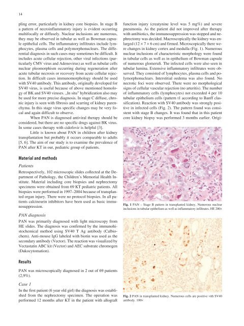
changes in kidney cortex and medulla (Fig. 1). Numerous<br />
nuclear inclusions of characteristic morphology were found<br />
in tubular cells as well as in epithelium of Bowman capsule<br />
of numerous glomeruli. The infected cells were also seen in<br />
tubular lumina. Extensive inflammatory infiltrates were observed.<br />
They consisted of lymphocytes, plasma cells and polymorphonuclears.<br />
Interstitial oedema was also found. No<br />
necrotic foci were observed. There were no morphological<br />
signs of cellular vascular rejection (no arteritis). The number<br />
of inflammatory cells (lymphocytes) not exceeded 4 per 10<br />
tubular epithelium cells (pattern t1 according to Banff classification).<br />
Reaction with SV40 antibody was strongly positive<br />
in infected cells (Fig. 2). The pattern found was consistent<br />
with stage B changes. It was found that in this patient<br />
core kidney biopsy was performed 3 months earlier. Origi-<br />
Material and methods<br />
Patients<br />
Retrospectively, 102 microscopic slides collected at the Department<br />
of Pathology, the Children’s Memorial Health Institute.<br />
Material including core biopsies and nephrectomy<br />
specimens were obtained from 69 KT pediatric patients. All<br />
biopsies were performed in 1997–2004 because of transplanted<br />
organ injury. There were no protocol biopsies. In all patients<br />
calcineurin inhibitors have been used as basic immunosuppression.<br />
PAN diagnosis<br />
PAN was primarily diagnosed with light microscopy from<br />
HE slides. The diagnosis was confirmed by the immunohistochemical<br />
method using SV40 T Ag antibody (Calbiochem).<br />
Anti-mouse IgG labeled with biotin was used as the<br />
secondary antibody (Vector). The reaction was visualized by<br />
Vectastatin ABC kit (Vector) and AEC substrate chromogen<br />
(Dakocytomation).<br />
Fig. 1 PAN – Stage B pattern in transplanted kidney. Numerous nuclear<br />
inclusions in tubular epithelium as well as inflammatory infiltrates. HE 200×<br />
Results<br />
PAN was microscopically diagnosed in 2 out of 69 patients<br />
(2,9%).<br />
Case 1<br />
In the first patient (6 year old girl) the diagnosis was established<br />
from the nephrectomy specimen. The operation was<br />
performed 12 months after KT in the patient with allograft<br />
Fig. 2 PAN in transplanted kidney. Numerous cells are positive vith SV40<br />
antibody. 100×