annals 1-2.qxd - Centrum Zdrowia Dziecka
annals 1-2.qxd - Centrum Zdrowia Dziecka
annals 1-2.qxd - Centrum Zdrowia Dziecka

You also want an ePaper? Increase the reach of your titles
YUMPU automatically turns print PDFs into web optimized ePapers that Google loves.
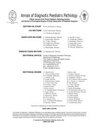
45<br />
A<br />
B<br />
Fig. 2<br />
A. Neuroblastoma. Specimen obtained by open tumor biopsy. Nests of small, primitive cells distributed between connective tissue are seen (hematoxylin and<br />
eosin, original magnification × 125).<br />
B. Histopathological examination of the specimen obtained by open tumor biopsy showed neuroblastoma: neuroblasts with hyperchromatic nuclei in smallfiber<br />
stroma consisted of unmyelinized neurons are seen (hematoxylin and eosin, original magnification × 225).<br />
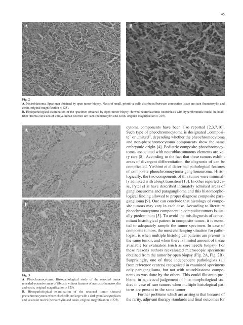
A<br />
B<br />
Fig. 3<br />
A. Pheochromocytoma. Histopathological study of the resected tumor<br />
revealed extensive areas of fibrosis without features of necrosis (hematoxylin<br />
and eosin, original magnification × 125).<br />
B. Histopathological examination of the resected tumor showed<br />
pheochromocytoma where chief cells are large with a dark granular cytoplasm<br />
and vesicular nuclei (hematoxylin and eosin, original magnification × 225).<br />
cytoma components have been also reported [2,3,7,10].<br />
Such type of pheochromocytoma is designated „composite”<br />
or „mixed”, depending whether the pheochromocytoma<br />
and non-pheochromocytoma components show the same<br />
embryonic origin [4]. Pediatric composite pheochromocytomas<br />
associated with neuroblastomatous elements are very<br />
rare [8]. According to the fact that these tumors exhibit<br />
areas of divergent differentiation, the diagnosis of can be<br />
complicated. Yoshimi et al described pathological features<br />
of composite pheochromocytoma-ganglioneuroma. Histologically,<br />
the two components of this tumor were minimally<br />
admixed with abrupt transition [13]. In other reported case,<br />
Pytel et al have described intimately admixed areas of<br />
ganglioneuroma and paraganglioma and this histomorphological<br />
finding allowed to proper diagnose composite paraganglioma<br />
[9]. One can conclude that histology of composite<br />
tumors may vary in each case. According to literature<br />
pheochromocytoma component in composite tumors is usually<br />
predominant [5]. To avoid the misdiagnosis of concomitant<br />
histological pattern in composite tumor, it is essential<br />
to adequately sample the tumor specimen. In case of<br />
composite tumors, the most challenging situation for pathologist,<br />
is when multiple histological patterns are present in<br />
the same tumor, and when there is limited amount of tissue<br />
available for evaluation (such as core needle biopsy). For<br />
these reasons authors reevaluated microscopic specimens<br />
obtained from the tumor by open biopsy (Fig. 2A, Fig. 2B).<br />
Surprisingly, one of three independent pathologists (all<br />
from reference centers) recognized in examined specimens<br />
only paraganglioma, but not with neuroblastoma components<br />
as was done by the others. This could illustrate problems<br />
in equivocal judgement of histomorphological studies<br />
in case of rare tumors when multiple histological patterns<br />
are present in the same tumor.<br />
Further problems which are arising is that because of<br />
the rarity, adjuvant therapy standards and final outcomes for