annals 1-2.qxd - Centrum Zdrowia Dziecka
annals 1-2.qxd - Centrum Zdrowia Dziecka
annals 1-2.qxd - Centrum Zdrowia Dziecka

Create successful ePaper yourself
Turn your PDF publications into a flip-book with our unique Google optimized e-Paper software.
10<br />
has also been reported in Turner syndrome [2, 50] and in<br />
Wiliams syndrome (8, 2%) [19].<br />
Based on retrospective studies the frequency of selective<br />
immunoglobulin (Ig) A deficiency in CD was between<br />
1,7% and 7,7% [6].<br />
There is strong evidence that first-degree relatives of<br />
diagnosed CD cases are at increased risk for CD with a prevalence<br />
of 4% to 5% [22]. The frequency of CD is lower in<br />
second-degree relatives.<br />
Diagnosis<br />
Although serological tests have been improved during last<br />
years, still biopsy of small intestine is necessary for CD recognition.<br />
According criteria established by European Society<br />
of Gastrology, Hepatology and Nutrition in 1990, CD diagnosis<br />
is based upon histological evidence of typical small<br />
intestinal mucosal abnormalities and clinical improvement<br />
after introduction of gluten free diet [62]. CD affects the mucosa<br />
of the proximal small intestine, with damage gradually<br />
decreasing in severity towards the distal small intestine. Because<br />
the histological changes in CD may be patchy, it is recommended<br />
that multiple biopsy specimens should be obtained<br />
from the jejunum and examined according modified<br />
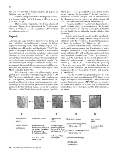
Marsh scale (Fig. 2) [38, 40].<br />
There is strong evidence that villous atrophy (Marsh<br />
grade III) is a characteristic histopathological feature of CD<br />
[22]. The presence of infiltrative changes with crypt hyperplasia<br />
(Marsh grade II) is compatible with CD but with less clear<br />
evidence. Diagnosis in these cases is dependent on positive<br />
serological tests. When serological tests are negative, other<br />
conditions for the intestinal changes should be considered.<br />
The presence of infiltrative intraepithelial lymphocytes alone<br />
(Marsh grade I) is not specific for CD. Concomitant positive<br />
serology increases the likehood of CD. In such cases it is recommended<br />
additional strategies, such as determination of<br />
the HLA antigens, repeat biopsy or a trial of treatment with<br />
a GFD and repeated measurement of antibodies [22].<br />
Thus, intestinal biopsy together with determination of<br />
specific antibodies in sera provides definitive CD diagnosis.<br />
As a GFD improved the intestinal lesions [64], it should be<br />
stressed that GF diet should not be introduced before planned<br />
biopsy.<br />
Serological tests are frequently used to identify individuals<br />
for whom the biopsy procedure. They are also useful<br />
for screening the population to determine the prevalence<br />
of CD and for monitoring of a GFD therapy [64].<br />
A comparison between several commercial available<br />
serological tests demonstrated that determination of anti-endomysial<br />
antibodies (EMA) by an indirect immunofluorescence<br />
technique (IIF) and anti-human recombinant tissue<br />
transglutaminase antibodies (hrtTG-Ab) by immunoenzymatic<br />
(ELISA) method are superior to anti-gliadin antibodies<br />
(AGA) and anti-guinea pig tissue transglutaminase antibodies<br />
(gTG-Ab) [63, 64]. The sensitivity and specificity<br />
of those tests range about 90%. Our studies showed that also<br />
anti-reticulin antibodies (ARA) determined by IIF are<br />
characterized by very high sensitivity (98%) and specificity<br />
(100%) [64].<br />
Thus, the autoantibodies produced against the same<br />
autoantigen, i.e. tissue transglutaminase [30], should be employed<br />
for diagnosis and screening of CD. Our observations<br />
confirm that in some patients EMA/ARA tests are negative,<br />
but hrtTG-Ab are present, so it seems that determination of<br />
autoantibodies by two tests performed by different methods<br />
are needed.<br />
Fig. 2 Histopathological lesions in celiac patients