Christoph Florian Schaller - FU Berlin, FB MI
Christoph Florian Schaller - FU Berlin, FB MI
Christoph Florian Schaller - FU Berlin, FB MI

You also want an ePaper? Increase the reach of your titles
YUMPU automatically turns print PDFs into web optimized ePapers that Google loves.
<strong>Christoph</strong> <strong>Schaller</strong> - STORMicroscopy 3<br />
1 Introduction<br />
The rst chapter illustrates the diculties of achieving nanometer resolution in microscopy and gives<br />
a brief abstract of the occuring phenomena. Finally the thesis is motivated and outlined.<br />
1.1 The diraction barrier<br />
In general the resolution of light microscopes is<br />
roughly limited by λ/2 ≈ 250 nm, which corresponds<br />
to half of the wavelength of the visible<br />
spectrum. This is caused by the fact that whenever<br />
we observe an object, we actually do not observe<br />
a point, but a distribution of photons. Usually<br />
the size of that distribution is negligible compared<br />
to the resolution. However when it comes<br />
to structures in the dimension of nanometers, we<br />
start to observe the so called Airy diraction pattern<br />
or Airy disk displayed in Figure 1.1. It is<br />
named after the English astronomer George Biddell<br />
Airy, who was the rst to theoretically treat<br />
this phenomenon in the year 1835 [2].<br />
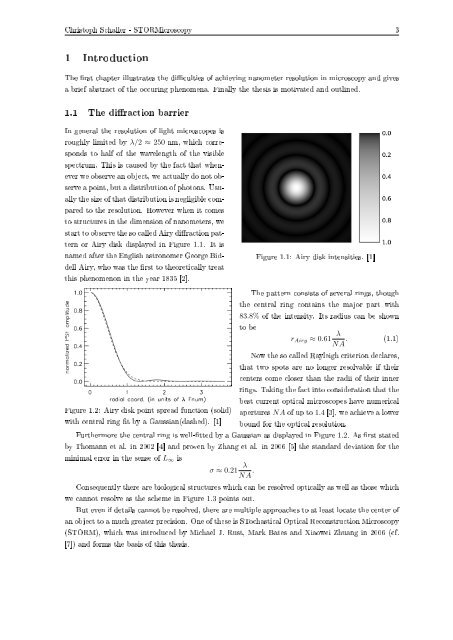
Figure 1.1: Airy disk intensities. [1]<br />
The pattern consists of several rings, though<br />
the central ring contains the major part with<br />
83.8% of the intensity. Its radius can be shown<br />
to be<br />
r Airy ≈ 0.61 λ<br />
NA . (1.1)<br />
Now the so called Rayleigh criterion declares,<br />
that two spots are no longer resolvable if their<br />
centers come closer than the radii of their inner<br />
rings. Taking the fact into consideration that the<br />
best current optical microscopes have numerical<br />
Figure 1.2: Airy disk point spread function (solid) apertures NA of up to 1.4 [3], we achieve a lower<br />
with central ring t by a Gaussian(dashed). [1] bound for the optical resolution.<br />
Furthermore the central ring is well-tted by a Gaussian as displayed in Figure 1.2. As rst stated<br />
by Thomann et al. in 2002 [4] and proven by Zhang et al. in 2006 [5] the standard deviation for the<br />
minimal error in the sense of L ∞ is<br />
σ ≈ 0.21 λ<br />
NA .<br />
Consequently there are biological structures which can be resolved optically as well as those which<br />
we cannot resolve as the scheme in Figure 1.3 points out.<br />
But even if details cannot be resolved, there are multiple approaches to at least locate the center of<br />
an object to a much greater precision. One of these is STochastical Optical Reconstruction Microscopy<br />
(STORM), which was introduced by Michael J. Rust, Mark Bates and Xiaowei Zhuang in 2006 (cf.<br />
[7]) and forms the basis of this thesis.