Christoph Florian Schaller - FU Berlin, FB MI
Christoph Florian Schaller - FU Berlin, FB MI
Christoph Florian Schaller - FU Berlin, FB MI

You also want an ePaper? Increase the reach of your titles
YUMPU automatically turns print PDFs into web optimized ePapers that Google loves.
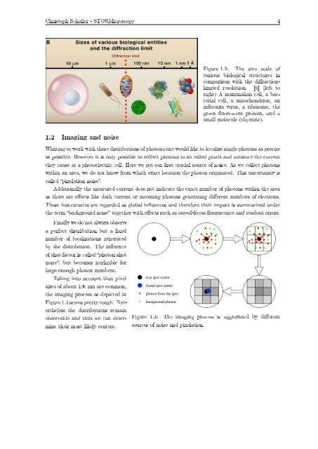
<strong>Christoph</strong> <strong>Schaller</strong> - STORMicroscopy 4<br />
Figure 1.3: The size scale of<br />
various biological structures in<br />
comparison with the diractionlimited<br />
resolution. [6] (left to<br />
right) A mammalian cell, a bacterial<br />
cell, a mitochondrion, an<br />
inuenza virus, a ribosome, the<br />
green uorescent protein, and a<br />
small molecule (thymine).<br />
1.2 Imaging and noise<br />
Wanting to work with these distributions of photons one would like to localize single photons as precise<br />
as possible. However it is only possible to collect photons in so called pixels and measure the current<br />
they cause at a photoelectric cell. Here we got our rst crucial source of noise: As we collect photons<br />
within an area, we do not know from which exact location the photon originated. This uncertainty is<br />
called pixelation noise.<br />
Additionally the measured current does not indicate the exact number of photons within the area<br />
as there are eects like dark current or incoming photons generating dierent numbers of electrons.<br />
Those inaccuracies are regarded as global inuences and therefore their impact is summarized under<br />
the term background noise together with eects such as out-of-focus uorescence and readout errors.<br />
Finally we do not always observe<br />
a perfect distribution but a xed<br />
number of localizations generated<br />
by the distribution. The inuence<br />
of that factor is called photon shot<br />
noise, but becomes negligible for<br />
large enough photon numbers.<br />
Taking into account that pixel<br />
sizes of about 100 nm are common,<br />
the imaging process as depicted in<br />
Figure 1.4 seems pretty rough. Nevertheless<br />
the distributions remain<br />
observable and thus we can determine<br />
their most likely centers.<br />
Figure 1.4: The imaging process is aggravated by dierent<br />
sources of noise and pixelation.