

Vol 41 # 3 September 2009 - Kma.org.kw
Vol 41 # 3 September 2009 - Kma.org.kw
Vol 41 # 3 September 2009 - Kma.org.kw

Create successful ePaper yourself
Turn your PDF publications into a flip-book with our unique Google optimized e-Paper software.
<strong>September</strong> <strong>2009</strong><br />
KUWAIT MEDICAL JOURNAL 231<br />
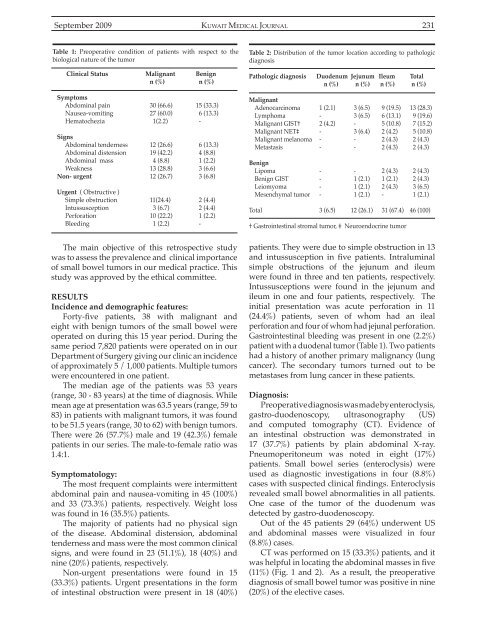
Table 1: Preoperative condition of patients with respect to the<br />
biological nature of the tumor<br />
Clinical Status Malignant Benign<br />
n (%) n (%)<br />
Symptoms<br />
Abdominal pain 30 (66.6) 15 (33.3)<br />
Nausea-vomiting 27 (60.0) 6 (13.3)<br />
Hematochezia 1(2.2) -<br />
Signs<br />
Abdominal tenderness 12 (26.6) 6 (13.3)<br />
Abdominal distension 19 (42.2) 4 (8.8)<br />
Abdominal mass 4 (8.8) 1 (2.2)<br />
Weakness 13 (28.8) 3 (6.6)<br />
Non- urgent 12 (26.7) 3 (6.8)<br />
Urgent ( Obstructive )<br />
Simple obstruction 11(24.4) 2 (4.4)<br />
Intussusception 3 (6.7) 2 (4.4)<br />
Perforation 10 (22.2) 1 (2.2)<br />
Bleeding 1 (2.2) -<br />
The main objective of this retrospective study<br />
was to assess the prevalence and clinical importance<br />
of small bowel tumors in our medical practice. This<br />
study was approved by the ethical committee.<br />
RESULTS<br />
Incidence and demographic features:<br />
Forty-five patients, 38 with malignant and<br />
eight with benign tumors of the small bowel were<br />
operated on during this 15 year period. During the<br />
same period 7,820 patients were operated on in our<br />
Department of Surgery giving our clinic an incidence<br />
of approximately 5 / 1,000 patients. Multiple tumors<br />
were encountered in one patient.<br />
The median age of the patients was 53 years<br />
(range, 30 - 83 years) at the time of diagnosis. While<br />
mean age at presentation was 63.5 years (range, 59 to<br />
83) in patients with malignant tumors, it was found<br />
to be 51.5 years (range, 30 to 62) with benign tumors.<br />
There were 26 (57.7%) male and 19 (42.3%) female<br />
patients in our series. The male-to-female ratio was<br />
1.4:1.<br />
Symptomatology:<br />
The most frequent complaints were intermittent<br />
abdominal pain and nausea-vomiting in 45 (100%)<br />
and 33 (73.3%) patients, respectively. Weight loss<br />
was found in 16 (35.5%) patients.<br />
The majority of patients had no physical sign<br />
of the disease. Abdominal distension, abdominal<br />
tenderness and mass were the most common clinical<br />
signs, and were found in 23 (51.1%), 18 (40%) and<br />
nine (20%) patients, respectively.<br />
Non-urgent presentations were found in 15<br />
(33.3%) patients. Urgent presentations in the form<br />
of intestinal obstruction were present in 18 (40%)<br />
Table 2: Distribution of the tumor location according to pathologic<br />
diagnosis<br />
Pathologic diagnosis Duodenum Jejunum Ileum Total<br />
n (%) n (%) n (%) n (%)<br />
Malignant<br />
Adenocarcinoma 1 (2.1) 3 (6.5) 9 (19.5) 13 (28.3)<br />
Lymphoma - 3 (6.5) 6 (13.1) 9 (19.6)<br />
Malignant GIST† 2 (4.2) - 5 (10.8) 7 (15.2)<br />
Malignant NET‡ - 3 (6.4) 2 (4.2) 5 (10.8)<br />
Malignant melanoma - - 2 (4.3) 2 (4.3)<br />
Metastasis - - 2 (4.3) 2 (4.3)<br />
Benign<br />
Lipoma - - 2 (4.3) 2 (4.3)<br />
Benign GIST - 1 (2.1) 1 (2.1) 2 (4.3)<br />
Leiomyoma - 1 (2.1) 2 (4.3) 3 (6.5)<br />
Mesenchymal tumor - 1 (2.1) - 1 (2.1)<br />
Total 3 (6.5) 12 (26.1) 31 (67.4) 46 (100)<br />
† Gastrointestinal stromal tumor, ‡ Neuroendocrine tumor<br />
patients. They were due to simple obstruction in 13<br />
and intussusception in five patients. Intraluminal<br />
simple obstructions of the jejunum and ileum<br />
were found in three and ten patients, respectively.<br />
Intussusceptions were found in the jejunum and<br />
ileum in one and four patients, respectively. The<br />
initial presentation was acute perforation in 11<br />
(24.4%) patients, seven of whom had an ileal<br />
perforation and four of whom had jejunal perforation.<br />
Gastrointestinal bleeding was present in one (2.2%)<br />
patient with a duodenal tumor (Table 1). Two patients<br />
had a history of another primary malignancy (lung<br />
cancer). The secondary tumors turned out to be<br />
metastases from lung cancer in these patients.<br />
Diagnosis:<br />
Preoperative diagnosis was made by enteroclysis,<br />
gastro-duodenoscopy, ultrasonography (US)<br />
and computed tomography (CT). Evidence of<br />
an intestinal obstruction was demonstrated in<br />
17 (37.7%) patients by plain abdominal X-ray.<br />
Pneumoperitoneum was noted in eight (17%)<br />
patients. Small bowel series (enteroclysis) were<br />
used as diagnostic investigations in four (8.8%)<br />
cases with suspected clinical findings. Enteroclysis<br />
revealed small bowel abnormalities in all patients.<br />
One case of the tumor of the duodenum was<br />
detected by gastro-duodenoscopy.<br />
Out of the 45 patients 29 (64%) underwent US<br />
and abdominal masses were visualized in four<br />
(8.8%) cases.<br />
CT was performed on 15 (33.3%) patients, and it<br />
was helpful in locating the abdominal masses in five<br />
(11%) (Fig. 1 and 2). As a result, the preoperative<br />
diagnosis of small bowel tumor was positive in nine<br />
(20%) of the elective cases.


