

Vol 41 # 3 September 2009 - Kma.org.kw
Vol 41 # 3 September 2009 - Kma.org.kw
Vol 41 # 3 September 2009 - Kma.org.kw

Create successful ePaper yourself
Turn your PDF publications into a flip-book with our unique Google optimized e-Paper software.
<strong>September</strong> <strong>2009</strong><br />
KUWAIT MEDICAL JOURNAL 251<br />
Fig. 1: CT brain, post-contrast showing cystic lesion in the right<br />
temporal lobe, just lateral to the right cerebral peduncle with<br />
adjacent enhancing nodules surrounded by vasogenic edema<br />
in the right hippocampus, paraventricular regions,<br />
frontal horns of lateral ventricles, left parietal lobe,<br />
and cerebellar hemispheres (Fig. 3). A thick nodular<br />
leptomeningeal enhancement at the right temporal<br />
lobe was also noted. These findings, together with<br />
the CXR were suggestive of neurosarcoidosis versus<br />
brain metastasis.<br />
A chest / abdomen CT scanning showed<br />
multiple enlarged pre-tracheal, para-tracheal,<br />
anterior mediastinal, aorto-pulmonary, sub-carinal,<br />
and hilar lymphadenopathy with few smaller ones<br />
in the anterior aorta adjacent to the celiac trunk.<br />
These nodes showed no evidence of caseation or<br />
calcification (Fig. 4).<br />
The differential diagnoses included: metastatic<br />
malignancy, lymphoma, leukemia, cysticercosis,<br />
tuberculosis, sarcoidosis, fungal or human immune<br />
deficiency virus infection (HIV). However, HIV,<br />
VDRL, and cystiocercosis serology, Mantoux test and<br />
blood film were all negative.At this stage, the seizure<br />
activity was well controlled by antiepileptic drugs.<br />
Therefore, video-assisted cervical mediastinoscopy<br />
was performed and multiple lymph node biopsies<br />
were obtained, the histopathological examination<br />
revealed extensive hyalinization with focally<br />
preserved lymphoid cells associated with multiple<br />
non-caseating compact granulomas (NCG) (Fig. 5).<br />
Stain for acid fast bacilli (AFB) and later the culture<br />
were negative.<br />
The patient was then extubated and became fully<br />
conscious. He was shifted to the general medical<br />
ward and commenced on oral corticosteroids (CS)<br />
in a dose of 1 mg / kg body weight (prednisolone<br />
80 mg daily). He showed marked improvement<br />
in his general condition and remained fit- free.<br />
Consequently, he was discharged home. Regular<br />
out-patient follow-up visits were arranged which<br />
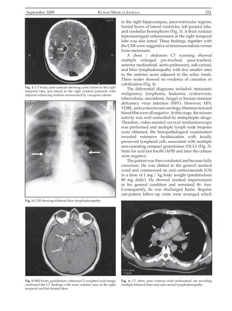
Fig. 2: CXR showing bilateral hilar lymphadenopathy<br />
Fig. 3: MRI brain, gadolinium- enhanced Tl weighted axial image<br />
confirmed the CT findings with more nodules seen at the right<br />
temporal and left frontal lobes<br />
Fig. 4: CT chest, post contrast axial mediastinal cut revealing<br />
multiple bilateral hilar and sub-carinal lymphadenopathy


