Paterson Institute for Cancer Research SCIENTIFIC REPORT 2005
Paterson Institute for Cancer Research SCIENTIFIC REPORT 2005
Paterson Institute for Cancer Research SCIENTIFIC REPORT 2005

Create successful ePaper yourself
Turn your PDF publications into a flip-book with our unique Google optimized e-Paper software.
RADIOCHEMICAL TARGETING AND IMAGING<br />
an improved toxicity profile, is currently being tested<br />
in phase I clinical studies. As part of this development<br />
we have designed, optimised and validated<br />
a clinical labelling strategy to label the drug with the<br />
positron emitting isotope 124 I. In the renal cancer<br />
patients treated so far, we have shown antigen-specific<br />
accumulation and retention of the labelled<br />
drug in their tumours see figure.<br />
Pharmacokinetics and pharmacodynamics of<br />
anti-angiogenic agents<br />
The aim of this project is to develop an 18 F-labelled<br />
anti-angiogenic agent with validated pharmacodynamic<br />
endpoints by comparing a biological<br />
response, related to the expression of a protein biomarker,<br />
with PET-derived intratumoral concentrations<br />
of the anti-angiogenic agent. As a biomarker<br />
we have synthesised an anti-integrin RGD-peptide<br />
which labelled with fluorine-18 using a novel fluorinating<br />
agent, [ 18 F]-Fluoroacetaldehyde. Unlike previously<br />
developed nucleophilic fluorinating agents,<br />
the advantage of this new compound in reductive<br />
N-alkylation reactions is that the conditions are<br />
mild, water and low temperature compatible, and<br />
can be used in protein radiolabelling.<br />
[ 18 F]Fluoroethyltosylate is produced, with a radioactive<br />
yield ranging from 75 to 90%, from ethylene dip-toluenesulfonate<br />
by fluorination with [ 18 F]FK. It<br />
is then oxidized to [ 18 F]Fluoroacetaldehyde which is<br />
distilled, radioactive yield 60-70%.<br />
[ 18 F]Fluoroacetaldehyde is thus produced in 30 minutes<br />
from [ 18 F]FH coming from the cyclotron target<br />
with an overall radioactive yield ranging from 45 to<br />
63%. This synthesis is easy to automate. Following<br />
the synthesis of the stable N-<br />
Fluoroethylbenzylamine derivative, the new compound<br />
has been characterised by mass spectroscopy<br />
and NMR and serves as a reference compound.<br />
The N-[ 18 F]Fluoroethylbenzylamine is analysed by<br />
HPLC under reverse phase conditions found to<br />
match the reference compound<br />
Radiotracer quantum dots<br />
We have initiated the development of quantum dot<br />
probes to target and image cellular pathways within<br />
tumours and their environment. We have achieved<br />
the synthesis of a new class of quantum dots,<br />
which we term “radiotracer quantum dots”. These<br />
are nanoparticles synthesized from tracer quantities<br />
of imaging and therapeutic radionuclides. We have<br />
demonstrated greater than 90% incorporation of<br />
radioactivity into dots, as well as retention of fluorescence,<br />
stability and other properties of stable<br />
dots. We have demonstrated no toxicity of CdSe<br />
dots at this tracer (nM) level. These multi-functional<br />
particles were localized to various tumour cell<br />
lines where 50-60% uptake was observed within 30<br />
minutes. The radiotracer dots were successfully<br />
injected into animals and the pattern of their localisation<br />
was studied. The non-conjugated dots were<br />
taken up by various tissues predominantly in accordance<br />
with blood flow. We are currently studying<br />
the pattern of accumulation of these nanoparticles<br />
in individual cells and organelles. Our analyses<br />
includes radioimaging, microautoradiography, optical<br />
detection and electron microscopy enabling<br />
there<strong>for</strong>e multi-modality detection and imaging.<br />
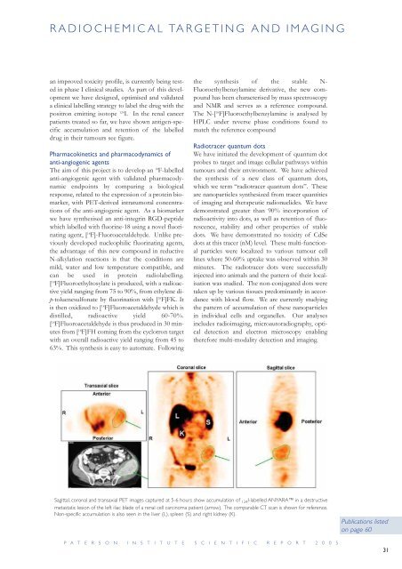
Sagittal, coronal and transaxial PET images captured at 5-6 hours show accumulation of 124I-labelled ANYARA in a destructive<br />
metastatic lesion of the left iliac blade of a renal cell carcinoma patient (arrow). The comparable CT scan is shown <strong>for</strong> reference.<br />
Non-specific accumulation is also seen in the liver (L), spleen (S) and right kidney (K).<br />
P A T E R S O N I N S T I T U T E S C I E N T I F I C R E P O R T 2 0 0 5<br />
Publications listed<br />
on page 60<br />
31