Paterson Institute for Cancer Research SCIENTIFIC REPORT 2005
Paterson Institute for Cancer Research SCIENTIFIC REPORT 2005
Paterson Institute for Cancer Research SCIENTIFIC REPORT 2005

You also want an ePaper? Increase the reach of your titles
YUMPU automatically turns print PDFs into web optimized ePapers that Google loves.
MEDICAL ONCOLOGY:<br />
PROTEOGLYCAN GROUP<br />
heparin ternary complexes with a 2:2:1 stoichiometry.<br />
We have recently extended our studies to<br />
FGF2, a potent stimulator of tumour angiogenesis.<br />
This revealed some similarities in the way that<br />
heparin promotes the activity FGF ligands. In common<br />
with FGF1, heparin dp6 was the shortest<br />
FGF2-binding saccharide, and dp8 the minimum<br />
needed <strong>for</strong> FGF2 dimerisation. Moreover, like<br />
FGF1, dimerisation appeared to be highly cooperative,<br />
because in the presence of excess saccharide<br />
a strong preference <strong>for</strong> the <strong>for</strong>mation of 2:1<br />
FGF2-heparin complexes was observed. Ternary<br />
complex <strong>for</strong>mation in the presence of FGF-receptor<br />
revealed a 2:2:1 FGF2-FGFR2c-dp12 assembly<br />
exactly as observed with FGF1. However<br />
FGF2 also gave rise to smaller “half ” complexes<br />
(1:1:1) that were not observed with FGF1 Previous<br />
studies showed that in contrast FGF1 heparin dp6dp12<br />
species failed to stimulate FGF2 mitogenesis<br />
through the FGFR2c receptor. Our binding data<br />
suggests that these saccharide species may <strong>for</strong>m<br />
some unproductive FGF2-FGFR combinations<br />
that may be more abundant on cell surfaces and<br />
compromise transmembrane FGFR signalling.<br />
Overall, these findings suggest that although FGF<br />
ligands engage heparin by a co-operative process<br />
different mechanisms of heparin-mediated <strong>for</strong>mation<br />
of signalling complexes may operate in the<br />
FGF family.<br />
Embryonic stem cells as a model system <strong>for</strong><br />
studying proteoglycan biology<br />
We have established the novel approach of using<br />
murine ES cells as potential in vitro model systems<br />
<strong>for</strong> studying HS during development. ES cells<br />
undergo self-renewal in culture whilst retaining the<br />
ability to differentiate into all cell lineages. Cell fate<br />
decisions are controlled by a complex and subtle<br />
interplay of secreted factors, cell-autonomous factors<br />
and cell-cell and cell-matrix interactions. Many<br />
of the signalling molecules are HS-dependent<br />
growth factors and morphogens. ES cells there<strong>for</strong>e<br />
present a tractable experimental system <strong>for</strong> assessing<br />
the role of HS in signalling processes in differentiation.<br />
This enables us to study the relationships<br />
between developmentally-regulated expression of<br />
HS-polymerase and sulphotransferase enzymes, and<br />
the structural and functional attributes of the<br />
resulting HS. This has become a hotly debated<br />
issue as evidence has emerged of the deleterious<br />
effects of enzyme mutations on both embryogenesis<br />
and HS function in the adult.<br />
Structural analysis of radiolabelled HS from mES<br />
cells grown in the presence of serum and LIF, to<br />
maintain pluripotency, demonstrated a very low<br />
level of sulphation (reflecting weak sulphotransferase<br />
activities), compared to HS from other<br />
mouse tissues. Using GFP reporter lines (i.e. Sox-<br />
1, a specific marker of neuroectodermal precursors,<br />
and brachyury, an early marker of mesoderm, in<br />
combination with Flk1 to identify haemangioblast<br />
specification) we were able to isolate discrete populations.<br />
Uniquely, in this study, we could directly<br />
compare HS structure with expression of a wide<br />
panel of relevant genes <strong>for</strong> FACS sorted populations.<br />
Biochemical analyses, and the use of ScFv<br />
antibodies (from Prof. Toin van Kuppevelt,<br />
Nijmegen) that recognise distinct HS epitopes,<br />
uncovered significant differences in HS structure<br />
between mES cells and their differentiated progeny.<br />
Neural differentiation is dependent on an initial<br />
response to FGF4 and, at a later stage, to other<br />
FGF family members. In contrast, initial mesodermal<br />
differentiation is critically dependent on FGF5,<br />
Wnt 3a/8a and BMP2/4, with VEGF and FGF-2<br />
becoming more significant later. We there<strong>for</strong>e suggest<br />
that the observed changes in HS sulphation<br />
pattern and distribution may regulate responses to<br />
these HS-dependent exogenous factors and thus<br />
play key roles in determining cell fate.<br />
P A T E R S O N I N S T I T U T E S C I E N T I F I C R E P O R T 2 0 0 5<br />
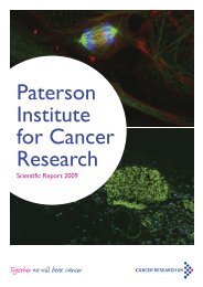
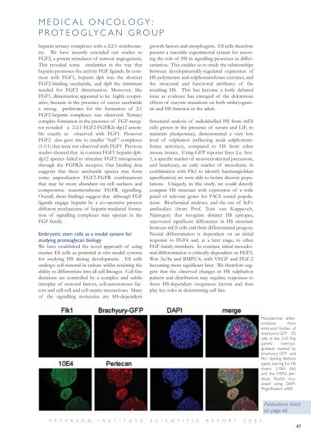
Mesodermal differentiation<br />
from<br />
embryoid bodies of<br />
brachyury-GFP ES<br />
cells at day 3.25. Top<br />
panels: haemangioblasts<br />
marked by<br />
brachyury-GFP and<br />
Flk1 staining. Bottom<br />
panel: staining <strong>for</strong> HS<br />
chains (10E4 Ab)<br />
and the HSPG perlecan.<br />
Nuclei visualised<br />
using DAPI.<br />
Magnification: x400<br />
Publications listed<br />
on page 66<br />
47