Paterson Institute for Cancer Research SCIENTIFIC REPORT 2005
Paterson Institute for Cancer Research SCIENTIFIC REPORT 2005
Paterson Institute for Cancer Research SCIENTIFIC REPORT 2005

Create successful ePaper yourself
Turn your PDF publications into a flip-book with our unique Google optimized e-Paper software.
50<br />
HEAD OF RESEARCH SERVICES<br />
Jenny Varley<br />
<strong>Research</strong> Services<br />
http://www.paterson.man.ac.uk/facilities/scifacs.jsp<br />
The Scientific <strong>Research</strong> Services continue to underpin all the<br />
research activities within the <strong>Paterson</strong> <strong>Institute</strong> as well as<br />
providing one CR-UK wide service (the Affymetrix<br />
Microarray service). It is essential that the services provide<br />
comprehensive state-of-the-art facilities which dovetail with<br />
the requirements of the users, and to this end each service<br />
has an associated user group which meets at regular intervals.<br />
These user groups carry significant weight, and can<br />
advise on the introduction of new services or cessation of<br />
redundant ones as well as equipment needs. We have continued<br />
to invest to ensure that all our services are optimally<br />
supported.<br />
____________________________________<br />
Advanced Imaging Facility<br />
Head: Steve Bagley<br />
http://www.paterson.man.ac.uk/facilities/advimg.jsp<br />
Since 1985 Laser Scanning Confocal Microscopy<br />
has facilitated the digitisation of data in three<br />
dimensions. This has been achieved by the use of a<br />
high intensity monochromatic light source (generally<br />
a laser) and an imaging aperture placed just<br />
be<strong>for</strong>e the detector leading to the imaging of those<br />
structures that were in-focus and excluding data<br />
that were above or below the plane of focus. The<br />
system was originally developed to assist imaging at<br />
depths inside embryos that normal microscopy<br />
could not resolve because of interference from<br />
in<strong>for</strong>mation above and below the plane of focus,<br />
however, the main advance <strong>for</strong> the broader biological<br />
research community was that 3D imaging<br />
became a routine process available to most laboratories.<br />
Over the last ten years there have been<br />
major advances in optical design, detection sensitivity<br />
and labelling technology which have allowed live<br />
cell imaging via widefield optics to become a viable<br />
possibility.<br />
Widefield imaging requires less light to <strong>for</strong>m an<br />
image hence it is less damaging to cellular processes.<br />
In the last four years the Advanced Imaging<br />
DEPUTY<br />
Caroline Chadwick<br />
Facility has been investigating, designing and developing<br />
techniques <strong>for</strong> the visualisation of live cellular<br />
processes on the micro- and macro-scale where<br />
the very act of describing real world, four dimensional<br />
objects in a digital manner disrupts biological<br />
processes as little as possible. This has led to a<br />
move away from laser scanning systems to less<br />
harmful and more sensitive techniques utilising<br />
standard optics and image processing algorithms.<br />
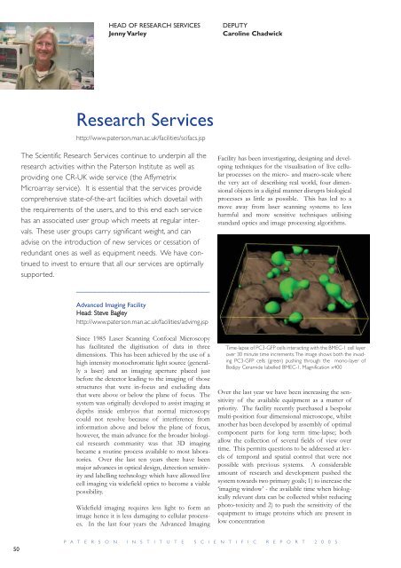
Time-lapse of PC3-GFP cells interacting with the BMEC-1 cell layer<br />
over 30 minute time increments.The image shows both the invading<br />
PC3-GFP cells (green) pushing through the mono-layer of<br />
Bodipy Ceramide labelled BMEC-1. Magnification x400<br />
Over the last year we have been increasing the sensitivity<br />
of the available equipment as a matter of<br />
priority. The facility recently purchased a bespoke<br />
multi-position four dimensional microscope, whilst<br />
another has been developed by assembly of optimal<br />
component parts <strong>for</strong> long term time-lapse; both<br />
allow the collection of several fields of view over<br />
time. This permits questions to be addressed at levels<br />
of temporal and spatial control that were not<br />
possible with previous systems. A considerable<br />
amount of research and development pushed the<br />
system towards two primary goals; 1) to increase the<br />
‘imaging window’ - the available time when biologically<br />
relevant data can be collected whilst reducing<br />
photo-toxicity and 2) to push the sensitivity of the<br />
equipment to image proteins which are present in<br />
low concentration<br />
P A T E R S O N I N S T I T U T E S C I E N T I F I C R E P O R T 2 0 0 5