2005 SAGES Abstracts
2005 SAGES Abstracts
2005 SAGES Abstracts

Create successful ePaper yourself
Turn your PDF publications into a flip-book with our unique Google optimized e-Paper software.
POSTER ABSTRACTS<br />
In the past we focused on robotic systems development, with<br />
which we were the first to experiment internationally. We continued<br />
research towards the development of a simple mechanical<br />
suturing device, enabling angulation and rotation of the<br />
tip.<br />
The instrument was developed by the company Tuebingen<br />
Scientific in Tuebingen, Germany. The handle system is<br />
designed ergonomically and it is hold by the whole hand.<br />
Angulating the handle means flection of the tip, stretching the<br />
handle means to put the tip in a straight position. Rotation of a<br />
knob at the tip of the handle allows rotation of the tip of the<br />
instrument.<br />
Following experimental evaluation which was highly successful,<br />
we started clinical application. Today we have performed<br />
the suturing of meshes on 10 patients in inguinal hernia. The<br />
technique is based on continuing suture of the mesh to the<br />
inguinal ligament. Also the fixation to the anterior abdominal<br />
wall together with the peritoneum is performed by the use of<br />
the suturing device.<br />
In 3 patients mesh was sutured to the anterior abdominal wall<br />
in patients with abdominal wall hernias. In 2 patients the<br />
mesocolon was closed following right colonic resection.<br />
Conclusions<br />
The new suturing device permits in an easy and ergonomic<br />
way sutures at the front of the tip of the instrument and<br />
sutures at the anterior abdominal wall. The principles of suturing<br />
can be compared to the robotic system DaVinci. Compared<br />
to this, the handling of the RADIUS surgical system is much<br />
more easy, does not need any time for installation of the technology<br />
and the price of the system is much less, compared to<br />
robotic systems.<br />
We are convinced that mechanical manipulators, as the<br />
RADIUS surgical system, will allow better and more precise<br />
manual suturing, compared to conventional straight instruments.<br />
P375–New Techniques<br />
SIS MESH FOR LAPAROSCOPIC INGUINAL HERNIA REPAIR- 5<br />
YEAR FOLLOW UP, David S Edelman MD, Laparoscopic<br />
Surgery Center, Baptist Hospital, Miami, Florida<br />
Intro: Synthetic mesh is routinely used for inguinal hernia<br />
repair. Porcine small intestine submucosa (SIS) mesh has been<br />
successfully tested and used in animal models with excellent<br />
results. This mesh is degradable, resorbable and had significant<br />
fibroblastic ingrowth equal to polypropylene mesh.<br />
Methods: Beginning August, 1999 a prospective study was<br />
begun using SIS mesh and laparoscopy in a pre-peritoneal<br />
approach to repair per-primum hernias. A 7x10 cm mesh was<br />
placed, uncut, over the myopectinate orifice and secured with<br />
5 tacks. Patients have were followed at 2 weeks, 6 weeks, 6<br />
months and yearly.<br />
Results: The surgeon has an experience of over 800 laparoscopic<br />
inguinal hernia operations. There were 50 patients having<br />
61 hernias studied. There were 16 direct, 42 indirect, 2 pantaloon<br />
and 1 femoral hernia repaired. Operative time averaged<br />
32 minutes. There were no major complications. Nine (9)<br />
patients developed seromas, 12 had pain lasting over 7 days<br />
requiring medication, 4 had swelling/orchitis and 5 patients<br />
(10%) developed a recurrent hernia.<br />
Conclusions: The recurrences were technical complications<br />
due to the small mesh size. It is unclear if the pain, seroma<br />
and swelling is a host versus graft reaction to the mesh which<br />
led to the hernia recurrences. The subgroup of 10 Sport?s<br />
Hernia patients did not have the same problems. However, it is<br />
concluded that at 5 years, SIS mesh can be used for inguinal<br />
hernia repairs and further technical modifications along with a<br />
prospective- randomized trial comparing SIS to other mesh is<br />
necessary.<br />
P376–New Techniques<br />
REPAIR OF A COMPLEX FOREGUT HERNIA AIDED BY NOVEL<br />
THREE-DIMENSIONAL SURGICAL RECONSTRUCTION,<br />
Stephen M Kavic MD, Ross D Segan MD,Patricia L Turner<br />
MD,Ivan M George,Adrian E Park MD, University of Maryland,<br />
Baltimore<br />
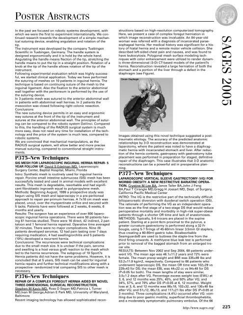
Recent imaging technology has allowed sophisticated reconstructions<br />
based on high-resolution computerized tomography.<br />
Here, we present a case of complex foregut herniation in<br />
which image reconstruction was invaluable. An 84-year-old<br />
woman was referred with a diagnosis of incarcerated paraesophageal<br />
hernia. Her medical history was significant for a history<br />
of hiatal hernia and a remote motor vehicle collision. She<br />
described left-sided chest pain and nausea, and was found to<br />
have leukocytosis. Polygonal mesh surface modeling techniques<br />
with color enhancement were utilized to render dynamic<br />
three-dimensional (3-D) CT-based models of the patient?s<br />
hernia. Reconstruction revealed a large herniation of both the<br />
stomach and a portion of the liver through a defect in the<br />
diaphragm (see Figure).<br />
Images obtained using this novel technique suggested a posttraumatic<br />
etiology. The accuracy of the predicted anatomic<br />
relationships by 3-D reconstruction was demonstrated at<br />
laparotomy, where the patient was noted to have a diaphragmatic<br />
hernia with incarcerated stomach and liver. After reduction<br />
of the hernia contents, gastropexy with gastrostomy tube<br />
placement was performed in preparation for staged, definitive<br />
repair of the diaphragm. This case illustrates that 3-D anatomic<br />
reconstructions can be a powerful aid in preoperative planning.<br />
P377–New Techniques<br />
LAPAROSCOPIC VERTICAL SLEEVE GASTRECTOMY (VG) FOR<br />
MORBID OBESITY: A NEW RESTRICTIVE BARIATRIC OPERA-<br />
TION, Crystine M Lee BA, Janos Taller BA,John J Feng<br />
BA,Paul T Cirangle MD,Gregg H Jossart MD, Dept. of Surgery,<br />
California Pacific Medical Center<br />
INTRO: The VG is the restrictive part of the technically difficult<br />
biliopancreatic diversion with duodenal switch operation (DS).<br />
The rationale of performing the VG as an independent operation<br />
was as the first stage of a two-stage DS that would reduce<br />
perioperative mortality and morbidity in high-risk super-obese<br />
patients through a shorter OR time and lack of anastomoses.<br />
METHODS: Typically, 5-6 trocars are placed in the supine<br />
patient. Starting at a point 6cm proximal to the pylorus, a<br />
greater curvature gastrectomy is performed along a 32 Fr<br />
bougie, using 5-7 firings of 45-60mm linear 3.5mm GI staplers,<br />
thus creating a 60-80ml gastric tube. Bioabsorbable<br />
Seamguards® are used to buttress the staple-line from the<br />
third firing onwards. A methlyene blue leak test is performed<br />
prior to removal of the bagged stomach from an enlarged trocar<br />
site.<br />
RESULTS: Between Nov 2002 and Sep 2004, 68 patients underwent<br />
VG. The mean age was 46.1±11.2 years and 72% were<br />
female. The mean preop weight and BMI was 335±89 lbs and<br />
53.2±11.9 kg/m2, respectively. Compared to 66 patients who<br />
underwent laparoscopic DS, the mean OR time was 102±29 vs<br />
229±43 for DS; the mean EBL was 44±22 cc vs 94±48 for DS<br />
(P