letters© 2010 Nature America, Inc. All rights reserved.abα-CD31 antibodyhVPr-GFP EBsd14 (−SB)10 510 410 3hESC derivedSorted hVPr-GFP+d14 (+SB)Sorted hVPr-GFP+d24 (+SB)CD31 +id1 low CD31 +10 2 10 2 10 3 10 4 10 5ld1 promoter: YFPUmbilical cord derivedEndothelium(HUVEC)Smooth musclecellsCord bloodCD34 + cells0.0 1.0GenesCXCR4EphrinB2EphB4Lyve-1Podoplaninc ld1:YFP dExpression0.5 2 3 4 5: Belowdetection thresholdProx-1VEGFR1 (Flt1)VEGFR2VEGFR3 (Flt4)CD34CD133 (Prominin1)E-SelectinP-SelectinPECAM (CD31)ThrombomodulinVE-cadherinvonWillebrand factorHOXA9ld1ld2ld3α-Smooth muscle actinld1:YFPefld1:YFP MFI (k)ld1 low (−SB)302418126IIIICD31 + ld1 low−SBI+SBd14 id1:YFP FACS3 Days (+SB)ld1 low (+SB)IVld1:YFP MFIII4 Daysld1 high (−SB)CD31 + ld1 high−SBVCD31 MFIII+SB5k 5k 11.2k 21.7k 18.3k 53.8kIII IV V VITotal cellsld1 high (+SB)VI352821147CD31 MFI (k)Figure 3 Molecular profiling of hESC-derived endothelial cells reveals a signature defined by high Id1 expression. Human VPr-GFP embryoid bodies andhighly purified hVPr-GFP + cells were compared to mature vascular cells by microarray analysis. (a) RNA was extracted for microarray analysis from humanVPr-GFP embryoid bodies cultured in the presence of recombinant cytokines alone until day 14; isolated endothelial cells (99.8% pure) from hVPr-GFPembryoid bodies cultured in the presence of recombinant cytokines and the TGFβ inhibitor SB431542 until day 14; isolated endothelial cells (>95%pure) from hVPr-GFP embryoid bodies cultured in the presence of recombinant cytokines and the TGFβ inhibitor SB431542 until day 14, followed by 10 dadditional culture in the presence of cytokines and SB431542; HUVECs; human umbilical vein smooth muscle cells; and CD34 + umbilical cord blood cells.Expressed factors are displayed in an ordered array, as labeled, with highly expressed factors shown in red, minimally expressed factors shown in blue andfactors for which transcripts are below a significant expression level shown in gray. (b–f) Following the endothelial cell differentiation protocol (Fig. 1d), Id1-YFP hESC-derived cells were sorted by FACS, separating the CD31 + population into Id1-YFP high -expressing cells (b (green) and c) and Id1-YFP low -expressingcells (b (red) and d). Insets, brightfield views on the day after isolation of Id1-YFP high (c) and Id1-YFP low (d) cells. (e) After 3 d culture in the presence ofSB431542, both populations were transferred to conditions with and without SB431542 for an additional 4 d (+SB and –SB, respectively). (f) Total cellsand mean fluorescence intensity (MFI) measurements of Id1:YFP (black) and CD31 + (white) were measured for: CD31 + Id1 low (I) and CD31 + Id1 high (II)populations upon isolation; and for four populations following culture conditions (as shown in e, III–VI). Scale bars, 100 µM.receptor decoys was used interchangeably to inhibit activation of theactivin/nodal branch of TGFβ superfamily signaling (SupplementaryFig. 5a–e). These results demonstrate that the effect of TGFβ inhibitionshown for the RUES1 line is applicable to other pluripotent cell lines.To define the vasculogenic transcriptional signature of hESC-derivedendothelial cells at different time points during phases 1 and 2, we carriedout Affymetrix microarray analyses of several hESC-derived populationsand mature cell types (Fig. 3a). The yield of freshly isolated phase 1endothelial cells in the absence of TGFβ inhibition was insufficient formicroarray analyses, underscoring the value of our approach for generatingsufficient expanding (phase 1) and vascular-committed (phase 2)endothelial cells for molecular profiling.Phase 1 hESC-derived endothelial cells showed increased levels of factorstypical of arterial-like endothelial cells (VEGFR2, VEGFR1, Id1, CD31,CD34, VE-cadherin, vWF, thrombomodulin, ephrinB2 and E-selectin) butnot of lymphatic endothelial cells (Prox1 and podoplanin). Markers ofvascular progenitor cells, including CD133 and Id1 (refs. 12–17), were alsohighly expressed in phase 1 endothelial cells and downregulated upon in vitroculture. Transcription factors expressed primarily in committed endothelialcells, including HoxA9 (ref. 18), were not expressed in phase 1 endothelialcells. Accordingly, we defined a comprehensive vasculogenic expressionprofile of the hESC-derived endothelial cell population as VE-cadherin+ VEGFR2 high Id1 high thrombomodulin high ephrinB2 + CD133 + HoxA9 − ,whereas mature endothelial cells were identified by a VE-cadherin + VEGFR2 low Id1 low ephrinB2 + CD133 − HoxA9 + phenotype.Id1 was one of numerous transcription factors upregulated in phase 1endothelial cells. Because it has been shown to modulate differentiationand maintenance of vascular cell fate 19 , we focused on Id1 as a potentialmediator of the pro-angiogenic effect of TGFβ-inhibition observed in ourstudy. To track Id1 expression in live hESC differentiation cultures, we used164 volume 28 number 2 february 2010 nature biotechnology
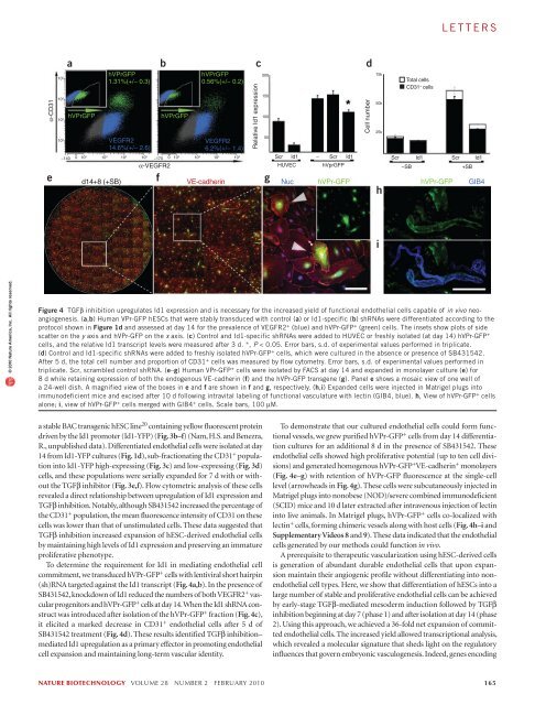
lettersα-CD31e10 510 410 3ahVPrGFPhVPrGFP1.31%(+/− 0.3)VEGFR214.6%(+/− 2.6)fbα-VEGFR2hVPrGFPhVPrGFP0.56%(+/− 0.2)VEGFR26.2%(+/− 1.4)10 2 −140 0 10 2 10 3 10 4 10 5 −170 0 10 2 10 3 10 4 10 5Relative Id1 expressionc20015010050gScrId1ScrId1d75k50k25khScrTotal cellsCD31 + cellsHUVEC hVprGFP −SB +SBd14+8 (+SB) VE-cadherin Nuc hVPr-GFP hVPr-GFP GIB4−Cell numberId1ScrId1i© 2010 Nature America, Inc. All rights reserved.Figure 4 TGFβ inhibition upregulates Id1 expression and is necessary for the increased yield of functional endothelial cells capable of in vivo neoangiogenesis.(a,b) Human VPr-GFP hESCs that were stably transduced with control (a) or Id1-specific (b) shRNAs were differentiated according to theprotocol shown in Figure 1d and assessed at day 14 for the prevalence of VEGFR2 + (blue) and hVPr-GFP + (green) cells. The insets show plots of sidescatter on the y axis and hVPr-GFP on the x axis. (c) Control and Id1-specific shRNAs were added to HUVEC or freshly isolated (at day 14) hVPr-GFP +cells, and the relative Id1 transcript levels were measured after 3 d. *, P < 0.05. Error bars, s.d. of experimental values performed in triplicate.(d) Control and Id1-specific shRNAs were added to freshly isolated hVPr-GFP + cells, which were cultured in the absence or presence of SB431542.After 5 d, the total cell number and proportion of CD31 + cells was measured by flow cytometry. Error bars, s.d. of experimental values performed intriplicate. Scr, scrambled control shRNA. (e–g) Human VPr-GFP + cells were isolated by FACS at day 14 and expanded in monolayer culture (e) for8 d while retaining expression of both the endogenous VE-cadherin (f) and the hVPr-GFP transgene (g). Panel e shows a mosaic view of one well ofa 24-well dish. A magnified view of the boxes in e and f are shown in f and g, respectively. (h,i) Expanded cells were injected in Matrigel plugs intoimmunodeficient mice and excised after 10 d following intravital labeling of functional vasculature with lectin (GIB4, blue). h, View of hVPr-GFP + cellsalone; i, view of hVPr-GFP + cells merged with GIB4 + cells. Scale bars, 100 µM.a stable BAC transgenic hESC line 20 containing yellow fluorescent proteindriven by the Id1 promoter (Id1-YFP) (Fig. 3b–f) (Nam, H.S. and Benezra,R., unpublished data). Differentiated endothelial cells were isolated at day14 from Id1-YFP cultures (Fig. 1d), sub-fractionating the CD31 + populationinto Id1-YFP high-expressing (Fig. 3c) and low-expressing (Fig. 3d)cells, and these populations were serially expanded for 7 d with or withoutthe TGFβ inhibitor (Fig. 3e,f). Flow cytometric analysis of these cellsrevealed a direct relationship between upregulation of Id1 expression andTGFβ inhibition. Notably, although SB431542 increased the percentage ofthe CD31 + population, the mean fluorescence intensity of CD31 on thesecells was lower than that of unstimulated cells. These data suggested thatTGFβ inhibition increased expansion of hESC-derived endothelial cellsby maintaining high levels of Id1 expression and preserving an immatureproliferative phenotype.To determine the requirement for Id1 in mediating endothelial cellcommitment, we transduced hVPr-GFP + cells with lentiviral short hairpin(sh)RNA targeted against the Id1 transcript (Fig. 4a,b). In the presence ofSB431542, knockdown of Id1 reduced the numbers of both VEGFR2 + vascularprogenitors and hVPr-GFP + cells at day 14. When the Id1 shRNA constructwas introduced after isolation of the hVPr-GFP + fraction (Fig. 4c),it elicited a marked decrease in CD31 + endothelial cells after 5 d ofSB431542 treatment (Fig. 4d). These results identified TGFβ inhibition–mediated Id1 upregulation as a primary effector in promoting endothelialcell expansion and maintaining long-term vascular identity.To demonstrate that our cultured endothelial cells could form functionalvessels, we grew purified hVPr-GFP + cells from day 14 differentiationcultures for an additional 8 d in the presence of SB431542. Theseendothelial cells showed high proliferative potential (up to ten cell divisions)and generated homogenous hVPr-GFP + VE-cadherin + monolayers(Fig. 4e–g) with retention of hVPr-GFP fluorescence at the single-celllevel (arrowheads in Fig. 4g). These cells were subcutaneously injected inMatrigel plugs into nonobese (NOD)/severe combined immunodeficient(SCID) mice and 10 d later extracted after intravenous injection of lectininto live animals. In Matrigel plugs, hVPr-GFP + cells co-localized withlectin + cells, forming chimeric vessels along with host cells (Fig. 4h–i andSupplementary Videos 8 and 9). These data indicated that the endothelialcells generated by our methods could function in vivo.A prerequisite to therapeutic vascularization using hESC-derived cellsis generation of abundant durable endothelial cells that upon expansionmaintain their angiogenic profile without differentiating into nonendothelialcell types. Here, we show that differentiation of hESCs into alarge number of stable and proliferative endothelial cells can be achievedby early-stage TGFβ-mediated mesoderm induction followed by TGFβinhibition beginning at day 7 (phase 1) and after isolation at day 14 (phase2). Using this approach, we achieved a 36-fold net expansion of committedendothelial cells. The increased yield allowed transcriptional analysis,which revealed a molecular signature that sheds light on the regulatoryinfluences that govern embryonic vasculogenesis. Indeed, genes encodingnature biotechnology volume 28 number 2 february 2010 165
- Page 3 and 4:
volume 28 number 2 february 2010COM
- Page 5 and 6:
in this issue© 2010 Nature America
- Page 7 and 8:
© 2010 Nature America, Inc. All ri
- Page 10 and 11:
NEWS© 2010 Nature America, Inc. Al
- Page 12 and 13:
NEWS© 2010 Nature America, Inc. Al
- Page 14 and 15:
NEWS© 2010 Nature America, Inc. Al
- Page 16 and 17:
© 2010 Nature America, Inc. All ri
- Page 18 and 19:
© 2010 Nature America, Inc. All ri
- Page 20 and 21: © 2010 Nature America, Inc. All ri
- Page 22 and 23: NEWS feature© 2010 Nature America,
- Page 24 and 25: uilding a businessComing to termsDa
- Page 26 and 27: uilding a business© 2010 Nature Am
- Page 28 and 29: correspondence© 2010 Nature Americ
- Page 30 and 31: correspondence© 2010 Nature Americ
- Page 32 and 33: correspondence© 2010 Nature Americ
- Page 34 and 35: correspondence© 2010 Nature Americ
- Page 36 and 37: case studyNever againcommentaryChri
- Page 38 and 39: COMMENTARY© 2010 Nature America, I
- Page 40 and 41: COMMENTARY© 2010 Nature America, I
- Page 42 and 43: patents© 2010 Nature America, Inc.
- Page 44 and 45: patents© 2010 Nature America, Inc.
- Page 46 and 47: news and viewsChIPs and regulatory
- Page 48 and 49: news and viewsFrom genomics to crop
- Page 50 and 51: news and views© 2010 Nature Americ
- Page 52 and 53: news and views© 2010 Nature Americ
- Page 54 and 55: e s o u r c eRational association o
- Page 56 and 57: e s o u r c e© 2010 Nature America
- Page 58 and 59: e s o u r c e© 2010 Nature America
- Page 60 and 61: e s o u r c e© 2010 Nature America
- Page 62 and 63: © 2010 Nature America, Inc. All ri
- Page 64 and 65: B r i e f c o m m u n i c at i o n
- Page 66 and 67: i e f c o m m u n i c at i o n sAUT
- Page 68 and 69: lettersa1.5 kb hVPrIntron 112.5 kbA
- Page 72 and 73: letters© 2010 Nature America, Inc.
- Page 74 and 75: l e t t e r sReal-time imaging of h
- Page 76 and 77: l e t t e r sFigure 2 Time-lapse li
- Page 78 and 79: l e t t e r s© 2010 Nature America
- Page 80 and 81: l e t t e r sRational design of cat
- Page 82 and 83: l e t t e r s© 2010 Nature America
- Page 84 and 85: l e t t e r s© 2010 Nature America
- Page 86 and 87: sample fluorescence was measured as
- Page 88 and 89: careers and recruitmentFourth quart


