
Create successful ePaper yourself
Turn your PDF publications into a flip-book with our unique Google optimized e-Paper software.
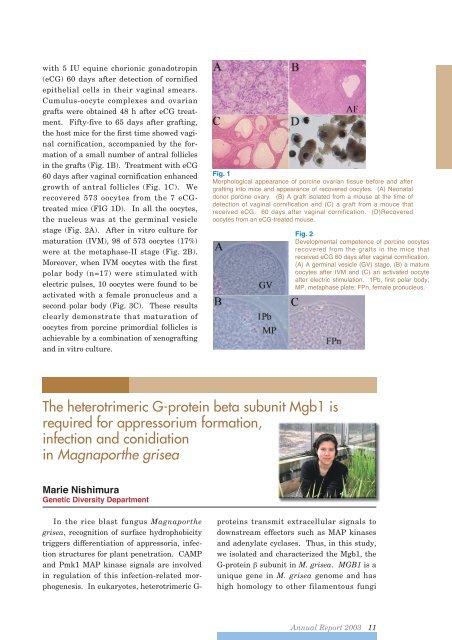
with 5 IU equine chorionic gonadotropin(eCG) 60 days after detection of cornifiedepithelial cells in their vaginal smears.Cumulus-oocyte complexes and ovariangrafts were obtained 48 h after eCG treatment.Fifty-five to 65 days after grafting,the host mice for the first time showed vaginalcornification, accompanied by the formationof a small number of antral folliclesin the grafts (Fig. 1B). Treatment with eCG60 days after vaginal cornification enhancedgrowth of antral follicles (Fig. 1C). Werecovered 573 oocytes from the 7 eCGtreatedmice (FIG 1D). In all the oocytes,the nucleus was at the germinal vesiclestage (Fig. 2A). After in vitro culture formaturation (IVM), 98 of 573 oocytes (17%)were at the metaphase-II stage (Fig. 2B).Moreover, when IVM oocytes with the firstpolar body (n=17) were stimulated withelectric pulses, 10 oocytes were found to beactivated with a female pronucleus and asecond polar body (Fig. 3C). These resultsclearly demonstrate that maturation ofoocytes from porcine primordial follicles isachievable by a combination of xenograftingand in vitro culture.Fig. 1Morphological appearance of porcine ovarian tissue before and aftergrafting into mice and appearance of recovered oocytes. (A) Neonataldonor porcine ovary. (B) A graft isolated from a mouse at the time ofdetection of vaginal cornification and (C) a graft from a mouce thatreceived eCG. 60 days after vaginal cornification. (D)Recoveredoocytes from an eCG-treated mouse.Fig. 2Developmental competence of porcine oocytesrecovered from the grafts in the mice thatreceived eCG 60 days after vaginal cornification.(A) A germinal vesicle (GV) stage, (B) a matureoocytes after IVM and (C) an activated oocyteafter electric stimulation. 1Pb, first polar body;MP, metaphase plate; FPn, female pronucleus.The heterotrimeric G-protein beta subunit Mgb1 isrequired for appressorium formation,infection and conidiationin Magnaporthe griseaMarie NishimuraGenetic Diversity DepartmentIn the rice blast fungus Magnaporthegrisea, recognition of surface hydrophobicitytriggers differentiation of appressoria, infectionstructures for plant penetration. CAMPand Pmk1 MAP kinase signals are involvedin regulation of this infection-related morphogenesis.In eukaryotes, heterotrimeric G-proteins transmit extracellular signals todownstream effectors such as MAP kinasesand adenylate cyclases. Thus, in this study,we isolated and characterized the Mgb1, theG-protein subunit in M. grisea. MGB1 is aunique gene in M. grisea genome and hashigh homology to other filamentous fungi<strong>Annual</strong> <strong>Report</strong> <strong>2003</strong> 11