
You also want an ePaper? Increase the reach of your titles
YUMPU automatically turns print PDFs into web optimized ePapers that Google loves.
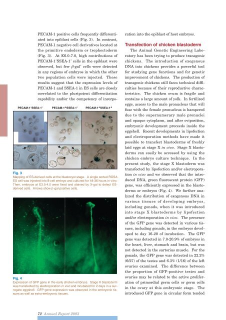
Fig. 3Mapping of ES-derived cells at the blastocyst stage. A single sorted ROSAES cell was injected into 8-cell embryo and cultured for 18-36 hours in vitro.Then, embryos at E3.5-4.0 were fixed and stained by X-gal to detect ESderivedcells. Arrows show -gal positive cells.Fig. 4Expression of GFP gene in the early chicken embryos. Stage X blastodermwas transfected by electroporation in vivo and incubated for 2 days in a surrogateeggshell. GFP gene expression was observed in the embryonic tissuesas well as extra-embryonic tissues.PECAM-1 positive cells frequently differentiatedinto epiblast cells (Fig. 3). In contrast,PECAM-1 negative cell derivatives located atthe primitive endoderm or trophectoderm(Fig. 3). At E6.0-7.0, high contributions ofPECAM-1 + SSEA-1 + cells in the epiblast wereobserved, but few -gal + cells were detectedin any regions of embryos in which the othertwo population cells were injected. Theseresults suggest that the expression levels ofPECAM-1 and SSEA-1 in ES cells are closelycorrelated to the pluripotent differentiationcapability and/or the competency of incorporationinto the epiblast of host embryos.Transfection of chicken blastodermThe Animal Genetic Engineering Laboratoryhas been trying to produce transgenicchickens. The introduction of exogenousDNA into chickens provides a powerful toolfor studying gene functions and for geneticimprovement of chickens. The production oftransgenic chickens still faces technical difficultiesbecause of their reproductive characteristics.The chicken ovum is fragile andcontains a large amount of yolk. In fertilizedeggs, access to the male pronucleus that willfuse with the female pronucleus is hampereddue to the supernumerary male pronucleiand opaque cytoplasm, and after oviposition,embryonic development proceeds inside theeggshell. Recent developments in lipofectionand electroporation methods have made itpossible to transfect blastoderms of freshlylaid eggs at stage X in vivo. Stage X blastodermscan easily be accessed by using thechicken embryo culture technique. In thepresent study, the stage X blastoderm wastransfected by lipofection and/or electroporationin vivo and we observed that the introducedDNA, green fluorescent protein (GFP)gene, was efficiently expressed in the blastodermsor embryos (Fig. 4). We further analyzedthe distribution of exogenous DNA invarious tissues of developing embryos,including gonads, when it was introducedinto stage X blastoderms by lipofectionand/or electroporation in vivo. The presenceof the GFP gene was detected in various tissues,including gonads, in the embryos developedto day 16-20 of incubation. The GFPgene was detected in 7.0-20.9% of embryos inthe heart, liver, stomach and brain, but wasnot detected in the sartorius muscle. For thegonads, the GFP gene was detected in 22.2%(6/27) of the testes and 6.3% (1/16) of the leftovaries examined. The difference betweenthe proportion of GFP-positive testes andovaries may be related to the active proliferationof primordial germ cells or germ cellsin the ovary at this embryonic stage. Theintroduced GFP gene in circular form tended72 <strong>Annual</strong> <strong>Report</strong> <strong>2003</strong>