

Volume 8 Issue 3 - Australasian Society for Ultrasound in Medicine
Volume 8 Issue 3 - Australasian Society for Ultrasound in Medicine
Volume 8 Issue 3 - Australasian Society for Ultrasound in Medicine

You also want an ePaper? Increase the reach of your titles
YUMPU automatically turns print PDFs into web optimized ePapers that Google loves.
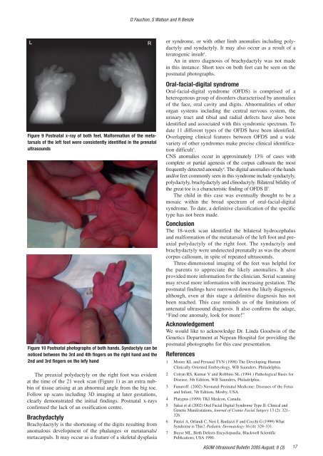
D Fauchon, S Watson and R Benzieor syndrome, or with other limb anomalies <strong>in</strong>clud<strong>in</strong>g polydactylyand syndactyly. It may also occur as a result of ateratogenic <strong>in</strong>sult 4 .An <strong>in</strong> utero diagnosis of brachydactyly was not made<strong>in</strong> this <strong>in</strong>stance. Short toes on both feet can be seen on thepostnatal photographs.Figure 9 Postnatal x-ray of both feet. Mal<strong>for</strong>mation of the metatarsalsof the left foot were consistently identified <strong>in</strong> the prenatalultrasoundsFigure 10 Postnatal photographs of both hands. Syndactyly can benoticed between the 3rd and 4th f<strong>in</strong>gers on the right hand and the2nd and 3rd f<strong>in</strong>gers on the lefy handThe preaxial polydactyly on the right foot was evidentat the time of the 21 week scan (Figure 1) as an extra nubb<strong>in</strong>of tissue aris<strong>in</strong>g at an abnormal angle from the big toe.Follow up scans <strong>in</strong>clud<strong>in</strong>g 3D imag<strong>in</strong>g at later gestations,clearly demonstrated the <strong>in</strong>itial f<strong>in</strong>d<strong>in</strong>gs. Postnatal x-raysconfirmed the lack of an ossification centre.BrachydactylyBrachydactyly is the shorten<strong>in</strong>g of the digits result<strong>in</strong>g fromanomalous development of the phalanges or metatarsals/metacarpals. It may occur as a feature of a skeletal dysplasiaOral-facial-digital syndromeOral-facial-digital syndrome (OFDS) is comprised of aheterogenous group of disorders characterised by anomaliesof the face, oral cavity and digits. Abnormalities of otherorgan systems <strong>in</strong>clud<strong>in</strong>g the central nervous system, theur<strong>in</strong>ary tract and tibial and radial defects have also beenidentified and associated with this syndromic spectrum. Todate 11 different types of the OFDS have been identified.Overlapp<strong>in</strong>g cl<strong>in</strong>ical features between OFDS and a widevariety of other syndromes make precise cl<strong>in</strong>ical identificationdifficult 5 .CNS anomalies occur <strong>in</strong> approximately 13% of cases withcomplete or partial agenesis of the corpus callosum the mostfrequently detected anomaly 6 . The digital anomalies of the handsand/or feet commonly seen <strong>in</strong> this syndrome <strong>in</strong>clude syndactyly,polydactyly, brachydactyly and cl<strong>in</strong>odactyly. Bilateral bifidity ofthe great toe is a characteristic f<strong>in</strong>d<strong>in</strong>g of OFDS II 7 .The child <strong>in</strong> this case was eventually thought to be amosaic with<strong>in</strong> the broad spectrum of oral-facial-digitalsyndrome. To date, a def<strong>in</strong>itive classification of the specifictype has not been made.ConclusionThe 18-week scan identified the bilateral hydrocephalusand mal<strong>for</strong>mation of the metatarsals of the left foot and preaxialpolydactyly of the right foot. The syndactyly andbrachydactyly were undetected prenatally as was the absentcorpus callosum, <strong>in</strong> spite of repeated ultrasounds.Three-dimensional imag<strong>in</strong>g of the feet was helpful <strong>for</strong>the parents to appreciate the likely anomalies. It alsoprovided more <strong>in</strong><strong>for</strong>mation <strong>for</strong> the cl<strong>in</strong>ician. Serial scann<strong>in</strong>gmay reveal more <strong>in</strong><strong>for</strong>mation with <strong>in</strong>creas<strong>in</strong>g gestation. Thepostnatal f<strong>in</strong>d<strong>in</strong>gs have narrowed down the likely diagnosis,although, even at this stage a def<strong>in</strong>itive diagnosis has notbeen reached. This case rem<strong>in</strong>ds us of the limitations ofantenatal ultrasound diagnosis. It also confirms the adage,“F<strong>in</strong>d one anomaly, look <strong>for</strong> more!”AcknowledgementWe would like to acknowledge Dr. L<strong>in</strong>da Goodw<strong>in</strong> of theGenetics Department at Nepean Hospital <strong>for</strong> provid<strong>in</strong>g thepostnatal photographs <strong>for</strong> this case presentation.References1 Moore KL and Persaud TVN (1998) The Develop<strong>in</strong>g HumanCl<strong>in</strong>ically Oriented Embryology, WB Saunders, Philadelphia.2 Cotran RS, Kumar V and Robb<strong>in</strong>s SL (1994 ) Pathological Basis <strong>for</strong>Disease, 5th Edition, WB Saunders, Philadelphia.3 Fanaroff: (2002) Neonatal-Per<strong>in</strong>atal Medic<strong>in</strong>e: Diseases of the Fetusand Infant, 7th Edition, Mosby, USA.4 Platypus (1999) TKI Medcon, Canada.5 Sakai et al (2002) Oral Facial Digital Syndrome Type II: Cl<strong>in</strong>ical andGenetic Manifestations, Journal of Cranio Facial Surgery 13 (2): 321–326.6 Patrizi A, Orlandi C, Neri I, Bardazzi F and Cocchi G (1999) WhatSyndrome is This?, Pediatric Dermatology 16 (4): 329–331.7 Buyse ML, Birth Defects Encyclopaedia, Blackwell ScientificPublications, USA 1990.ASUM <strong>Ultrasound</strong> Bullet<strong>in</strong> 2005 August; 8 (3)17


