

Volume 8 Issue 3 - Australasian Society for Ultrasound in Medicine
Volume 8 Issue 3 - Australasian Society for Ultrasound in Medicine
Volume 8 Issue 3 - Australasian Society for Ultrasound in Medicine

Create successful ePaper yourself
Turn your PDF publications into a flip-book with our unique Google optimized e-Paper software.
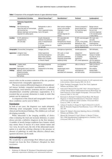
Fetal upper abdom<strong>in</strong>al masses; a prenatal diagnostic dilemmaTable 1 Comparison of the sonographic features of upper abdom<strong>in</strong>al massesIntraabdom<strong>in</strong>al Extralobar Adrenal Haemorrhage 2–4 Neuroblastoma 3–5 Teratoma 6 Lymphangioma 7Pulmonary Sequestration(IEPS) 1Pathology Non-function<strong>in</strong>g lung Pathogenesis <strong>in</strong> utero is Most common malignant Tumours composed of Benign tumourstissue that does not communicate unknown. tumour <strong>in</strong> the neonate. tissues derived from of the lymphatic vesselwith bronchial tree. Total or segmental <strong>in</strong>volve- Orig<strong>in</strong>at<strong>in</strong>g <strong>in</strong> neural the 3 germ<strong>in</strong>al layers that are mostBetween diaphragm and liver/kidney, ment of the gland. crest cells. of the embryo. commonly diagnosedseparate from adrenal gland. Prenatal diagnosis 32 <strong>in</strong> the neonatal periodweeks+.or early <strong>in</strong>fancy.Location 8–10% abdomen or Usually <strong>in</strong>tracapsular. 50% associated with 60–65% sacrococcygeal 95% head, neckretroperitoneum. Extension <strong>in</strong>to peritoneal Adrenal gland. region. and axilla.90% left sided cavity or retroperitoneum Retroperitoneal, thoracic 10–20% <strong>in</strong> the gonads. 63% of abdom<strong>in</strong>alif ruptures. or cervical paravertebral Mediast<strong>in</strong>um, naso- lymphangiomas.3–4 x more common on regions. pharynx head and neck, occur on the left side.the right side.and the retroperitoneum.Sonographic Circumscribed, homogenous Changes over time. Homogenous hyperechoic, Variable appearance Well del<strong>in</strong>eated,appearance echogenic mass, Initially hyperechoic 50% cystic, or mixed rang<strong>in</strong>g from hypoechoic cystic± cystic components. becom<strong>in</strong>g heterogenous echo pattern. completely anechoic masses of variablebe<strong>for</strong>e f<strong>in</strong>ally anechoic. Occasional calcification to completely size. May be uni orMaybe round, triangular Frequently encapsulated hyperechoic. multiloculatedor crescent shaped. displac<strong>in</strong>g kidney. 60% mixed appearance. with f<strong>in</strong>e septations.May conta<strong>in</strong> debris.Vascularity ± feeder vessel Rim shaped peripheral Low impedance Little or no flow on Little or no flow onfrom aorta. vascularity. wave<strong>for</strong>m. colour Doppler.Associated Diaphragmatic hernia, bronchial Displacement of the kidney Hepatomegaly and/or Polyhydramnios fetal Sk<strong>in</strong> oedema,f<strong>in</strong>d<strong>in</strong>gs agenesis, colonic duplication and caudally. fetal hydrops hydronephrosis hydrops fetalisvertebral anomalies. associated with metastases. and fetal hydrops. and polyhydramnios.Oligo/polyhydramnios.fetal hydrops.masses relies on the accurate evaluation of the size, positionand characteristic sonographic appearances.The most common differential diagnosis of <strong>in</strong>traabdom<strong>in</strong>almasses <strong>in</strong>cludes congenital neuroblastoma or adrenalhaemorrhage, renal tumours, teratomas and less commonly<strong>in</strong>traabdom<strong>in</strong>al extralobar pulmonary sequestration. It isessential that an accurate dist<strong>in</strong>ction is made <strong>in</strong> order thatpatient care be optimised.A comparison of the cl<strong>in</strong>ical and sonographic features ofthese conditions can be seen <strong>in</strong> Table 1.ConclusionIn both of our cases, the diagnosis was made ultimatelyfollow<strong>in</strong>g serial sonographic review correlated with thecl<strong>in</strong>ical and biochemical f<strong>in</strong>d<strong>in</strong>gs. The children are well anddevelop<strong>in</strong>g normally.While ultrasound is the imag<strong>in</strong>g modality of choicewhen evaluat<strong>in</strong>g the fetal and neonatal abdomen, it cannotalways provide a def<strong>in</strong>itive diagnosis <strong>in</strong> the first <strong>in</strong>stance.A number of mostly benign conditions have features whichmay mimic those of masses of a more s<strong>in</strong>ister nature andvice versa. In such cases, the role of the ultrasound exam<strong>in</strong>ationis to alert the referr<strong>in</strong>g cl<strong>in</strong>ician to the presence orabsence of pathology <strong>in</strong> order that effective cl<strong>in</strong>ical managementdecisions can be made.AcknowledgementsWe would like to acknowledge Prof Albert Lam and MrJoseph Lui from the New Children’s Hospital <strong>for</strong> theirassistance.References1 Pumberger W, Moroder W, Wiesbauer P. Intraabdom<strong>in</strong>al extralobarpulmonary sequestration exhibit<strong>in</strong>g cystic adenomatoid mal<strong>for</strong>ma-tion: prenatal diagnosis and characterization of a left suprarenal mass<strong>in</strong> the newborn. Abdom Imag<strong>in</strong>g 2001; 26: 28–31.2 Seigel. Paediatric Sonography 3rd ed. New York: Lipp<strong>in</strong>cottWilliams and Wilk<strong>in</strong>s; 2002.3 Schwarzler P, Bernard JP, Senat MV, Ville Y. Prenatal Diagnosisof Fetal Adrenal Masses: Differentiation between Hemorrhage andSolid Tumour by Colour Doppler Sonography. <strong>Ultrasound</strong> ObstetGynaecol 1999; 13: 351–355.4 Kesrouani A, Duchatel F, Seilanian M and Muray JM. PrenatalDiagnosis of Adrenal Neuroblastoma by <strong>Ultrasound</strong>: A Reportof Two Cases and Review of the Literature. <strong>Ultrasound</strong> ObstetGynaecol. 1999; 13: 446–449.5 Acharya S, Jayabose S, Kogan SJ, Tugal O, Beneck D, Leslie D,Slim M. Prenatally Diagnosed Neuroblastoma. Cancer 1997; 80 (2):304–309.6 Dahnert W. Dahnert’s Electronic Radiology Review [CD-ROM],Williams and Wilk<strong>in</strong>s, 1998.7 Mentzel HJ, Schramm D, Vogt S, Reuter A, Mentzel T, KaiserWA, (1998) Intraabdom<strong>in</strong>al Lymphangioma <strong>in</strong> a Newborn. J Cl<strong>in</strong><strong>Ultrasound</strong> 1998; 26 (6): 320–322.ASUM <strong>Ultrasound</strong> Bullet<strong>in</strong> 2005 August; 8 (3)21


